
Microstructure fiber biochip and manufacturing method thereof

A microstructure optical fiber and biochip technology, which is applied in biochemical equipment and methods, enzymology/microbiology devices, and determination/inspection of microorganisms, etc. effect and other problems, to achieve the effect of low cost of detection instruments, realization of stimulated emission amplification, and improvement of fluorescence conversion efficiency

Active Publication Date: 2015-09-23
NANKAI UNIV
View PDF7 Cites 5 Cited by
  • Summary
  • Abstract
  • Description
  • Claims
  • Application Information

AI Technical Summary

Problems solved by technology

However, the diameter of such microbeads is usually very large, which has a strong scattering effect on the excitation light, which brings serious background noise to the final optical detection.
More importantly, this microbead-based probe immobilization technology cannot place the probe within 100 nanometers of the fiber core ring wall to form effective optical isolation, let alone produce microcavity effects to form stimulated radiation amplification, and the detection sensitivity is difficult. improve

Method used


Image

  • Microstructure fiber biochip and manufacturing method thereof
  • Microstructure fiber biochip and manufacturing method thereof
  • Microstructure fiber biochip and manufacturing method thereof

Examples


Embodiment 1

[0025] HLA-DRB Oligonucleotide Microstructure Optical Fiber Biochip and Its Preparation

[0026] 1) Preparation of optical fiber

[0027] Take a section of microstructured optical fiber, and use a selective filling method to block the peripheral air holes, leaving only the central air hole unblocked.

[0028] 2) Preparation of nanomagnetic beads

[0029] Take 1 mg of carboxylated magnetic beads with a diameter of 15 nanometers, wash 3 times, adjust the pH value to the isoelectric point, and fully disperse the nano magnetic beads.

[0030] 3) Coupling nano magnetic beads with probes

[0031] The designed and synthesized HLA-DRB probes are fully mixed and reacted with nano-magnetic beads, and the unreacted reagents are eluted.

[0032] 4) Fabrication of Microstructure Optical Fiber Biochip

[0033] The reacted nano-magnetic bead-molecular probe solution is added to the gravity perfusion device 12, and flows into the core hole of the microstructured optical fiber 15 through t...

Embodiment 2

[0039] Microstructure fiber optic biochip for HbsAg detection

[0040] In the implementation step 3 of the above coupling of nano-magnetic beads and probes, the sandwich-like magnetic immunoassay method is used, with the help of magnetic particles as solid-phase carriers, and through the combination of streptavidin and biotin, quantum Points are markers. There is a special force between streptavidin and biotin, the binding force between them is extremely strong, the binding speed is fast and the binding efficiency is high. Firstly, the magnetic beads are coupled with streptavidin, the anti-HbsAg is coupled with biotin, and the magnetic beads and anti-HbsAg are immobilized together through the combination of streptavidin and biotin. In step 5 of the above sample pretreatment, the test sample is marked with quantum dots, and then the presence of the test substance in the sample is detected by antigen-antibody reaction.


PUM

PropertyMeasurementUnit
diameteraaaaaaaaaa

Abstract

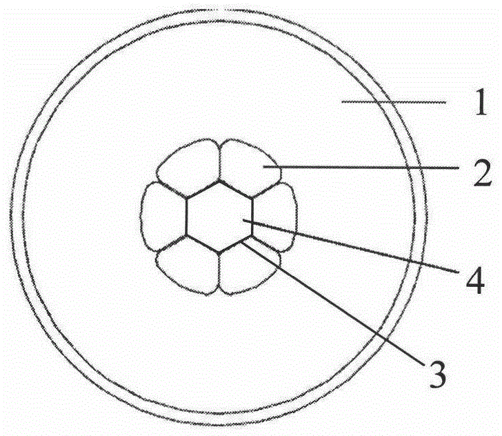
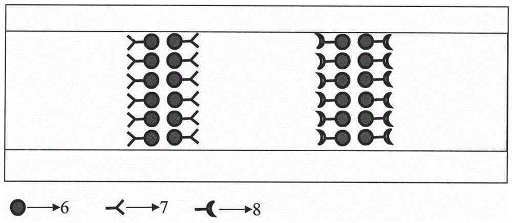
The invention relates to a one-dimensional biochip, that biomolecular probes are immobilized on the inner surface of fiber core annular wall of a hollow-core microstructure fiber by utilizing nano magnetic beads in a segmented mode, and a manufacturing method thereof, used for detecting protein, nucleic acid and like biological molecules. The microstructure fiber biochip is composed of a hollow-core microstructure fiber, nano magnetic beads and a probe. The hollow-core microstructure fiber comprises a central air hole and at least one circle of peripheral air holes; the annular wall thickness of the central air hole is on nano scale; the diameter of the nano magnetic bead is less than 100nm; the biomolecular probe can comprises DNA, RNA, antigen or antibody and the like. The manufacturing method comprises steps of 1) preprocessing the nano magnetic beads and the fiber; 2) coupling the nano magnetic beads to the molecular probe; 3) sequentially immobilizing the nano magnetic beads on the fiber core annular wall of the microstructure fiber in a segmented mode by virtue of a magnetic field and a fluid; 4) modifying, cleaning and curing; and 5) storing. The biochip disclosed by the invention has the main advantages that the biochip can be used for detecting with stimulated radiation fluorescence and can greatly improve the sensitivity of detection.

Description

Technical field [0001] The invention relates to the field of biochips, in particular to microstructure optical fiber biochips. technical background [0002] Biochip technology is a new technology developed in the field of life science in recent years, and the emergence of biochip has aroused widespread international attention. Common biochips include gene chips, protein chips, lab-on-a-chip, cell chips, tissue chips, sugar chips and other types of biochips. Biochip technology is a biochemical analysis system that combines microprocessing technology, microelectronics technology and molecular biology to build a microarray structure on the surface of a solid medium. By "hybridizing" the unknown target to be tested with the known probe on the chip , to determine unknown molecules through spatial resolution. Compared with traditional analysis techniques, biochip technology has obvious advantages. Thousands of densely arranged molecular probe arrays are integrated on the biochi...

Claims


Application Information

Patent Timeline
Patent Type & Authority Applications(China)
IPC IPC(8): G01N33/543C12M1/34
CPCC12Q1/6837G01N33/54326G01N33/54346C12Q2563/143C12Q2563/155C12Q2565/501
Inventor 周文远晏敏李志莉刘艳格田建国
Owner NANKAI UNIV
Learn More