Stem cell preparation for treating retinosis and use method of stem cell preparation

A technology of stem cell preparation and retinal degeneration, which is applied in the fields of regenerative medicine and stem cell biology, can solve the problems of stem cells losing their microenvironment, achieve the effects of reducing immunity, accurate drug delivery, and inhibiting the release of inflammatory factors

Active Publication Date: 2015-12-02
奥思达干细胞有限公司
View PDF2 Cites 2 Cited by
  • Summary
  • Abstract
  • Description
  • Claims
  • Application Information

AI Technical Summary

Problems solved by technology

At present, although the transplantation method of directly injecting the cell suspension into the subretinal space is simple and feasible, the stem cells lose the microenvironment that maintains their survival at the same time.

Method used


Image

  • Stem cell preparation for treating retinosis and use method of stem cell preparation

Examples


Embodiment 1

[0041] Example 1 Preparation of Stem Cell Preparation for Treating Retinal Degeneration

[0042] A stem cell preparation for treating retinal degeneration, specifically comprising the following preparation steps:

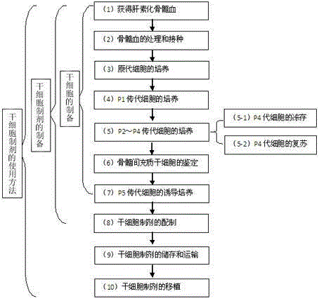
[0043] (1) Obtain heparinized bone marrow blood;

[0044] (2) Treatment and inoculation of bone marrow blood: add LG-DMEM culture medium containing 10% FBS at a volume ratio of 1:2, mix well, and inoculate directly into the culture bottle;

[0045] (3) Cultivation of primary cells: Place the above culture flask in 5% CO 2 , 37 ℃ incubator for culture, 48h for the first time without cleaning the direct change of medium, after that, change the medium every 2 to 3 days, and wash with buffer solution PBS before changing the medium, all change the medium in full; the cells grow to 80% to 90% After fusion, the primary cells were harvested by digestion with 0.25% trypsin, which were recorded as P0 generation cells;

[0046] (4) Culture of P1 generation cells: the above ...

Embodiment 2

[0054] Example 2 Identification of Retinal Neural-like Precursor Cells by Flow Cytometry

[0055] 1. The identification steps are as follows:

[0056] a) Collect the P4 generation cells before induction and differentiation in step 4 of the above-mentioned embodiment 1 and the P5 generation cells after induction in step 7 respectively, place them in a centrifuge tube, centrifuge at 1000 rpm for 5 min, and discard the supernatant;

[0057] b) Add 6ml of PBS to resuspend the cells, divide into 6 flow tubes, centrifuge at 1000rpm for 5min, and discard the supernatant;

[0058] c) Add 4% paraformaldehyde to fix at room temperature for 30min, centrifuge at 1000rpm for 5min, and discard the supernatant;

[0059] d) Add PBS to resuspend the cells, shake well, centrifuge at 1000rpm for 5min, and discard the supernatant;

[0060] e) Add 10% goat serum blocking solution dropwise, block at room temperature for 30min, centrifuge at 1000rpm for 5min, and discard the supernatant;

[0061]...

Embodiment 3

[0072] Embodiment 3 animal model experiments

[0073] 1. Establishment of choroidal neovascularization (CNV) animal model

[0074] Krypton red laser was used to photocoagulate the experimental retina of mice. The laser power, photocoagulation spot diameter and exposure time were respectively 300mW, 50μm and 0.060s to induce CNV animal model.

[0075] 2. Cell preparation transplantation test

[0076] 1) Take the stem cell preparation prepared in Example 1, and the cell concentration is 5×10 9 pcs / ml, put the prepared stem cell preparation in EP tubes, and put them in a refrigerated and insulated box at 2-15°C for use. During the experiment, the cells will precipitate after standing still, and pay attention to mixing before use;

[0077] 2) Take one experimental rat and weigh it;

[0078] 3) Anesthetized by intraperitoneal injection of 3% pentobarbital sodium 30mg / kg;

[0079] 4) 0.5% dicaine was instilled as a corneal surface anesthetic, and compound tropicamide was instilled...


PUM


Abstract

The invention provides a stem cell and a preparation for treating retinosis. Through optimum culture and induced differentiation on mesenchymal stem cells, the efficiency for differentiating the cell into the retina nerve precursor cell is improved; the similar effect to that of the retina precursor cell is achieved in the aspect of the transplantation effect, so that the retina function in the transplantation region is effectively improved; an ideal cell source is provided for the treatment of retinosis diseases through stem cell transplantation; high superiority is realized. A preparation method of the stem cell preparation provided by the invention has the advantages that the preparation process of the stem cell product obtained through separation, culture, posterity pass-down and induction is simple and reliable; the repeatability is good. A use method of the stem cell preparation for treating retinosis provided by the invention belongs to a cell replacing method with the advantages that the operation is simple, the popularization is convenient, and the injury is small. Wide application prospects are realized. The method belongs to the best path for treating the retinosis type degenerative diseases and rescuing the vision.

Description

technical field [0001] The invention relates to the fields of regenerative medicine and stem cell biotechnology, and relates to a stem cell preparation for treating retinal degeneration and a use method thereof. Background technique [0002] The thickness of the human retina is about 0.1-0.5 mm. Histologically, it is divided into 10 layers, of which there are mainly 4 layers of cells, which are pigment epithelial cell layer, visual cell layer, bipolar cell layer and ganglion from outside to inside. cell layer. The retinal pigment epithelium is closely connected with the choroid and is composed of pigment epithelial cells (retinalpigmentepitheium, RPE). The normal RPE is a single layer of cells with polarity, located in the outermost layer of the retina. It is a highly metabolically active layer of cells that supports and nourishes photoreceptor cells, shades, dissipates heat, and regenerates and repairs. Its main functions include transshipment of nutrients, recycling of r...

Claims


Application Information

Patent Timeline
Patent Type & Authority Applications(China)
IPC IPC(8): C12N5/0775C12N5/0797A61K35/30A61P27/02
Inventor 周萱王云娟
Owner 奥思达干细胞有限公司
Why Eureka
Try Eureka