
Magnetic particulate chemiluminescent micro-fluidic chip for quantitatively detecting myohemoglobin

A microfluidic chip and myoglobin technology, which is applied in the field of clinical medical testing, can solve problems such as the difficulty in realizing micro-sample detection, insufficient mixing of the reaction system, and the complexity of the microfluidic system, so as to reduce non-specific interference and cost Low, the effect of improving the efficiency of immune response

Active Publication Date: 2015-12-30
SHENZHEN HUAMAIXINGWEI MEDICAL TECH CO LTD
View PDF11 Cites 15 Cited by
  • Summary
  • Abstract
  • Description
  • Claims
  • Application Information

AI Technical Summary

Problems solved by technology

The microfluidic chip system has the characteristics of fast, efficient, and integrated. The combination of the two will become a new type of high-performance detection method to solve the problems of low sensitivity, complicated detection process, and difficulty in achieving trace amounts in current detection methods. The problem of sample detection is expected to further promote the development of clinical testing instruments towards portability and miniaturization
[0006] The biological microfluidic chip of immune magnetic beads is an analysis and detection method that integrates magnetic particle technology and immune analysis on the microfluidic chip. At present, the main difficulties of this comprehensive detection method are as follows: For the intelligent control of internal micro-flow, the method commonly used at present is to set multiple micro-pumps and micro-valves inside the chip, which makes the micro-fluidic system more complicated; 2) Insufficient mixing of the reaction system leads to insufficient reaction; 3) The degree of integration is not high, resulting in high non-specific background

Method used


Image

  • Magnetic particulate chemiluminescent micro-fluidic chip for quantitatively detecting myohemoglobin
  • Magnetic particulate chemiluminescent micro-fluidic chip for quantitatively detecting myohemoglobin
  • Magnetic particulate chemiluminescent micro-fluidic chip for quantitatively detecting myohemoglobin

Examples


Embodiment 1

[0054] Embodiment 1: horseradish peroxidase-luminol (HRP-luminol) system is used for the detection of myoglobin

[0055] 1. Fabrication of microfluidic chip

[0056] 1) Antibody labeling: i) Enzyme-labeled antibody: Weigh 5 mg of HRP and dissolve in 1 ml of distilled water; add 0.2 ml of freshly prepared 0.1M NaIO4 solution to the supernatant, and stir for 20 minutes at room temperature in the dark; put the above solution into a dialysis bag , dialyze against 1mM pH4.4 sodium acetate buffer, overnight at 4°C; add 20μl of 0.2M pH9.5 carbonate buffer to raise the pH of the above hydroformylated HRP to 9.0-9.5, then immediately add 10mg of myoglobin antibody , in 1ml of 0.01M carbonate buffer, stir gently at room temperature for 2 hours in the dark; add 0.1ml of newly prepared 4mg / ml NaBH4 solution, mix well, and then place at 4°C for 2 hours; put the above solution into a dialysis bag , dialyze against 0.15MpH7.4PBS, overnight at 4°C; add an equal volume of saturated ammonium s...

Embodiment 2

[0064] Embodiment 2: alkaline phosphatase-adamantane (ALP-AMPPD) system is used for the detection of myoglobin

[0065] 1. Fabrication of microfluidic chip

[0066]1) Antibody labeling: i) Enzyme-labeled antibody: 2.5mgALP (50IU / mg), add 200uL of 100mM PB (pH6.8) containing 1.25% glutaraldehyde, mix well, and react overnight at room temperature; Stir, dialyze to 50mMPBS (pH7.2), 12 hours, change the medium 4 times; dissolve 1.5mg myoglobin antibody in 100uL 1M carbonate solution (pH9.0); add the activated AP into the prepared protein liquid , mix well, react at 4°C for 24 hours, add 10 μL of 200mM lysine solution, mix well, react at 22°C for 2 hours; times; centrifuge, take the supernatant, with 50mMTB7.4+0.6%BSA+0.05%NaN 3 Dilute to the desired concentration and store at -20°C. ii) Magnetically labeled antibody: accurately pipette 30 μl of streptavidin-labeled magnetic beads with a concentration of 1 mg / ml, wherein the functionalized magnetic particles are Fe 3 o 4 As th...

Embodiment 3

[0074] Example 3: Magnetic Particle Size Screening

[0075] The particle size of magnetic microspheres is small, the specific surface area is large, and the surface contains active groups, so the coupling capacity is large, but the size of magnetic particles is too small to be conducive to magnet collection, so the magnetic particle size screening is carried out.

[0076] Refer to Example 2 for other experimental conditions, and the particle size of the magnetic particles is determined according to the following scheme.

[0077] Magnetic particle sizes of 0.1 μm, 0.5 μm, 1.8 μm, 2 μm, 3 μm, and 10 μm were selected to label the anti-C-reactive protein antibody. The permanent magnet whose magnetic size has been optimized is used in the detection to fix the height of the magnet.

[0078] The experimental results are as follows:

[0079] The particle size of magnetic particles increases sequentially from 0.1μm, 0.5μm, 1.8μm, 2μm, and 3μm. The interference increases at 3μm and de...


PUM

PropertyMeasurementUnit
volumeaaaaaaaaaa
widthaaaaaaaaaa
depthaaaaaaaaaa

Abstract

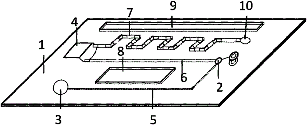
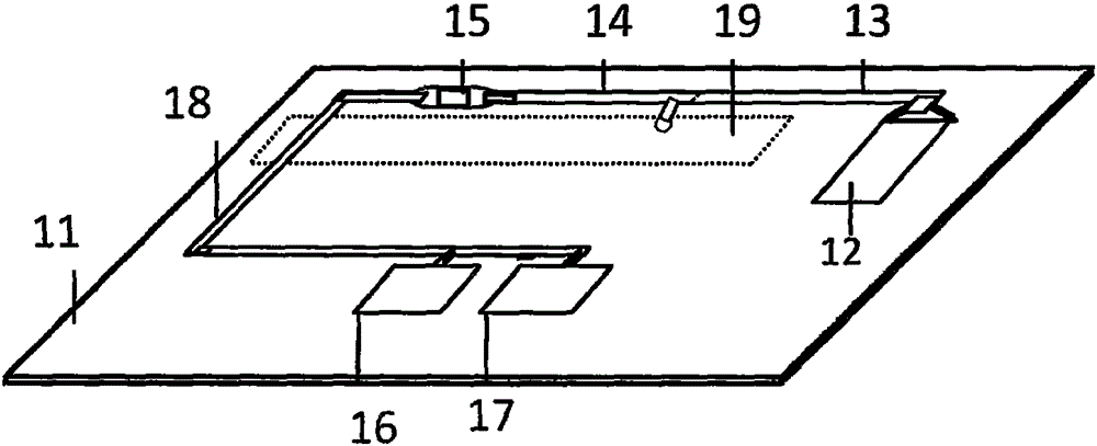
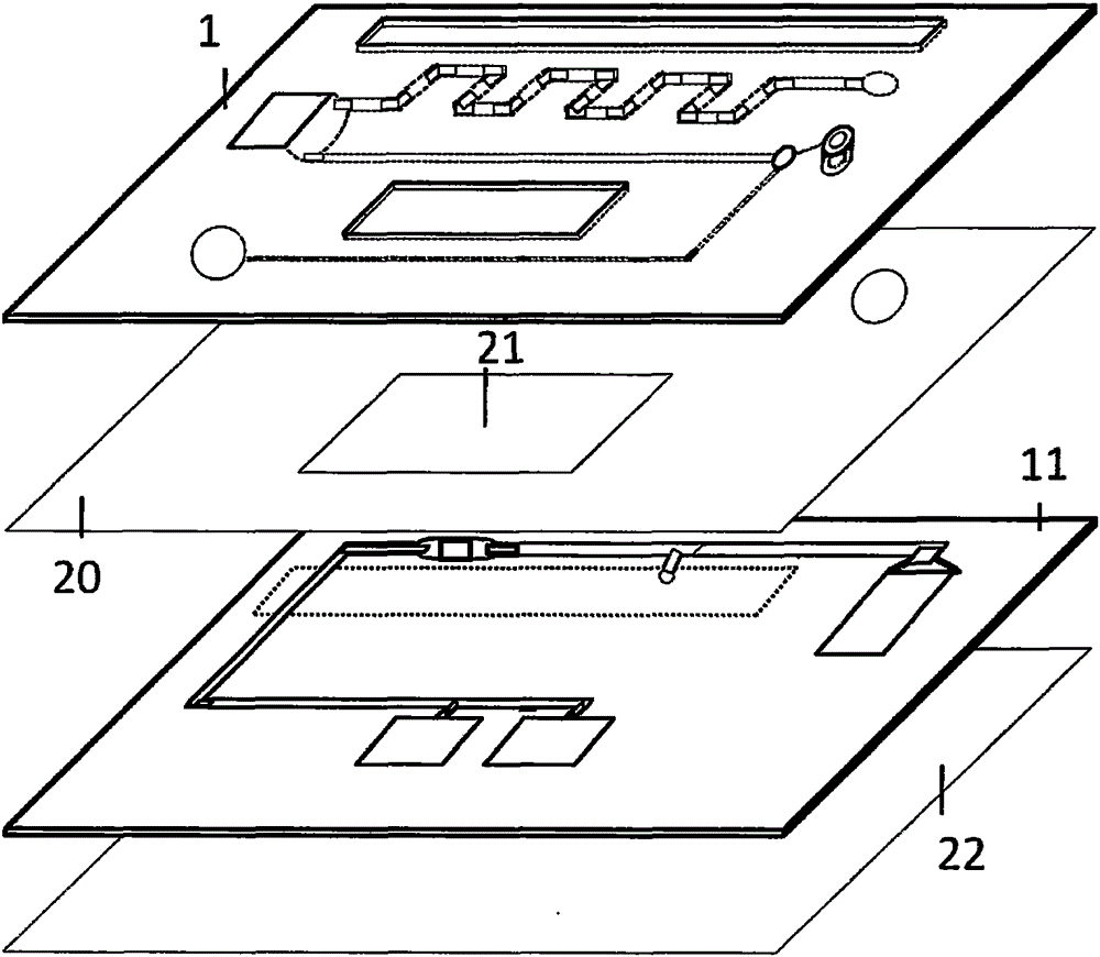
The invention discloses a magnetic particulate chemiluminescent micro-fluidic chip for quantitatively detecting myohemoglobin. The structure of the magnetic particulate chemiluminescent micro-fluidic chip mainly comprises a cover piece (1) and a bottom piece (11), wherein an air pump (3), an airflow micro-channel (5), a sample feeding hole (2), a sample liquid flowing channel (6), a first biological marker storage tank (4), a micro-mixer (7) and a transition region (10) on the cover piece (1) are connected in sequence; a filter (12), a reaction tank (13), a washing tank (14), a detection tank (15) and a solution releasing channel (18) on the bottom piece (11) are connected in sequence; the detection tank (15) on the bottom piece (11) is connected with a washing liquid storage tank (16) and a luminescent liquid storage tank (17) through the solution releasing channel (18).

Description

technical field [0001] The invention relates to the field of clinical medical examination, in particular to a magnetic particle chemiluminescence microfluidic chip for quantitative detection of myoglobin, which can realize quantitative detection of myoglobin in biological samples within a few minutes, and the detection method has the advantages of simple operation , high sensitivity, low cost and so on. technical background [0002] Myoglobin (MYO, Mb) is a binding protein composed of a peptide chain and a heme prosthetic group. It is a protein that stores oxygen in muscle. Its oxygen saturation curve is hyperbolic. Oxygen-carrying protein in muscle, consisting of 153 amino acid residues, molecular weight 16.7KD, containing heme, homologous to hemoglobin, and oxygen-binding ability between hemoglobin and cytochrome oxidase, which can help muscle cells convert Oxygen transport to mitochondria. Myoglobin is the main protein that composes skeletal muscle and cardiac muscle. W...

Claims


Application Information

Patent Timeline
Patent Type & Authority Applications(China)
IPC IPC(8): B01L3/00G01N21/76G01N33/553
Inventor 范玉霞李泉
Owner SHENZHEN HUAMAIXINGWEI MEDICAL TECH CO LTD
Learn More