Cell membrane particle expressing parafusin and preparation and application of particle

A cell membrane and membrane protein technology, applied in the field of synthetic biology, can solve the problems of low transfer efficiency, time-consuming and labor-intensive biological safety, and inapplicable batch preparation, etc., to achieve the effect of improving efficiency, complete form, and improving transfer efficiency

Active Publication Date: 2016-01-20
SHANTOU UNIV
View PDF0 Cites 4 Cited by
  • Summary
  • Abstract
  • Description
  • Claims
  • Application Information

AI Technical Summary

Problems solved by technology

Studies have found that mitochondria can spontaneously transfer between cells through membrane nanotubes (membranenanotubes), and can restore the aerobic respiration function of mitochondria-deficient cells; mitochondrial microinjection can also achieve mitochondrial transfer, but strict operating requirements and special The equipment is not suitable for batch preparation; in addition, the use of actinomycin D (actinomycinD) can make cells functionally "enucleated" and become cytoplasts containing mitochondria, but some cells are not affected by drugs and still retain nuclear function ;Recent studies have reported that the use of cell-penetrating peptides (CPP) can also effectively deliver mitochondria isolated in vitro
But the transmission efficiency is relatively low
[0005] Due to the problems of low delivery efficiency, time-consuming and labor-intensive delivery and biological safety in the current delivery system for delivering biomacromolecules and organelles, it is of great significance to establish a method for efficiently delivering biomacromolecules and organelles for the treatment of various diseases

Method used


Image

  • Cell membrane particle expressing parafusin and preparation and application of particle
  • Cell membrane particle expressing parafusin and preparation and application of particle
  • Cell membrane particle expressing parafusin and preparation and application of particle

Examples


Embodiment 1

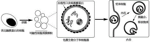
[0041] A preparation of membrane granules (PMVs) expressing fusion membrane proteins. per 1x10 6 Ad293 cells were transfected with 1 mg of a plasmid expressing vesicular stomatitis virus protein (pLP-VSVG).

[0042] Preparation methods such as figure 1 Shown: Add 1μg of pLP-VSVG plasmid to a 1.5ml centrifuge tube, add 50μl of serum-free culture medium, mix well; add 3μl of Polyjet transfection reagent and 50ml of serum-free culture medium into a new 1.5ml centrifuge tube, and Add it to the plasmid solution, mix well to obtain a transfection mixture, let it stand at room temperature for 5-10min, absorb the culture medium in the culture plate cells, and add 1ml of fresh culture medium, and transfer the transfection mixture immediately Add to cells and shake gently. After 12 hours, the culture medium was replaced with a new one, and after 48 hours, the cell suspension was digested and centrifuged at 800 g for 3 minutes. Under normal circumstances, in order to obtain more cell...

Embodiment 2

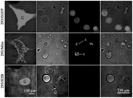
[0048] Characterization of cell membrane granules expressing fusin.

[0049] Ad293 cells were transfected with 1ugpEGFP-N1 (a plasmid expressing green fluorescent protein), pMito-EGFP, and pH2B-EGFP (a plasmid expressing nucleoprotein H2B and green fluorescent protein fusion protein), 48 hours after transfection, and passed the above The cell membrane particles obtained by the extrusion method used in Example 1 were observed under a confocal microscope. In addition, it was characterized by DNA separation technology, real-time PCR technology and western blotting. Cell membrane granules obtained from cells transfected with pEGFP-N1, pMito-EGFP, pH2B-EGFP, these three plasmids mark cell cytoplasm, cell mitochondria and nucleus respectively. From figure 2 It can be seen that membrane granules contain mitochondrial DNA, cellular RNA and miRNA, cytoplasmic proteins and membrane proteins, but not nuclear DNA, nuclear proteins. Membrane particles therefore enclose various biologic...

Embodiment 3

[0051] Monitoring and functional validation of proteins delivered by membrane granules expressing fusin.

[0052] Ad293 cells were co-transfected with 1mgpLP-VSVG and 1ugpDsRed-Expressed (plasmid expressing red fluorescent protein). 48 hours after transfection, VSV-G cell membrane particles were obtained by extrusion in Example 1 above, and added to 1x10 4 Remove the Ad293 cells, digest the cells after 5h, put them on a 35mm confocal culture dish, stain the nuclei with DAPI at 2h and 12h, and observe under a confocal microscope. Ad293 cells were co-transfected with 1mgpLP-VSVG and 1mgpCMV (CMC promoter)-CreER (Cre recombinase and estrogen receptor fusion protein). 48h after transfection, VSV-G cell membrane particles were obtained by the above extrusion membrane, and added to 1x10 4 In pCMV-DsRed / loxP2 / DsRed (reporter gene of the Cre-Loxp system) recipient cells, 10 mM 4-hydroxytamoxifen (4-HT) was added to the culture medium. 48h for fluorescence microscope observation. Fr...


PUM

PropertyMeasurementUnit
pore sizeaaaaaaaaaa

Abstract

The invention relates to a cell membrane particle expressing parafusin and preparation and application of the particle. VSVG is expressed on the cell membrane particle. Biomacromolecules and organelles are wrapped in the cell membrane particle. The cell membrane particle does not have a cell nucleus. The preparation method includes the steps that transient transfection is conducted on Ad293 cells through expression vesicular stomatits virus protein plasmids; the cells are digested through pancreatic enzymes 48 hours after transfection is carried out, and then centrifugation is carried out for 3 min; an obtained cell suspension is placed in a syringe, the syringe is installed in a syringe filter provided with a polycarbonate membrane, the syringe is pushed so that the cell suspension can be pushed from one side of the filter to the other side of the filter, and the cell membrane particle expressing VSVG is obtained. The cell membrane particle expressing parafusin is applied to biomacromolecule and organelle transfer. The membrane particle is larger, can wrap biomacromolecules and mitochondria, and transfers protein and the mitochondria in the aspect of function. Extra chemical reagents are not added, activity of VSVG is effectively maintained, mitochondrion release efficiency of the membrane particle is improved after the membrane particle enters a body, and accordingly mitochondrion transfer efficiency is improved.

Description

technical field [0001] The invention relates to synthetic biology technology, in particular to a fusion vesicular stomatitis virus glycoprotein membrane particle and its preparation and application in delivering biological macromolecules and organelles. Background technique [0002] Studies have found that some diseases, such as phenylketonuria, have abnormal symptoms due to the lack of related proteases; while other diseases, such as neurodegenerative Alzheimer's disease, commonly known as Alzheimer's disease, are related to mitochondrial dysfunction There is a great relationship. Although significant progress has been made in the use of drug delivery systems for therapeutic purposes, however, these systems have not been able to effectively solve existing problems. [0003] A variety of drug delivery systems have been established. For example, nanoparticles can deliver therapeutic proteins and peptides through packaging or conjugation. However, complex production processes...

Claims


Application Information

Patent Timeline
Patent Type & Authority Applications(China)
IPC IPC(8): C12N5/10C12N15/85C12N15/63
Inventor 魏炽炬林浩鹏郑德锦
Owner SHANTOU UNIV
Why Eureka
Try Eureka