Tissue engineering cartilage composite scaffold and preparation method thereof

A composite scaffold and tissue engineering technology, applied in medical science, prosthesis, etc., can solve the problems of cell loss, long recovery period, extracellular matrix loss, etc., to promote chondrogenic differentiation, good biocompatibility, good mechanics the effect of strength

Active Publication Date: 2016-02-03
GENERAL HOSPITAL OF PLA +1
View PDF5 Cites 15 Cited by
  • Summary
  • Abstract
  • Description
  • Claims
  • Application Information

AI Technical Summary

Problems solved by technology

And when in use, it is necessary to digest the seed cells from the microcarriers with trypsin, which will inevitably lead to the loss of extracellular matrix and the loss of cells.
[0005] Second, the material selection and construction method of the scaffold need to be improved; the three-dimensional porous scaffold often encounters the "hollow" phenomenon in the process of compound cell culture
That is, the cells on the surface of the porous scaffold are more likely to obtain nutrients and excrete metabolites and proliferate in large quantities, while the cells in the deep layer of the scaffold are less in number due to nutritional difficulties, which obviously affects the construction quality of tissue engineered cartilage; in addition, studies have shown that extracellular The cartilage repair effect of tissue engineering scaffolds constructed of natural biomaterials such as matrix, collagen, and alginic acid is significantly better than that of chemically synthesized scaffolds, but most natural bioscaffolds have poor mechanical properties and cannot provide sufficient stress support for tissue engineered cartilage, which will lead to The poor integration between the repair tissue and the normal cartilage will cause the degeneration of the surrounding cartilage, thus affecting the cartilage repair effect
[0006] Third, after the existing tissue engineering cartilage repairs cartilage defects, a long-term mechanism secretion and shaping process is required, which makes the rehabilitation period of the existing cartilage tissue engineering repair technology long after implantation and affects the quality of life of patients

Method used


Image

  • Tissue engineering cartilage composite scaffold and preparation method thereof
  • Tissue engineering cartilage composite scaffold and preparation method thereof
  • Tissue engineering cartilage composite scaffold and preparation method thereof

Examples


Embodiment 1

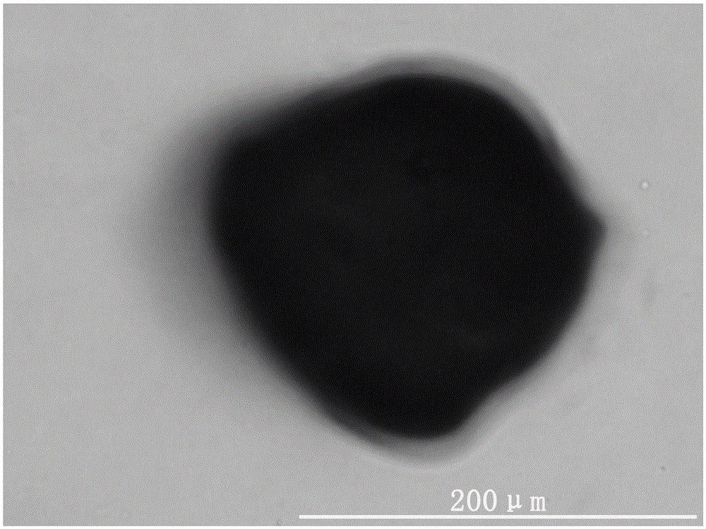
[0045] Example 1: Preparation of cartilage extracellular matrix-derived microcarriers

[0046] 500 g of the obtained fresh porcine articular cartilage was rinsed repeatedly with sterile physiological saline, mixed with a certain amount of sterile three-distilled water, and placed in a pulverizer for pulverization. Pass through stainless steel sieves with sieve diameters of 100 μm and 500 μm respectively to obtain cartilage extracellular matrix-derived microparticles with a diameter of 100 μm-500 μm. Add 1% SDS solution at 4°C, place in a shaker, and keep shaking for 8 hours. After washing away the remaining SDS solution with sterile three-distilled water, add the microparticles to 5ml DNAse (50U / mL) and RNase (1U / mL) solution, shake on a shaker at 37°C for 4 hours, remove cellular components. The decellularized cartilage extracellular matrix-derived microparticles are thoroughly washed to remove residual decellularized fluid. After packaging, place it at 25kGyC O 60 Radia...

Embodiment 2

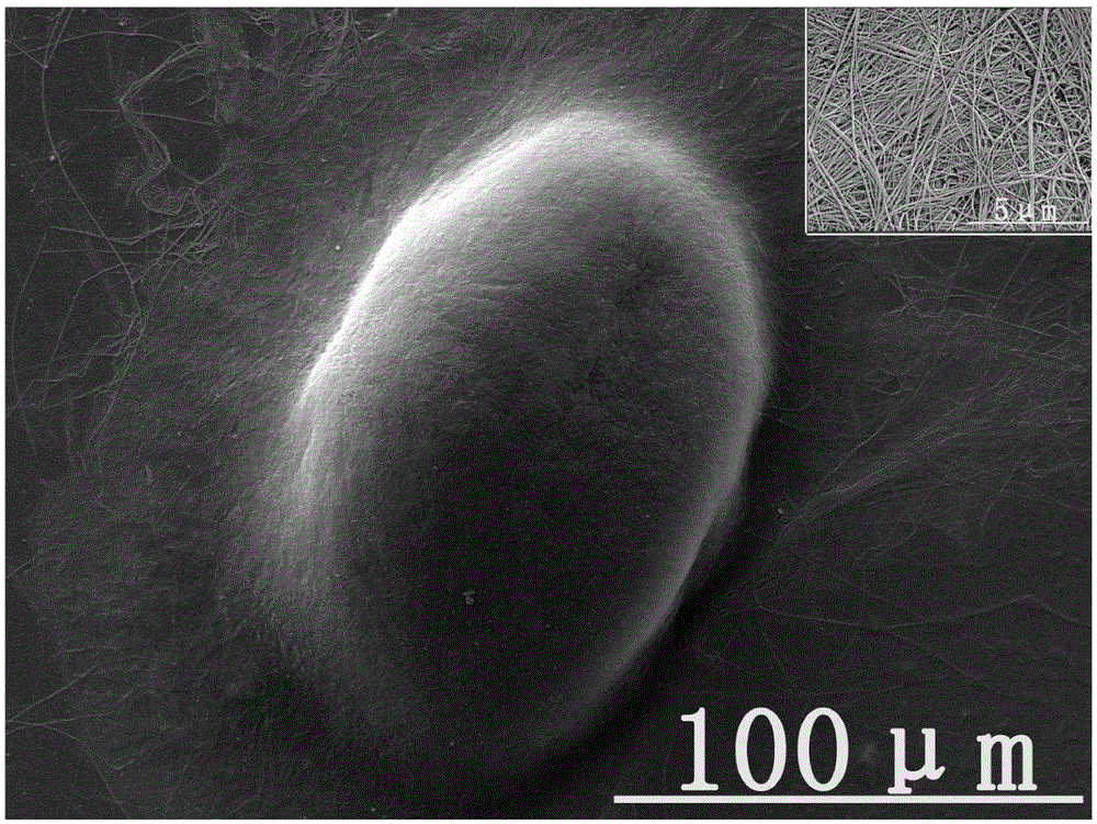
[0054] Cartilage extracellular matrix-derived microcarriers with a diameter of 100 μm-500 μm were prepared according to the method in Example 1.

[0055] 100mg cartilage extracellular matrix-derived microcarriers and 50ml containing 1 × 10 6 The DMEM cell suspension of rat bone marrow mesenchymal stem cells was mixed and placed in the reaction chamber of a rotary three-dimensional bioreactor (Rotary Cell Culture Systems, RCCS-D) for co-cultivation. In order to facilitate the attachment of cells to the microparticles, the rotation speed of the bioreactor was set at 30 rpm, with 1 minute per rotation and 30 minutes of rest. After 24 hours, the rotation speed was set to 80rpm and the rotation was continued. Bioreactor placed at 37 °C 5% CO 2 condition. The medium was changed every 2-3 days.

[0056] The 3D printed PLGA porous scaffold with a pore diameter of about 600 μm was placed in a petri dish and immersed in a 1.8% (w / v) sodium alginate solution. The chondrocyte extrace...

Embodiment 3

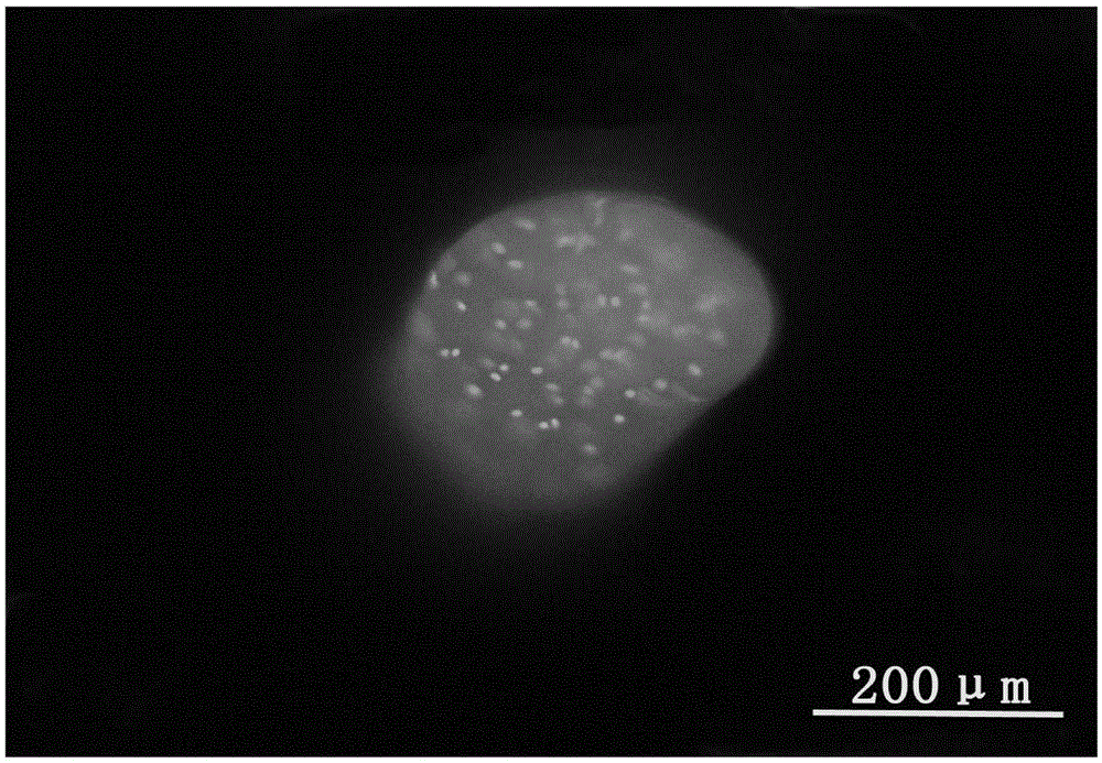
[0058] Cartilage extracellular matrix-derived microcarriers with a diameter of 100 μm-500 μm were prepared according to the method in Example 1.

[0059] 100mg cartilage extracellular matrix-derived microcarriers and 50ml containing 8 × 10 6 The DMEM cell suspension of rat chondrocytes was mixed and placed in the reaction chamber of a rotary three-dimensional bioreactor (Rotary Cell Culture Systems, RCCS-D) for co-cultivation. In order to facilitate the attachment of cells to the microparticles, the rotational speed of the bioreactor was set at 50 rpm, with 1 minute per revolution and 30 minutes of rest. After 24 hours, the rotation speed was set to 100rpm and the rotation was continued. Bioreactor placed at 37 °C 5% CO 2 condition. The medium was changed every 2-3 days.

[0060] A porous demineralized bone matrix scaffold with a pore diameter of about 2000 μm was placed in a culture dish and immersed in a 2.4% (w / v) fibrinogen solution. The chondrocyte extracellular matr...


PUM

PropertyMeasurementUnit
diameteraaaaaaaaaa
diameteraaaaaaaaaa
particle diameteraaaaaaaaaa

Abstract

The invention belongs to the field of cartilage tissue engineering. The invention discloses a tissue engineering cartilage composite scaffold, which is constructed based on an extracellular matrix-derived cartilage micro-tissue, and a preparation method of the composite scaffold. Specifically, a cartilage extracellular matrix-derived micro-carrier is prepared from fresh cartilage through such processes as wet pulverization, sieving, decellularizing and the like; and the prepared micro-carrier can be used as a seed cell amplification vector. By virtue of a bioreactor, cartilage cells are rapidly amplified by the micro-carrier and the phenotype of the cartilage cells is kept; and meanwhile, the induced differentiation of stem cells towards the cartilage is also promoted and the cartilage micro-tissue is formed. By filling the cartilage micro-tissue in pores of a three-dimensional porous scaffold which is good in mechanical performance, the tissue engineering cartilage composite scaffold is constructed. The composite scaffold is good in mechanical performance, rich in natural cartilage extracellular matrix components and conducive to the induced differentiation of the stem cells towards the cartilage, and the composite scaffold is capable of providing a good microenvironment for the growth of seed cells and is expected to accelerate the in vivo construction speed of the tissue engineering cartilage.

Description

technical field [0001] The invention relates to the field of cartilage tissue engineering. Specifically relates to a tissue engineering cartilage composite scaffold constructed based on extracellular matrix-derived cartilage microtissues and a preparation method thereof. Background technique [0002] Articular cartilage damage caused by trauma, inflammation, degeneration and other reasons is a common disease in orthopedics. Because articular cartilage lacks the innervation of blood vessels and nerves, its ability to regenerate and self-repair is extremely limited. Once damaged, it is difficult to repair itself. As the disease continues to develop, osteoarthritis will inevitably occur. This not only seriously affects the quality of life of patients, but also brings heavy economic burden to patients' families. Therefore, it is very important to provide effective treatment for early articular cartilage damage, prevent further development of cartilage damage, and reduce the in...

Claims


Application Information

Patent Timeline
Patent Type & Authority Applications(China)
IPC IPC(8): A61L27/38A61L27/36A61L27/54
Inventor 彭江尹合勇王玉汪爱媛卢世璧全琦孙振孙逊许文静郭全义朱昀刘舒云
Owner GENERAL HOSPITAL OF PLA
Why Eureka
Try Eureka