
A 3D printed porous scaffold with antibacterial functional coating and its preparation method

A porous stent, 3D printing technology, applied in the direction of coating, prosthesis, tissue regeneration, etc., can solve the problems of cumbersome and limited sterilization effect of metal ions, and achieve the effect of simplifying the preparation steps, improving long-term stability and uniform thickness

Active Publication Date: 2019-04-12
HUAZHONG UNIV OF SCI & TECH
View PDF9 Cites 0 Cited by
  • Summary
  • Abstract
  • Description
  • Claims
  • Application Information

AI Technical Summary

Problems solved by technology

However, the purpose of this method is to improve the biological activity of the scaffold and slow down the corrosion rate of the matrix, and the bactericidal effect of the metal ions is very limited, and the process of realizing the bactericidal function is too cumbersome.

Method used


Image

  • A 3D printed porous scaffold with antibacterial functional coating and its preparation method
  • A 3D printed porous scaffold with antibacterial functional coating and its preparation method
  • A 3D printed porous scaffold with antibacterial functional coating and its preparation method

Examples


preparation example Construction

[0036] The embodiment of the present invention also provides a method for preparing a 3D printed porous scaffold with an antibacterial functional coating, the method mainly includes the following steps:

[0037] 1) Establish a three-dimensional model of the porous scaffold through computer modeling software, and then import it into the SLM device for slicing;

[0038] 2) Prepare the metal powder and determine the optimized process parameters to form the porous scaffold:

[0039] Specifically select the following process parameters: laser power 180W-300W, laser spot diameter 0.05mm-0.1mm, scanning distance 50μm-70μm, scanning speed 200mm / s-400mm / s, powder layer thickness 20μm-30μm, using the above process parameters, It can form Co-Cr alloy entities with better surface quality.

[0040] 3) Prepare silk fibroin / gentamycin mixed solution:

[0041] First, add silkworm cocoons to 0.02M sodium carbonate solution, boil for 20 minutes, and then rinse with ultrapure water; secondly, ...

Embodiment 1

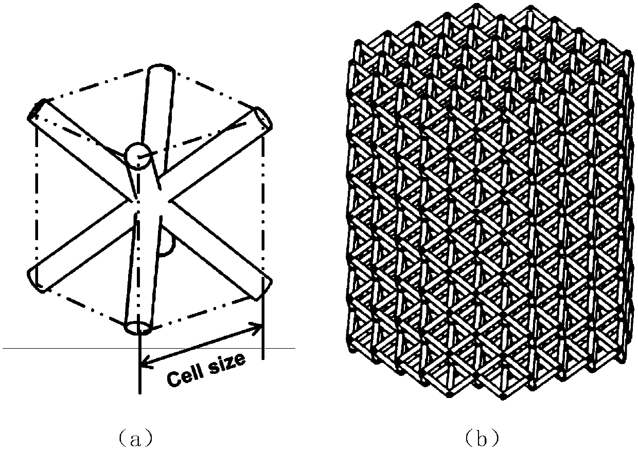
[0048] The three-dimensional model of the porous scaffold is established by computer modeling software. The model is a cylindrical porous scaffold with an outer diameter of 8 mm and a height of 10 mm. The porous scaffold is formed by stacking body-centered cubic units. The size of the topological unit is 1250 μm, and the diameter of the pillar is 200 μm. Then import the model into SLM equipment for slicing processing; use Co-Cr alloy powder, set the process parameters as laser power 220W, scanning distance 70μm, scanning speed 300mm / s, powder layer thickness 20μm, and form Co-Cr porous scaffold.

Embodiment 2

[0050] Add silkworm cocoons to 0.02M sodium carbonate solution, boil for 20 minutes, and then rinse with ultrapure water; secondly, after drying the extracted silk fibroin, add 9.3M lithium bromide solution to dissolve in 60°C water bath for 4 hours; The obtained solution was poured into a dialysis belt, and dialyzed in ultrapure water at 4°C for 72 hours; finally, the solution was centrifuged at 1500rpm at 4°C for 40 minutes to remove the insoluble part, and finally diluted to a concentration of 20mg / mL silk fibroin solution. Gentamicin was prepared into a solution with a concentration of 1 mg / mL, and added to the silk fibroin solution to prepare a silk fibroin / gentamicin mixed solution.


PUM

PropertyMeasurementUnit
heightaaaaaaaaaa
thicknessaaaaaaaaaa
diameteraaaaaaaaaa

Abstract

The invention discloses a 3D printing porous scaffold with antibacterial functional coating and a preparation method thereof. The porous scaffold includes a scaffold body and an antibacterial coating. The scaffold body is shaped by SLM (selective laser melting) technology, and is formed by body-centered cubic unit accumulation. The antibacterial coating is constructed on the scaffold body surface by electrodeposition technology, and is a composite coating of silk fibroin composite and gentamicin. The method comprises the steps of: establishing a porous scaffold three-dimensional model, introducing the model into SLM equipment to conduct slicing; preparing metal powder to form a porous scaffold with optimized process parameters; preparing a silk fibroin / gentamicin mixed solution; carrying out electrodeposition of the antibacterial coating on the porous scaffold surface; taking out the porous scaffold and performing air drying so as to obtain the required porous scaffold. The method provided by the invention utilizes electrodeposition to continuously construct the silk fibroin / gentamicin composite coating evenly on the inner surface and outer surface of a Co-Cr porous scaffold formed by SLM, simplifies the preparation steps, reduces the cost, and can reach the purpose of preventing infection after joint replacement.

Description

technical field [0001] The invention belongs to the field of advanced manufacturing technology, and more specifically relates to a 3D printed porous bracket with an antibacterial functional coating and a preparation method thereof. Background technique [0002] The temporomandibular joint is one of the joints that people use most every day. Factors such as joint ankylosis, trauma, and tumors often cause morphological defects and loss of function of the temporomandibular joint, which seriously affects people's speech, chewing and swallowing activities, and brings serious physiological and psychological problems to patients. Psychological trauma, the complexity of the morphological structure and physiological functions of the temporomandibular joint itself have brought great challenges to its artificial joint replacement surgery and reconstruction. [0003] 3D printing is based on the layer-by-layer additive manufacturing method, which can form personalized structures with com...

Claims


Application Information

Patent Timeline
Patent Type & Authority Patents(China)
IPC IPC(8): A61L27/56A61L27/34A61L27/28A61L27/54B33Y10/00
CPCA61L27/28A61L27/34A61L27/54A61L27/56A61L2300/232A61L2300/252A61L2300/404A61L2430/02B33Y10/00C08L89/00
Inventor 魏青松韩昌骏程岘王倩陈莉莉史玉升周燕
Owner HUAZHONG UNIV OF SCI & TECH
Learn More