Photoelectric chemical detection method of zearalenone based on TiO2 mesocrystal
A technology of zearalenone and mesoscopic crystals, applied in the direction of electrochemical variables of materials, can solve the problems of low photoelectric conversion efficiency and large forbidden band width, and achieve the effect of improving specificity and sensitivity
- Summary
- Abstract
- Description
- Claims
- Application Information
AI Technical Summary
Problems solved by technology
Method used
Image
Examples
Embodiment 1
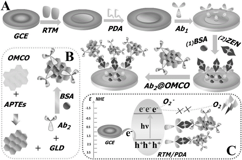
[0030] A TiO-based 2 The preparation method of the photoelectrochemical sensor of mesotube crystal (such as figure 1 shown):
[0031] (1) Pretreatment of the glassy carbon electrode: the glassy carbon electrode is first mechanically polished and polished on the suede covered with alumina powder, washed with secondary water to remove the residual powder on the surface, and then moved into an ultrasonic water bath for cleaning until it is cleaned, and finally Wash thoroughly with ethanol, dilute acid and water;
[0032] (2) Add dropwise 4 μL of RTM suspension with a concentration of 3 mg / ml on the surface of a clean glassy carbon electrode, dry it under infrared light, and cool to room temperature;
[0033] (3) Immerse the electrode in 0.5 mg / ml PDA solution for 30 min, and dry it at room temperature;
[0034] (4) Put the modified electrode in 5 μLAb 1 (2 mg / m) solution and incubated at 37°C for 1h, then washed with pH 7.5 phosphate buffer solution to remove excess Ab 1 The...
Embodiment 2
[0037] Rutile TiO 2 Preparation of mesoscopic crystal (RTM) materials:
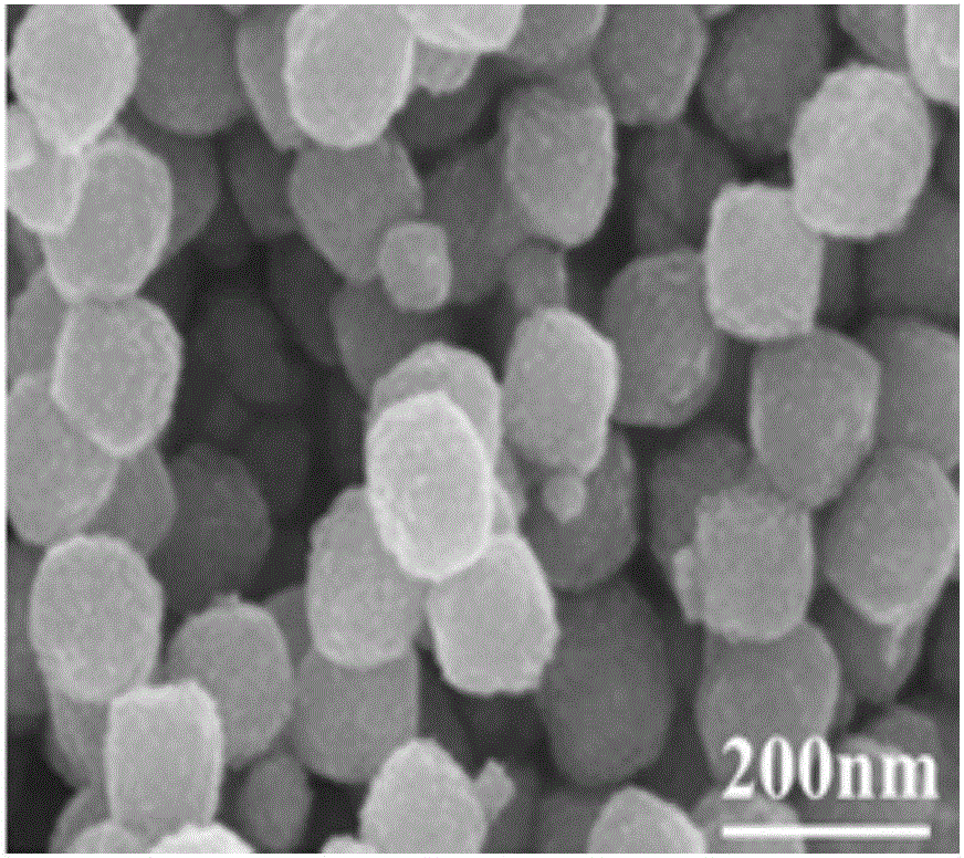
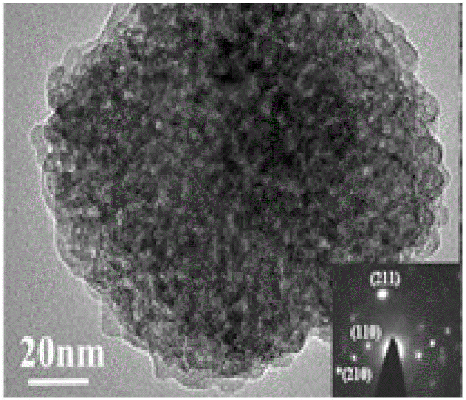
[0038] 0.5 g sodium dodecylbenzenesulfonate (SDBS) dissolved in 25 mL 2.2 mol / ml HNO 3 solution, stirred for 15 minutes. Then 0.5 mL titanium(IV) isopropoxide was added and stirred at 80 °C for 48 h. Subsequently, the obtained product was centrifuged, washed 4-5 times with ultrapure water and ethanol, and dried overnight at 60 °C. The above product was calcined in the air at 400 °C for 1 h to remove residual organic matter to obtain rutile TiO 2 mesoscopic crystals. Electron emission scanning electron microscope (SEM) image of RTM, as shown in Figure 2A, RTAM is a porous structure with a size of 90-130 nm. Transmission electron microscope (TEM) image and selected area electron diffraction (SAED) image of RTM, as shown in Figure 2 B and Figure 2B As shown in the inset, it is shown that the rutile TiO 2 The formation of mesogens.
Embodiment 3
[0040] Preparation of Mesoporous Cobalt Tetroxide (OMCO) Solution:
[0041] Preparation of mesoporous cobalt tetroxide (OMCO): 3 g of KIT-6 molecular sieve was added to 30 mL of 0.84 mol / L Co(NO 3 ) 2 •6H 2 O in ethanol and evaporated to dryness at 80 °C. Repeat the above steps. Finally, the material was calcined at 450 °C for 6 h. KIT-6 hard template was removed with 2 mol / L NaOH solution, sodium silicate was removed by centrifugation, and the sample was dried at 100°C to obtain 3d-Co 3 o 4 powder. The electron emission scanning electron microscope (SEM) image of OMCO, shown in Fig. 2C, shows that OMCO has a periodic mesoporous network structure. Transmission electron microscopy (TEM) images of OMCO, such as Figure 2D As shown, it shows that OMCO has a regular and ordered mesoporous structure, which can immobilize more biomacromolecules, so that it can effectively improve the sensitivity of PEC biosensors.
[0042] Mesoporous cobalt tetroxide labeled secondary antib...
PUM
 Abstract
Description
Claims
Application Information
 - R&D
- Intellectual Property
- Life Sciences
- Materials
- Tech Scout
- Unparalleled Data Quality
- Higher Quality Content
- 60% Fewer Hallucinations
Browse by: Latest US Patents, China's latest patents, Technical Efficacy Thesaurus, Application Domain, Technology Topic, Popular Technical Reports.
© 2025 PatSnap. All rights reserved.Legal|Privacy policy|Modern Slavery Act Transparency Statement|Sitemap|About US| Contact US: help@patsnap.com


