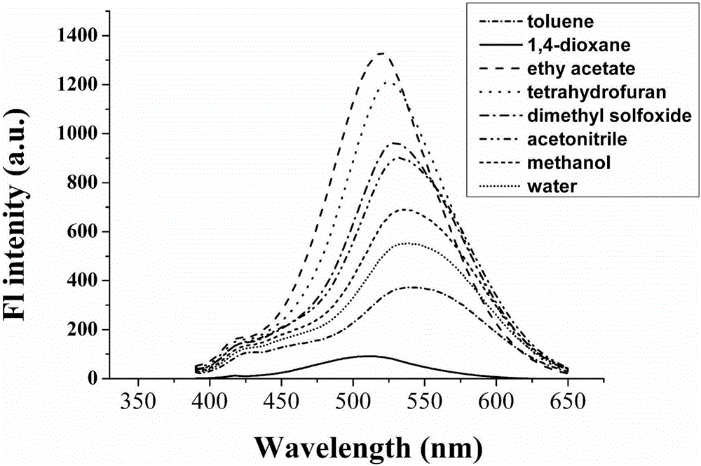
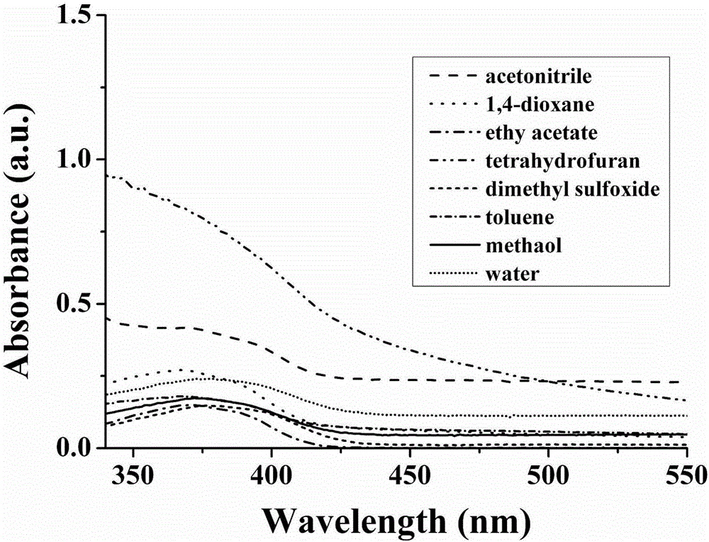
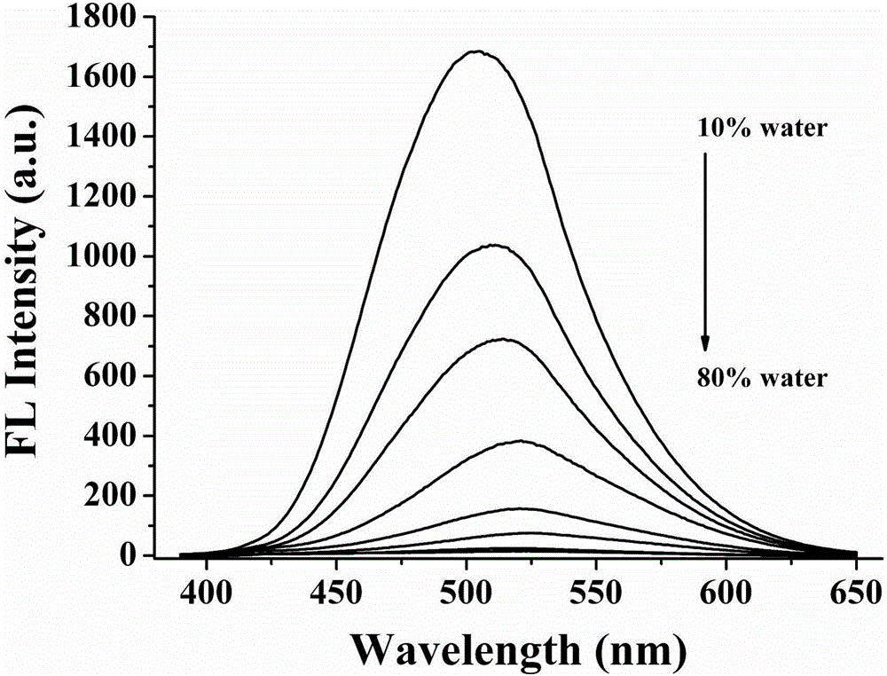
Cell autophagy monitoring probe, preparation method therefor and use of cell autophagy monitoring probe
A cell and autophagy technology, applied in the fields of biochemical equipment and methods, chemical instruments and methods, measuring devices, etc., can solve the problems of autophagy monitoring is not special, cannot monitor the autophagy state of living cells, and is expensive. , to achieve the effect of simple structure, simple operation and low cytotoxicity
- Summary
- Abstract
- Description
- Claims
- Application Information
AI Technical Summary
Problems solved by technology
Method used
Image
Examples
Embodiment 1
[0033] Embodiment 1: the synthesis of fluorescent probe molecule Lyso-OSC
[0034]7-((4-methoxyphenyl)ethylene)-3-carboxylic acid-coumarin (1g, 3mmol), N,N-diisopropylethylamine (1.2g, 9mmol), 1-hydroxy Benzotriazole (0.65g, 4.5mmol), EDC·HCl (0.65g, 3.3mmol) and 2-morphine-1-ethylamine (1.2g, 9mmol) were added to the Shrek bottle, under anhydrous and oxygen-free conditions Add 30mL of N,N-dimethylformamide, stir and react at room temperature for 24h; after the reaction is completed, add 50mL of dichloromethane to the reaction solution to dissolve the reactant, and then extract with 30mL of water (3 times) to obtain an organic phase. Through column chromatography on 200-300 mesh silica gel (the eluent is composed of ethyl acetate and petroleum ether mixed at a volume ratio of 1:1), 0.35 g of the target product was obtained with a yield of 40%.
[0035] 1 H NMR (400MHz, CDCl 3 )δ9.13(s,1H),8.86(s,1H),7.63(d,J=8.0Hz,1H),7.53(s,1H),7.50(s,1H),7.49(s,1H), 7.46(s,1H),6.95(d,J=8...
Embodiment 2
[0037] Example 2: Two-photon test of fluorescent probe molecule Lyso-OSCC
[0038] Using two-photon measurement technology, test the two-photon absorption cross section of fluorescent probe molecules, from Figure 8 It can be seen that the maximum effective absorption cross section of the fluorescent probe molecule is 93GM, and the two-photon excitation wavelength is 760nm. The two-photon absorption of fluorescent probe molecules was verified. Under the two-photon excitation wavelength of 760nm, the voltage was gradually adjusted from 200w to 800w. Two-photon excitation is possible.
Embodiment 3
[0039] Example 3: Cytotoxicity Test
[0040] The MTT (3-(4,5-dimethylthiazole-2)-2,5-diphenyltetrazolium bromide) experiment is based on the reported literature, and the cytotoxicity test is done. Add 0, 10, 20, and 30 μM fluorescent probe molecules to the same batch of MCF-7 cells, and the conditions are 37 ° C, 5% CO 2 Incubate in a cell incubator for 24 hours, according to the formula of cell viability: cell viability%=OD 570 (sample) / OD 570 (control group)×100, the cell survival rate can be calculated ( Figure 5 ). From Figure 5 We can see that when the concentration is 5 μM, the cell survival rate is about 98%, and when the probe concentration reaches 15 μM, the cell survival rate is still about 82%, which shows that the fluorescent probe molecule of the present invention has basically no toxic effect on cells , so it can be used for cell detection and monitoring of polarity changes in lysosomes.
PUM
 Abstract
Description
Claims
Application Information
 - R&D
- Intellectual Property
- Life Sciences
- Materials
- Tech Scout
- Unparalleled Data Quality
- Higher Quality Content
- 60% Fewer Hallucinations
Browse by: Latest US Patents, China's latest patents, Technical Efficacy Thesaurus, Application Domain, Technology Topic, Popular Technical Reports.
© 2025 PatSnap. All rights reserved.Legal|Privacy policy|Modern Slavery Act Transparency Statement|Sitemap|About US| Contact US: help@patsnap.com


