Biosensor for detecting kanamycin and preparation method thereof

A biosensor, kanamycin technology, applied in the field of biosensors, can solve the problems of high cost, low specificity and sensitivity, and achieve the effects of simple electrode, improved sensitivity and stable performance

Inactive Publication Date: 2017-06-30
UNIV OF JINAN
View PDF3 Cites 14 Cited by
  • Summary
  • Abstract
  • Description
  • Claims
  • Application Information

AI Technical Summary

Problems solved by technology

[0004] In order to solve the problems of relatively low specificity and sensitivity and high cost of the method for detecting kanamycin in the above prior art, the present invention provides a...

Method used


Image

  • Biosensor for detecting kanamycin and preparation method thereof
  • Biosensor for detecting kanamycin and preparation method thereof
  • Biosensor for detecting kanamycin and preparation method thereof

Examples


preparation example Construction

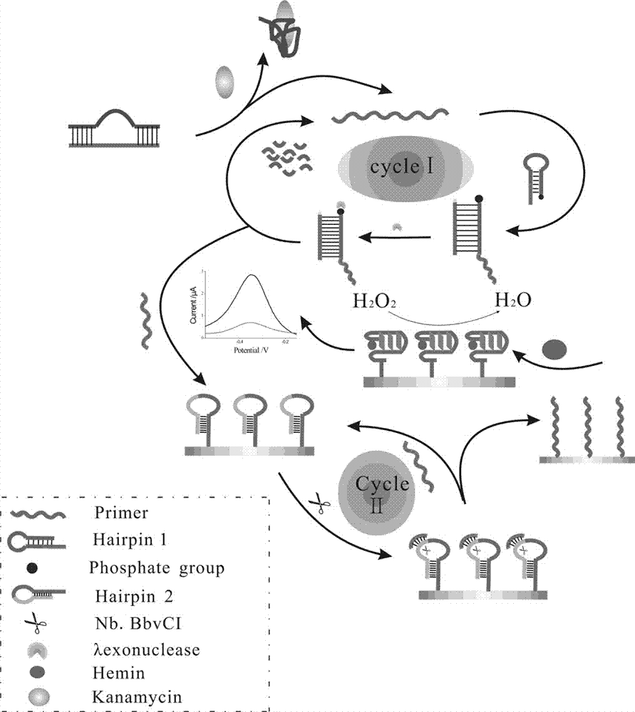
[0045] The preparation method of described biosensor comprises the following steps:

[0046] (1) Pretreatment of the electrodes;

[0047](2) Modification of HAP2 to the electrode surface;

[0048] (3) Modification of the homogeneous reaction product layer onto the electrode surface.

[0049] In the preparation method, the operation steps of modifying HAP2 onto the electrode surface are preferably as follows: 10 μL of HAP2 is added dropwise to the pretreated electrode surface, and incubated at 37° C. for 2 hours.

[0050] In the preparation method, the preferred steps of modifying the homogeneous reaction product onto the electrode surface are as follows:

[0051] (1) Add sterilized water, 10× buffer buffer, HAP1, Primer, Helper, Lambda exonuclease, and the target to be tested into a centrifuge tube, shake for 30 seconds, and incubate in a 37°C incubator for 2 hours;

[0052] (2) Place the incubated mixed solution in an incubator at 75°C for 5 minutes to inactivate Lambda ex...

Embodiment 1

[0059] The main steps of the electrode modification process are as follows:

[0060] a. The gold electrode is first polished in 0.3 and 0.05 µm alumina slurry until it becomes a mirror surface, and then rinsed repeatedly with PBS and secondary water;

[0061] b. Add 10 μL of HAP2 (10 μM) dropwise to the electrode surface and incubate at 37°C for 2h. Fix the sulfhydryl chains to the electrode surface through Au-S bonds;

[0062] So far, the modification process of the electrode has come to an end. The following describes the reaction in the homogeneous solution and the main steps in the homogeneous reaction:

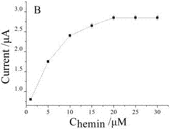
[0063] a. Mix 3 μL sterilized water, 5 μL 10× buffer buffer, 4 μL different concentrations of HAP1 (5 μM, 10 μM, 25 μM, 50 μM, 75 μM, 100 μM), 2 μL Primer (10 μM), lambda exonucleic acid Enzyme (1 μL), 2 μL Helper (50 μM), and 2 μL target substance to be tested were added to a centrifuge tube, shaken for 30 seconds, and incubated in a 37°C incubator for 2 hours;

[006...

Embodiment 2

[0076] The main steps of the electrode modification process are as follows:

[0077] a. The gold electrode is first polished in 0.3 and 0.05 µm alumina slurry until it becomes a mirror surface, and then rinsed repeatedly with PBS and secondary water;

[0078] b. Add 10 μL of HAP2 (10 μM) dropwise to the surface of the electrode and incubate at 37°C for 2 hours. Fix the sulfhydryl chains to the electrode surface through Au-S bonds;

[0079] So far, the modification process of the electrode has come to an end. The following describes the reaction in the homogeneous solution and the main steps in the homogeneous reaction:

[0080] a. Mix 3 μL sterilized water, 5 μL 10× buffer buffer, 4 μL different concentrations of HAP1 (50 μM), 2 μL Primer (10 μM), lambda exonuclease (1 μL), 2 μL Helper (100 μM), and Add 2 μL of the target substance to be tested into a centrifuge tube, shake for 30 seconds, and incubate in a 37°C incubator for 2 hours. b. Incubate the incubated mixed solutio...


PUM


Abstract

The invention relates to the technical field of the biosensor and in particular to a biosensor for detecting kanamycin based on nucleic acid aptamer comformational change induced by a target object. The specific preparation method comprises the following steps: pre-processing an electrode; decorating an HAP2 layer on the surface of the electrode; and decorating homogeneous reaction mixed liquid on the surface of the electrode. The biosensor is capable of using the specific recognition of the nucleic acid aptamer, using the aptamer of the kanamycin as a recognition substance for high specific detection of the target object kanamycin, using nucleic acid tool enzyme, and realizing the cyclic utilization of the target object, and has the function of amplifying the signal.

Description

technical field [0001] The invention relates to the technical field of biosensors, in particular to a biosensor for detecting kanamycin based on target-induced nucleic acid aptamer conformation change and lambda exonuclease, and also relates to a preparation method thereof. Background technique [0002] Kanamycin is an aminoglycoside antibiotic. For most Enterobacteriaceae bacteria, such as Escherichia coli, Klebsiella, Proteus, Enterobacter, Shigella, Salmonella, Citrobacter, Providencia, Yale Mori bacteria, etc. have a good effect; influenza bacilli, Brucella, meningococcus, gonorrhea, etc. are also mostly sensitive to this product, and are ineffective against Pseudomonas aeruginosa. It also has a certain effect on methicillin-sensitive strains of Staphylococcus and Mycobacterium tuberculosis. Other Gram-positive bacteria such as hemolytic streptococci, Streptococcus pneumoniae, enterococci and anaerobic bacteria are mostly resistant to this product. However, kanamyc...

Claims


Application Information

Patent Timeline
IPC IPC(8): C12Q1/44G01N27/327G01N27/48
CPCC12Q1/005G01N27/327G01N27/48G01N2333/922
Inventor 李慧韩聪黄加栋刘素王玉
Owner UNIV OF JINAN
Why Eureka
Try Eureka