Exosome-compounded collagen biological scaffold with directional releasing function as well as preparation method and application thereof

A collagen biology and exosome technology, applied in the field of medical biomaterials, can solve the problems of difficulty in forming a target, and MSC-Exo cannot be administered in a concentrated area, so as to achieve effective retention of activity and improve targeted repair and therapeutic effects. , the preparation method is simple and easy to implement

Active Publication Date: 2018-01-09
龙乾发 +1
4 Cites 7 Cited by

AI-Extracted Technical Summary

Problems solved by technology

However, there are still many problems in the use of MSC-Exo in the treatment of diseases. The most important point is that the existing methods of drug delivery, such as intravenous injection, intranasal administration, and intracerebroventricular administration, are easy to spre...

Abstract

The invention provides an exosome-compounded collagen biological scaffold with a directional releasing function as well as a preparation method and application thereof. The exosome-compounded collagenbiological scaffold comprises nano collagen and exosomes dispersed in the nano-collagen. The preparation method of the biological scaffold comprises the following steps: separating collagen from animal tissue, carrying out ball milling till the collagen is nanoscale collagen; separating exosomes from culture of bone marrow or umbilical cord mesenchymal stem cells; mixing the exosomes with the collagen, freezing and drying. The preparation method of the biological scaffold is used for dispersing the exosomes in the nanoscale collagen to form a biological scaffold material with a function of directionally releasing exosomes; the targeted repairing and treating effects of the exosomes can be obviously improved; the development tendency of accurate drug administration can be met; the dosage forms are various; the biological scaffold has extensive clinical application values.

Application Domain

Technology Topic

Bone marrowNanoscopic scale +8

Image

  • Exosome-compounded collagen biological scaffold with directional releasing function as well as preparation method and application thereof
  • Exosome-compounded collagen biological scaffold with directional releasing function as well as preparation method and application thereof
  • Exosome-compounded collagen biological scaffold with directional releasing function as well as preparation method and application thereof

Examples

  • Experimental program(1)

Example Embodiment

[0032] The present invention will be described in further detail below in conjunction with the accompanying drawings and embodiments.
[0033] 1. Main reagents and instruments
[0034] Pepsin, Chloroform, Glacial Acetic Acid, NaHCO 3 , NaCl, high-speed centrifuge, freeze dryer, deep-low temperature refrigerator, meat grinder, dialysis membrane, liquid nitrogen planetary ball mill.
[0035] 2. Method
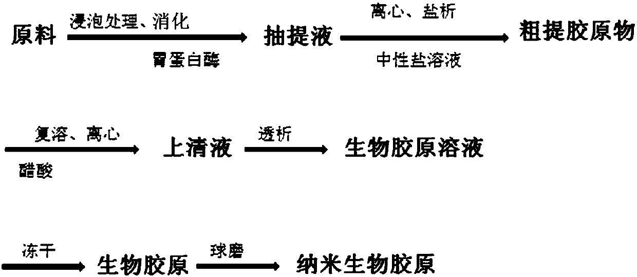
[0036] 2.1 Collagen Preparation
[0037] (1) Pretreatment: take fresh bovine (pig, sheep or other animal) Achilles tendon, bovine (pig, sheep or other animal) skin, bovine (pig, sheep or other animal) articular cartilage and other biological materials (for example, take skin), wash and remove subcutaneous fat and dermis, remove fascia, remove hair, and chop finely.
[0038] (2) Degreasing: The shredded biological material is washed with double distilled water, minced in a meat grinder, soaked in a mixed solvent of chloroform-methanol (for example, volume ratio 1:1) at 2-6°C (for example, 4°C) for degreasing , The soaking time is 3 to 6 hours (for example, 4 hours), during which the stirring is interrupted.
[0039] (3) Washing: the above-mentioned degreased biological material was dissolved in NaCl and NaHCO 3 Mixed aqueous solution (NaCl mass fraction is 2% to 30%, NaHCO 3 The mass fraction is 1% to 10%, for example, both are 10%) and soaked in 2 to 6°C (for example, 4°C), and the soaking time is 3 to 8h (for example, 4h), stirring continuously during this period; then washing with double distilled water for 3 to 5 times (for example, 3 times), then centrifuge at 1000-20000r/min (for example, 20000r/min) for 5-60min (for example, 15min), pour off the liquid, and use a 300-800 mesh (for example, 800 mesh) sieve to remove the solid Impurities, serum and other components, the resulting precipitate (residue after sieving) is the raw material for collagen extraction.
[0040] (4) extract collagen ( figure 1 ):
[0041] Extraction: Submerge the raw material in 0.5-1M acetic acid (for example, 1M) at 2-6°C (for example, 4°C) for 10-72h (for example, 48h), then add pepsin for extraction (digestion), in 2-6°C 4°C (for example, placed in a 4°C refrigerator) for a total of 1 to 7 days (for example, 2 to 3 days) of extraction, during which the magnetic stirrer continued to stir to obtain the extract; the mass ratio of pepsin: raw materials was 0.1 to 5 :10~80 (for example, 1:10).
[0042] Centrifugation: centrifuge the above extract at 1,000-20,000 r/min (eg, 20,000 r/min) for 5-60 min (eg, 15 min) to remove the precipitate, and the obtained supernatant is crude collagen extract.
[0043] Salt-out: Add the crude collagen extract to 2%-30% NaCl aqueous solution (for example, 10%) by mass fraction until a large amount of white precipitates are precipitated, and stand at 2-6°C (for example, 4°C) for 5-60min (for example, 30min) , centrifuge at 1000-20000 r/min (eg 20000 r/min) for 3-60 min (eg, 15 min), discard the supernatant, and precipitate as crude collagen.
[0044] Reconstitution: redissolve the crude collagen in 0.5-10M acetic acid (for example, 1M), centrifuge at 1000-20000r/min (for example, 20000r/min) for 3-60min (for example, 15min), remove insoluble impurities, and keep the supernatant liquid.
[0045] Dialysis: Put the supernatant obtained in the reconstitution step into a dialysis membrane (for example, 14000 Da) with a molecular weight cut-off of 8000-20000 Da, immerse in double-distilled water at 2-6°C (for example, 4°C) and dialyze for 24-72h (for example, 48h), The solution (double distilled water) was changed every 1 to 2 hours, and the refined collagen solution was obtained after dialysis (the liquid wrapped by the dialysis membrane was the refined collagen solution), that is, the biological collagen solution.
[0046] Freeze-drying: Put the refined collagen solution into a large petri dish, freeze-dry in a freeze dryer (-70°C slowly rise to -20°C, and increase the temperature by 1-2°C every 2-4 hours), and you can get a sponge collagen( figure 2 ).
[0047] Ball milling: ball mill the spongy collagen in a liquid nitrogen planetary ball mill (-70°C to -20°C) for 24h to 72h (for example, 48h) to obtain nano-collagen.
[0048] (5) Identification and activity analysis
[0049] Co 60 Or ethylene oxide and other methods for disinfection, and then accurately identify its composition, purity, and biocompatibility.
[0050] Table 1. Comparison table of collagen components
[0051]
[0052] Comparing the prepared spongy collagen with the amino acid composition of the type I collagen standard product, it can be seen that the amino acid composition of the two is basically the same (Table 1). The electrophoretic patterns were compared, and the results were consistent. From top to bottom, they were gamma chain (molecular weight: 130,000), beta chain (molecular weight: 98,000), and alpha chain (molecular weight: 97,200). High type I collagen.
[0053] After being identified as type I collagen (or other types of collagen), ball milling is carried out, and the nano-collagen after ball milling is made into a solid-phase nano-collagen scaffold by redissolving and freeze-drying, and a solid-phase standard made of collagen standard (non-nano) The collagen scaffolds were inoculated with bone marrow mesenchymal stem cells for bioactivity (toxicity) testing. Bone marrow mesenchymal stem cells seeded on two different scaffolds were detected by MTT method. With the growth of seeding time, the number of cells seeded on the two scaffolds increased continuously, and it was found that the nano-collagen scaffold had higher histocompatibility and the cells grew faster ( image 3 In the figure, the black solid is the result of solid-phase nano-collagen scaffold, and the round hollow is the result of solid-phase standard collagen scaffold, * indicates significant difference).
[0054] 2.2 Exosome isolation and identification
[0055] The 3rd to 5th passage bone marrow or umbilical cord MSCs were cultured until 70% to 80% confluence, rinsed with PBS, added chemically defined and protein medium (chemically defined and protein medium, CDPF) and continued to incubate for 6 hours (37°C), and then continued to replace with fresh CDPF Cultivate for 42h. Collect the culture medium by high-speed centrifugation (2,565×g) for 15 minutes to remove cell debris, and then collect the suspension and add it to an ion column containing anion exchange resin (Catalog#4079302, Whatman). The ion column is pretreated with an equilibrium solution (mobile phase), and the flow rate is set to 4mL/min, after the ion column filtration is completed, the balance solution rinses the ion column exchange resin, and finally elutes with 50mM tromethamine buffer and collects the eluate (exosomes), which is stored at -20°C after quantification by the BCA kit. Under the condition of ℃, spare.
[0056] Enzyme linked immunosorbent assay (enzyme linked immunosorbent assay, ELISA) or FCM was used to detect the expression of exosomal CD63, CD81, CD9, CD13 and other antigens. The number and size of exosomes were further analyzed by nanoparticle tracking analysis (NTA), and the equipment (Nanosight LM10; Malvern) was standardized using polystyrene latex microspheres (NTA4088, 100 nm). The expression rate of CD63 and CD81 ≥ 95%, the expression rate of CD9 and CD13 ≤ 2%, the number of exosomes ≥ 10×10 11 /mL (10~15×10 11 /mL), the diameter of exosomes is 50-150nm. Freeze-dried for later use.
[0057] 2.3 Composite
[0058] Dissolve nano-collagen in double-distilled water, add exosomes, and stir evenly with a magnetic stirrer to prepare a mixed solution with a collagen mass fraction of 0.1% to 10%. After standing still, there is no obvious precipitation. After the mixed solution is obtained, put it in a large After freeze-drying in a petri dish in a freeze dryer, a new type of medical collagen sponge (that is, MSC-Exo composite nano-collagen bioscaffold in the form of a spongy solid) can be obtained. After testing, the exosomes in The dispersion concentration in collagen is 3%-5% (w/w).
[0059] 3. Activity experiment of MSC-Exo composite nano-collagen bioscaffold
[0060] Experiment 1 Inhibitory effect of MSC-Exo composite nano-collagen bioscaffold on glial cell activation
[0061] 8-10 week SD rats were intraperitoneally injected with lipopolysaccharides (Lipopolysaccharides, LPS) (5mg/kg) to establish animal models of neuroinflammation, respectively received 10μL of PBS (control group I, subdural injection, n=10), about 6mm 2 MSC-Exo collagen composite bioscaffold (control group II, containing 10×10 9 MSC-Exo, implanted subdurally, n=10), about 6mm 2 MSC-Exo composite nano-collagen biological scaffold (experimental group, containing 10×10 9 MSC-Exo, implanted subdurally, n=12) were treated, samples from each group were collected on the 4th to 6th day of the peak inflammation period, and normal SD rats of the same age were used as negative controls. Brain tissue sections were taken for immunohistochemical staining of Iba1 (microglial marker) and GFAP (astrocyte marker), and Image J software was used to analyze the results.
[0062] Experiment 1 immunohistochemical staining results showed that the hippocampus and cortex GFAP+ and Iba1+ cells were seen in the control group I ( Figure 4 A) The cell body is large and the protrusions are obvious, suggesting that the intracranial glial cells of SD rats were significantly activated after intraperitoneal injection of LPS, and neuroinflammation was obviously induced; in the control group II (exosomes + type I non-nanocollagen), GFAP+ and GFAP+ in the hippocampus and cortex were seen in SD rats Iba1+ cells ( Figure 4 B) The protrusions of the cell body are retracted, suggesting that the activation of glial cells is inhibited; the experimental group sees GFAP+ and Iba1+ cells in the hippocampus and cortex ( Figure 4 C) The morphology of the cell body and protrusions was not much different from that of the negative control, suggesting that the MSC-Exo composite nano-collagen bioscaffold significantly inhibited the glial cell activation of neuroinflammation compared with the control group II. Image J analysis results showed that the area percentages of positive cells in control group I, control group II and experimental group were GFAP+: 64.3±8.9%, Iba1+: 57.7±4.2%; GFAP+: 37.3±2.9%, Iba1+: 27.5±4.9% ; GFAP+: 24±1.7%, Iba1+: 19.7±3.2% (P<0.01, experimental group vs. control group I or control group II).
[0063] According to the conclusion of Experiment 1, it is suggested that the MSC-Exo composite nano-collagen bioscaffold can be used to inhibit the activation of glial cells (ie, neuroinflammation) induced by craniocerebral injury, stroke, and epilepsy.
[0064] Experiment 2 Neuroprotective effect of MSC-Exo composite nano-collagen bioscaffold
[0065] SD rats at 8-10 weeks received 3-5 times (each interval 45min) intraperitoneal injection of kainic acid to induce grade 4 epileptic seizures for 2 hours to establish a model of hippocampal nerve injury and received 10 μL PBS (control group I, subdural Injection, n=10), about 6mm 2 MSC-Exo collagen composite bioscaffold (control group II, containing 10×10 9 MSC-Exo, implanted subdurally, n=10), about 6mm 2 MSC-Exo composite nano-collagen biological scaffold (experimental group, containing 10×10 9 MSC-Exo, implanted subdurally, n=12) were treated, and the samples of each group were collected after 1 week of nerve injury model induction treatment, and normal SD rats of the same age were used as negative controls. Brain tissue sections were taken for immunohistochemical staining for NeuN (neuron marker), and the experimental results were quantitatively analyzed by stereology.
[0066] Experiment 2 result shows: control group 1 sees NeuN+ cell in hippocampus CA1 area ( Figure 5 A) and dentate gyrus hilus (DH) were significantly reduced compared with the normal group, hippocampal CA1 ( Figure 5 B) and DH area neuron loss (NeuN+ cells) were improved compared with control group I, the experimental group saw NeuN+ cells in the hippocampal CA1 area ( Figure 5 C) and dentate gyrus hilus (DH) were significantly increased compared with control group II. Stereological quantitative analysis of the amount of NeuN+ cells in each group was: control group I CA1: 1.4±0.15×10 5 , DH: 0.9±0.11×10 4; control group II CA1: 2.2 ± 0.48 × 10 5 , DH: 1.4±0.24×10 4; Experimental group CA1: 2.5±0.32×10 5 , DH: 1.7±0.18×10 4 (P<0.05, experimental group vs. control group I or control group II). It is comprehensively suggested that MSC-Exo composite nano-collagen bioscaffold has better neuroprotective effect on nerve injury than MSC-Exo collagen composite bioscaffold subdural implantation.
[0067] According to the conclusion of Experiment 2, it is suggested that MSC-Exo composite nano-collagen bioscaffold can be used to treat epilepsy, Alzheimer's disease, craniocerebral injury, stroke and other hippocampal neuron loss diseases.
[0068] The above conclusions also apply to other types of collagen.
[0069] According to other experimental results, bioscaffolds in different forms such as injections, liquids, membranes, external sprays, and oral liquids can be used to treat one or more complex system diseases such as nerves, skin, and soft tissues.
[0070] In a word, the present invention proves that MSC-Exo composite nano-collagen bioscaffolds in different forms can be used for the repair of nerve damage, cartilage damage, skin and subcutaneous tissue damage, and the treatment of one or more complex system diseases, and can be used as food , pharmaceuticals, health care products and cosmetics materials, used in medical, beauty, rehabilitation and other fields.

PUM

no PUM

Description & Claims & Application Information

We can also present the details of the Description, Claims and Application information to help users get a comprehensive understanding of the technical details of the patent, such as background art, summary of invention, brief description of drawings, description of embodiments, and other original content. On the other hand, users can also determine the specific scope of protection of the technology through the list of claims; as well as understand the changes in the life cycle of the technology with the presentation of the patent timeline. Login to view more.

Similar technology patents

Classification and recommendation of technical efficacy words

  • Strong combination
  • Good targeting

Method for processing high-temperature alloy furnace tube

ActiveCN101565807AStrong combinationMitigation of catalytic cokingSolid state diffusion coatingProduct gasMetallurgy
Owner:CHINA UNIV OF PETROLEUM (BEIJING)

Preparation method of macroporous aluminum oxide with diplopore distribution

InactiveCN101214454AStrong combinationOvercoming low mechanical strengthCatalyst carriersCarbon blackPetroleum
Owner:SHANGHAI SECOND POLYTECHNIC UNIVERSITY

User directed donation system and method

InactiveUS20140278861A1Good targetingFundraising managementPayment architectureRegistered userDatabase
Owner:4ME 4WE

Chimeric antigen receptor resistant to placenta chondroitin sulfate and application of chimeric antigen receptor

ActiveCN105753991AGood targetingReduce damageAntibody medical ingredientsHybrid peptidesReceptor for activated C kinase 1Antigen recognition
Owner:BLUE ELEGANT BIOTECH CO LTD

Temperature-sensitive liposome preparation of vinblastine medicament and preparation method thereof

InactiveCN102688193AExtend cycle timeGood targetingOrganic active ingredientsAntineoplastic agentsPh gradientSide effect
Owner:INST OF PHARMACOLOGY & TOXICOLOGY ACAD OF MILITARY MEDICAL SCI P L A
Why Eureka
Try Eureka