Preparation of CHO cell expressed infectious bovine rhinotracheitis virus protein gD and subunit vaccine thereof and application
What is AI technical title?
AI technical title is built by PatSnap AI team. It summarizes the technical point description of the patent document.
A subunit vaccine, rhinotracheitis virus technology, applied in vaccines, viruses, viral peptides, etc., can solve problems such as economic losses, and achieve the effects of high safety, good immunogenicity, and sufficient supply
Pending Publication Date: 2018-05-01
NOVO BIOTECH CORP
View PDF5 Cites 3 Cited by
Summary
Abstract
Description
Claims
Application Information
AI Technical Summary
This helps you quickly interpret patents by identifying the three key elements:
Problems solved by technology
Method used
Benefits of technology
Problems solved by technology
Epidemiological surveys show that the average infection rate of IBRV in the 14 major cattle raising provinces in my country is 33.3%, which has caused huge economic losses to our cattle raising industry
Method used
Image
Click on the blue label to locate the original text in one second.
Reading with bidirectional positioning of images and text.
Examples
Embodiment 1
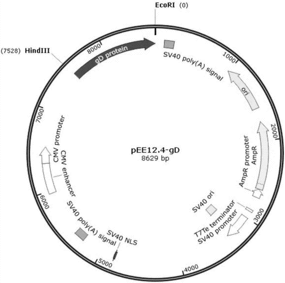
[0032] Example 1: Codon optimization of gD protein of bovine infectious rhinotracheitis virus and construction of pEE12.4-OPTI-gD recombinant plasmid
[0033] By codon-optimizing the nucleotide sequence of the gD protein of bovine infectious rhinotracheitis virus, the OPTI-gD sequence was obtained, as shown in SEQ ID NO.2, and this work was commissioned to Nanjing GenScript Biotechnology Co., Ltd.
Embodiment 2
[0034] Example 2: Construction of pEE12.4-OPTI-gD recombinant plasmid
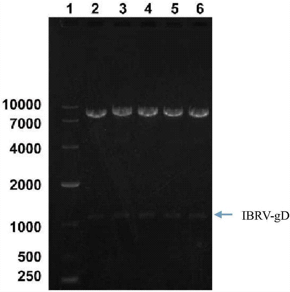
[0035] 2.1 PCR amplification of the target fragment OPTI-gD
[0045] (1) Mark the sample collection EP tube, adsorption column and collection tube;
[0046] (2) Take the weight of the marked empty EP tube, and record the value;
[0047] (3) Carefully cut out a single target DNA band from the agarose gel with a scalpel on a gel cutter and put it into a clean 1.5mL centrifuge tube;
[0048] (4) Add 600 μL PC buffer to the 1.5mL centrifuge tube in step (3), place in a 50°C water bath for about 5 minutes, and gently turn...
Embodiment 3
[0112] Example 3: Establishment of transfection of pEE12.4-OPTI-gD recombinant plasmid into CHO-K1 cells and monoclonal screening
[0114] (1) Preparation: UV sterilization in a biological safety cabinet for 30 minutes; DMEM / F12 (containing 10% serum, 1% double antibody), DMEM / F12 and PBS were placed in a 37°C water bath and preheated to 37°C.
[0115] (2) Take out the cells (10 cm cell culture dish) from the incubator at 37° C., discard the supernatant medium, wash the cells once with pre-warmed 8 mL PBS, and discard the PBS.
[0116] (3) Add 1-2mL 0.25% trypsin-EDTA to each 10cm cell culture dish, digest at room temperature for about 2 minutes, observe under the microscope that the cells shrink and become round, and appear as single cells.
[0117] (4) Add 4 mL of DMEM / F12 (containing 10% serum, 1% double antibody) to terminate the digestion reaction, and blow the cells away with a pipette.
[0118] (5) Transfer the digested cells to a...
PUM
Abstract
The invention discloses preparation of CHO cell expressed recombinant infectious bovine rhinotracheitisvirusprotein gD and a subunit vaccine thereof and an application and belongs to the technical fields of animal vaccines and veterinary biologicals. The condition that the vaccine can generate relatively high humoral immunity in bovine bodies is proven. The object of the invention is to providea preparation method capable of industrially producing the infectious bovine rhinotracheitisvirus recombinant subunit vaccine on a large scale. The reparation method for the recombinant subunit vaccine comprises the following steps: 1) cloning an eukaryotic expression vector containing a protein gD coding gene; 2) transfecting CHO cells, and obtaining suspending CHO cell strains, which stably andefficiently express the protein gD, in a selecting, screening and acclimatizing manner; 3) subjecting the cell strains obtained in the step 2) to fermented culture, and carrying out purification, soas to obtain recombinant protein gD; and 4) uniformly mixing the recombinant protein gD and ISA 201 VG thoroughly, thereby obtaining the recombinant subunit vaccine. According to the method provided by the invention, target protein can be obtained from cell culture supernatant, the yield reaches up to 2g / L to 3g / L, the protein purification time is shortened, the vaccine production steps are simplified, and the vaccine production cost is greatly reduced.
Application Information
Patent Timeline
Application Date:The date an application was filed.
Publication Date:The date a patent or application was officially published.
First Publication Date:The earliest publication date of a patent with the same application number.
Issue Date:Publication date of the patent grant document.
PCT Entry Date:The Entry date of PCT National Phase.
Estimated Expiry Date:The statutory expiry date of a patent right according to the Patent Law, and it is the longest term of protection that the patent right can achieve without the termination of the patent right due to other reasons(Term extension factor has been taken into account ).
Invalid Date:Actual expiry date is based on effective date or publication date of legal transaction data of invalid patent.

