

Preparation method of urinary protein and detection method for urinary proteome
A urine protein and protein technology, which is applied in the detection field of urine proteome, can solve the problems of low throughput of quantitative deep urine proteome, etc., and achieve the effects of improving accuracy and repeatability, short mass spectrometry detection time, and simple preparation process
- Summary
- Abstract
- Description
- Claims
- Application Information
AI Technical Summary
Problems solved by technology
Method used
Image

Examples
Embodiment 1
[0055] Embodiment 1 urine protein preparation method
[0056] (1) Centrifuge 1ml of urine sample at 20°C for 75 minutes with a centrifugal force of 200,000g, discard the supernatant, and keep the precipitate;
[0057] (2) Transfer the precipitate to a 1.5ml centrifuge tube, add 400 μl of resuspension buffer (50mM Tris, 250mM sucrose, pH 8.5) to the centrifuge tube, let it stand at room temperature for 30 minutes, and blow the weight with a pipette. suspended sediment;
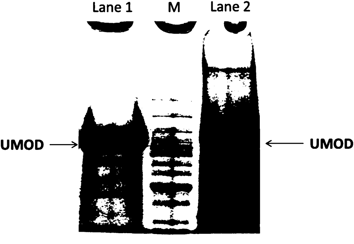
[0058] (3) Dithiothreitol was added to the above resuspended pellet to a final concentration of about 100mM, and heated at 65°C for 30 minutes to remove most of the uromodulin (UMOD, see figure 1 );
[0059] (4) Add cleaning buffer (10mM triethanolamine, 100mM sodium chloride, pH7.4) to 4ml, then centrifuge at 20°C for 75 minutes with a centrifugal force of 200000g, discard the supernatant, and leave the precipitate;
[0060] (5) Redissolve the precipitate with 30 μl of digestion buffer (such as 10 mM Tris o...
Embodiment 2
[0064] Embodiment 2 urine protein preparation method
[0065] (1) Centrifuge 10ml of urine sample at 4°C for 20 minutes at a centrifugal force of 100,000, discard the supernatant and keep the precipitate;
[0066] (2) Transfer the above precipitate to a centrifuge tube, add 60ul of resuspension buffer (50mM Tris, 250mM sucrose, pH8.5) into the centrifuge tube, let it stand at room temperature for 10 minutes, and blow the resuspended precipitate fully with a pipette ;
[0067] (3) Add dithiothreitol to the above resuspended pellet to a final concentration of 50 mM, and heat at 80°C for 10 minutes to remove most of the uromodulin protein in the sample;
[0068] (4) Add cleaning buffer (10mM triethanolamine, 100mM sodium chloride, pH7.4) to 400ul, then centrifuge for 20 minutes under 4 conditions with a centrifugal force of 100000, discard the supernatant, and leave the precipitate;
[0069] (5) Use one-dimensional electrophoresis (SDS-PAGE) for protein separation, dissolve the...
Embodiment 3
[0070] Embodiment 3 A method for enriching urinary protein,
[0071] Including the following steps:
[0072] (1) Add 10 mg of diatomaceous earth to 1 ml of urine and shake well;
[0073] (2) Heating at a temperature between 37-100°C for 5 minutes, then cooling to room temperature;
[0074] (3) Rotate and mix at room temperature for 30 minutes, centrifuge at 12,000 rpm for 5 minutes, and discard the supernatant;
[0075] (4) Add 30-100 microliters of digestion buffer to carry out in-solution digestion of enriched urinary protein.
PUM
| Property | Measurement | Unit |
|---|---|---|
| Diameter | aaaaa | aaaaa |
| The inside diameter of | aaaaa | aaaaa |
| Diameter | aaaaa | aaaaa |
Abstract
Description
Claims
Application Information