Method for culturing human umbilical cord mesenchymal stem cells

A technology of stem cells and culture methods, applied in cell dissociation methods, cell culture active agents, animal cells, etc., can solve problems such as cell damage, introduction of bovine-derived and pig-derived viruses, easy residual bovine serum albumin, etc., to achieve Broad clinical application prospects, the effect of stable cell properties

Inactive Publication Date: 2018-09-07
SICHUAN UNIV +1
View PDF6 Cites 4 Cited by
  • Summary
  • Abstract
  • Description
  • Claims
  • Application Information

AI Technical Summary

Problems solved by technology

[0007] The technical problem to be solved by the present invention is: the existing method for isolating human umbilical cord mesenchymal stem cells is easy to cause cell d

Method used


Image

  • Method for culturing human umbilical cord mesenchymal stem cells
  • Method for culturing human umbilical cord mesenchymal stem cells
  • Method for culturing human umbilical cord mesenchymal stem cells

Examples


Embodiment 1

[0054] Example 1 Primary isolation and culture of HUMSC

[0055] The umbilical cords of healthy fetuses delivered by cesarean section at term were collected. Before the umbilical cord is collected, the puerpera needs to be tested for HIV antibody, hepatitis B virus antibody, hepatitis C virus antibody, Treponema pallidum antibody, etc., all of which are qualified to ensure safety before collection.

[0056] (1) Sterile environment Take an umbilical cord with a length of about 10-15 cm, rinse it repeatedly with sterilized PBS buffer, cut it into 2-3 cm long pieces, and place it in a basal medium (plus 1 % penicillin and streptomycin) in a Petri dish.

[0057] (2) Remove blood vessels, and wash in culture medium until no obvious blood stains.

[0058] (3) In a small amount of culture medium, cut the umbilical cord tissue into small pieces, and use ophthalmic scissors to cut them into pieces of about 1 mm×1 mm.

[0059] (4) Move the fragments to a 50mL centrifuge tube, add 0.3...

Embodiment 2

[0062] Example 2 Subculture of HUMSC

[0063] The specific operation process is as follows:

[0064] (1) When the P0 generation cells in Example 1 are to be fused to 80% to 90%, they are digested with trypsin substitute and centrifuged to remove the supernatant;

[0065] (2) Resuspend the cell pellet with serum-free medium (containing 10% of the total amount of cytokines and chemokines), reseet it in a new culture dish at a ratio of 1:3, and store it in 5% CO 2 , Continue culturing in a 37°C incubator, and the cells are at the P1 generation;

[0066] (3) When the confluence of the P1 generation cells reaches 80-90%, repeat the above operations (1) and (2) to obtain the P2 generation cells.

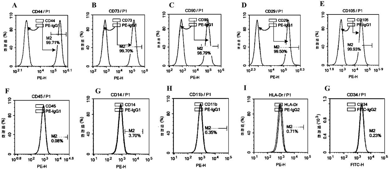
[0067] The reason for using a petri dish here: the purity of the P0-P2 generation cells gradually increases, and the cell surface markers at the P2 generation are consistent with the detection standards.

Embodiment 3

[0068] Example 3 HUMSC expanded culture

[0069] After the P2 generation cells obtained in Example 2 were digested, they were inoculated in spinner bottles for adherent culture.

[0070] (1) When the P2 generation cells in Example 2 are fused to 80-90%, digest with trypsin substitute, obtain a single cell suspension and centrifuge to remove the supernatant;

[0071] (2) resuspend the cell pellet with serum-free medium (containing 10% of the total amount of cytokines and chemokines);

[0072] (3) After counting the cells, they were reseeded in a spinner bottle and placed in 5% CO 2 , Continue culturing in a 37°C incubator until the confluence of the cells reaches 80-90%, and obtain P3 generation cells;

[0073] (4) Steps (1) (2) (3) above were repeated until the P5 generation of human umbilical cord mesenchymal stem cells was obtained.

[0074] After the cell pellet was resuspended, counted and re-inoculated into a new spinner bottle, the P5 generation cells were obtained by...


PUM

PropertyMeasurementUnit
Lengthaaaaaaaaaa

Abstract

The invention belongs to the technical field of cell culture and in particular relates to a method for culturing human umbilical cord mesenchymal stem cells. Aiming at the problems that conventional separated human umbilical cord mesenchymal stem cells have residual bovine serum, bovine-derived and swine-derived viruses, and the like, the invention provides the method for culturing the human umbilical cord mesenchymal stem cells. The method comprises the following steps: a, carrying out primary culture on human umbilical cord tissue with a serum-free medium till a P0 generation; b, carrying out subculture on cells of the P0 generation till a P2 generation in a culture dish; c, carrying out amplification culture on cells of the P2 generation till a fifth generation in a spinner bottle, thereby obtaining human umbilical cord mesenchymal stem cells after multiplication culture. By adopting the method provided by the invention, a serum-free culture mode is carried out in the whole process,artificially prepared cell factors and chemotactic factors are added, and the culture medium is low in sensitization and small in potential hazard factor; due to adoption of amplification culture inthe spinner bottle, the method has the advantages of rapid multiplication, short cycle, low cost, considerable multiplication cell number, and the like, thereby having wide clinical application prospects.

Description

technical field [0001] The invention belongs to the technical field of stem cell culture, and in particular relates to a culture method for human umbilical cord mesenchymal stem cells. Background technique [0002] Mesenchymal stem cells (MesenchymalStemCell, MSC) are a group of stem cells with strong proliferation ability and multi-lineage differentiation potential, and have remarkable effects in immune regulation and anti-inflammatory response. Therefore, MSCs have shown very good clinical prospects in the treatment of many major diseases such as graft-versus-host reaction (GVHD), autoimmune diseases, severe liver diseases, and diabetes. [0003] MSCs have a wide range of tissue sources, such as umbilical cord, bone marrow, fat and other tissues rich in MSCs. Among them, the umbilical cord is a cord-like structure connecting the umbilicus of the embryo and the placenta, and is rich in various stem / progenitor cells such as hematopoietic, mesenchymal, neural, and endothelia...

Claims


Application Information

Patent Timeline
IPC IPC(8): C12N5/0775
CPCC12N5/0668C12N2500/90C12N2501/20C12N2501/21C12N2509/00
Inventor 邓洪新魏于全宋海峰
Owner SICHUAN UNIV
Why Eureka
Try Eureka