Method for separating free fetal cells from peripheral blood of pregnant women

A technology for maternal peripheral blood and fetal cells, applied in chemical instruments and methods, analytical materials, material excitation analysis, etc., can solve the problems of small size, only identification, low enrichment rate of fetal cells, and reduce non-specific adsorption. , Improve the probability of acquisition, and improve the effect of capturing purity

Active Publication Date: 2018-09-14
WISDOM HEALTHY BIOTECH XIAMEN CO LTD
View PDF6 Cites 10 Cited by
  • Summary
  • Abstract
  • Description
  • Claims
  • Application Information

AI Technical Summary

Problems solved by technology

[0003] At present, the main problems of fetal cell separation and capture include: 1. The capture efficiency is very low
The cell loss rate of magnetic bead separation and flow cytometry is very high. The capture of fetal cells often requires more than 10 ml of peripheral blood, and the highest even requires 30 ml. Such a high sample demand is difficult for all pregnant women to accept, and The number of captured fetal cells is still less than 10, and the enrichment rate is too low
The cell smear method is based on the traditional nucleated red blood cell detection method to analyze fetal cells in peripheral blood. The cell smear method mainly relies on huma...

Method used


Image

  • Method for separating free fetal cells from peripheral blood of pregnant women
  • Method for separating free fetal cells from peripheral blood of pregnant women
  • Method for separating free fetal cells from peripheral blood of pregnant women

Examples


Embodiment 1

[0035] Example 1 Preparation of fetal cell capture microfluidic chip

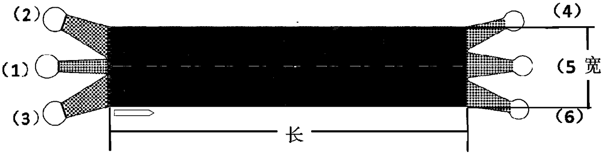
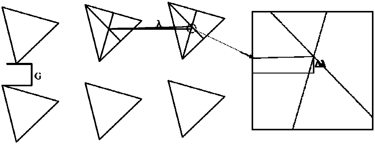
[0036] see figure 1, to make a microfluidic chip. The chip consists of two layers of PDMS and a layer of glass chip from bottom to top. The PDMS thick block, PDMS channel layer, and slide glass are sequentially used to form a complete chip by plasma bonding. The chip is equipped with three sample inlets (1), (2), (3) and three sample outlets (4), (5), (6), with a triangular microarray between the sample inlet and the sample outlet, The microarray arrangement adopts the DLD design principle, such as figure 1 shown.

[0037] In this embodiment, three sample inlets are located on the left side of the chip, and three sample outlets are located on the right side of the chip, and a 0.7mm punch pen is used to prepare the inlet and outlet ports.

[0038] In this embodiment, the chip size is designed to be 1 cm wide and 4.5 cm long.

[0039] In this embodiment, the horizontal distance G between the pillars is se...

Embodiment 2

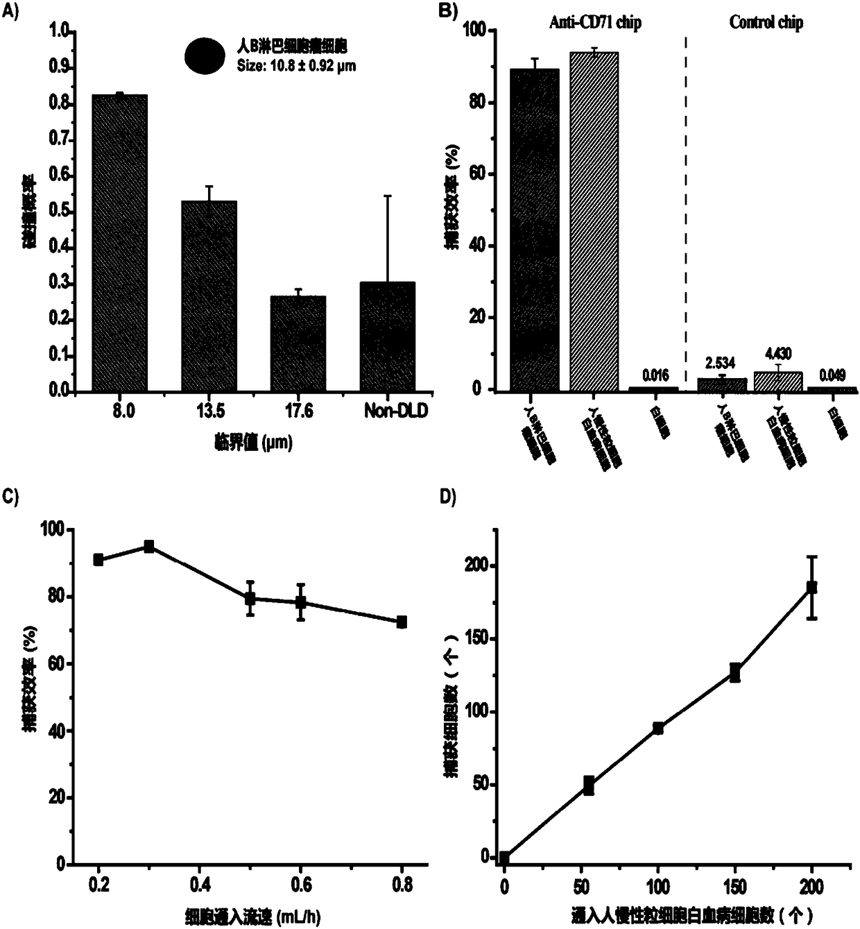
[0041] Example 2 Simulated Fetal Cell Capture Characterization

[0042] The condition parameters and specific steps are as follows:

[0043] 1) Fluorescence pre-stained target cells (Ramos&K562) or control cells (WBCs) were resuspended in PBS buffer solution after cell counting;

[0044] 2) The sample is injected from the injection hole (1) through the injection system, and the buffer solution is injected through (2) and (3);

[0045] 3) Inject PBS buffer solution through the injection port (1, 2, 3) to clean the chip;

[0046] 4) The cells are counted and counted, and divided by the number of injected cells to calculate the capture efficiency;

[0047] The transferrin CD-71 antibody is bound to the chip, and the efficiency of capturing target cells is greater than 80%, and the white blood cells are 0.016%.

[0048] In this example, the result is as image 3 shown.

[0049] image 3 Statistics for the capture efficiency of simulated fetal cells by the microfluidic chip. ...

Embodiment 3

[0051] Embodiment 3 contains the preparation of the mononuclear cell suspension of fetal cell

[0052] Two milliliters of peripheral blood from pregnant women was taken, diluted to 4 milliliters by adding PBS buffer, and the above sample was processed by percoll gradient centrifugation to obtain a mononuclear cell layer, washed, and resuspended to obtain a mononuclear cell suspension. The percoll density is 1.090, the centrifugal force is 400g, and the time is 30 minutes. Figure 5 Schematic diagram of gradient centrifugation of peripheral blood in pregnant women.


PUM

PropertyMeasurementUnit
Critical diameteraaaaaaaaaa
Diameteraaaaaaaaaa

Abstract

The present invention relates to a method for separating free fetal cells from peripheral blood of a pregnant woman. The method utilizes the cell separation function and the specific affinity recognition principle of a microfluidic chip to realize high-efficiency and high-purity capture and separation of the free fetal cells (fetal nucleated red blood cells, trophoblast cells, and the like) in theperipheral blood of the pregnant woman. Within the microfluidic chip, a microarray structure with a specific geometric arrangement is designed, and the microarray structure is modified with moleculargroups having an affinity recognition function such as a nucleic acid aptamer, an antibody and a polypeptide. By adjusting parameters of the microarray structure, the collision efficiency of different sizes of fetal cells and microarrays can be controlled, and the capture and enrichment of the fetal cells of different sizes can be realized.

Description

technical field [0001] The invention relates to a method for separating free fetal cells from peripheral blood of pregnant women. Background technique [0002] In recent years, with the liberalization of the two-child policy, the number of elderly mothers has continued to increase, and the birth defect rate is on the rise. In the tertiary prevention of birth defects, the second-level "prenatal screening and diagnosis" is the most important means of preventing birth defects. Amniocentesis is the main method of antenatal antenatal drainage for high-risk puerperas, but it is highly invasive and has high risks of infection and miscarriage. Noninvasive prenatal testing based on fetal cell-free DNA has been widely used in the screening of autosomal aneuploidy, but there are shortcomings such as a high false positive rate and the inability to rule out parental chromosomal abnormalities. The fetal cells in the maternal peripheral blood, discovered in 1969, have complete fetal gene...

Claims


Application Information

Patent Timeline
IPC IPC(8): G01N21/64B01L3/00
CPCB01L3/502707B01L3/502761G01N21/6486
Inventor 杨朝勇张惠敏杨园园李星锐施远志朱志
Owner WISDOM HEALTHY BIOTECH XIAMEN CO LTD
Why Eureka
Try Eureka