3D printed Ti-PDA-PLGA microsphere bone defect repair stent

A 3D printing and PLGA technology, applied in microcapsules, coatings, medical science, etc., can solve the problems of unguaranteed biological safety, high price, pathogen transmission, etc., and achieve good bone ingrowth characteristics and biological safety , Improved hydrophilicity and biocompatibility, and the effect of good biocompatibility

Inactive Publication Date: 2018-11-23
南京冬尚生物科技有限公司
View PDF6 Cites 12 Cited by
  • Summary
  • Abstract
  • Description
  • Claims
  • Application Information

AI Technical Summary

Problems solved by technology

However, biologically active factors have the possibility of immune rejection, pathogen transmission, allergic reaction, potential tumorigenicity, and teratogenicity, and biological safety cannot be guaranteed, and clinical-grade biological factors such as rhBMP-2 and TGF-β often need to be imported, and The price is expensive, and the purchase price per milligram reaches nearly a thousand dollars

Method used


Image

  • 3D printed Ti-PDA-PLGA microsphere bone defect repair stent
  • 3D printed Ti-PDA-PLGA microsphere bone defect repair stent
  • 3D printed Ti-PDA-PLGA microsphere bone defect repair stent

Examples


Embodiment 1

[0046] 1. Preparation of 3D printed Ti scaffolds

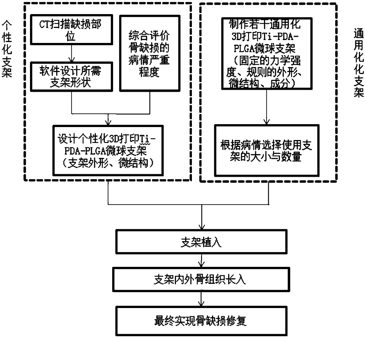
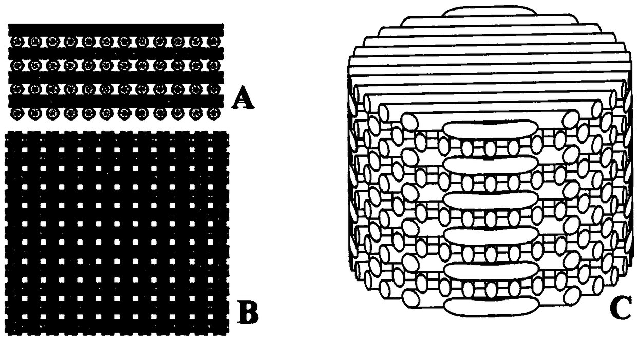
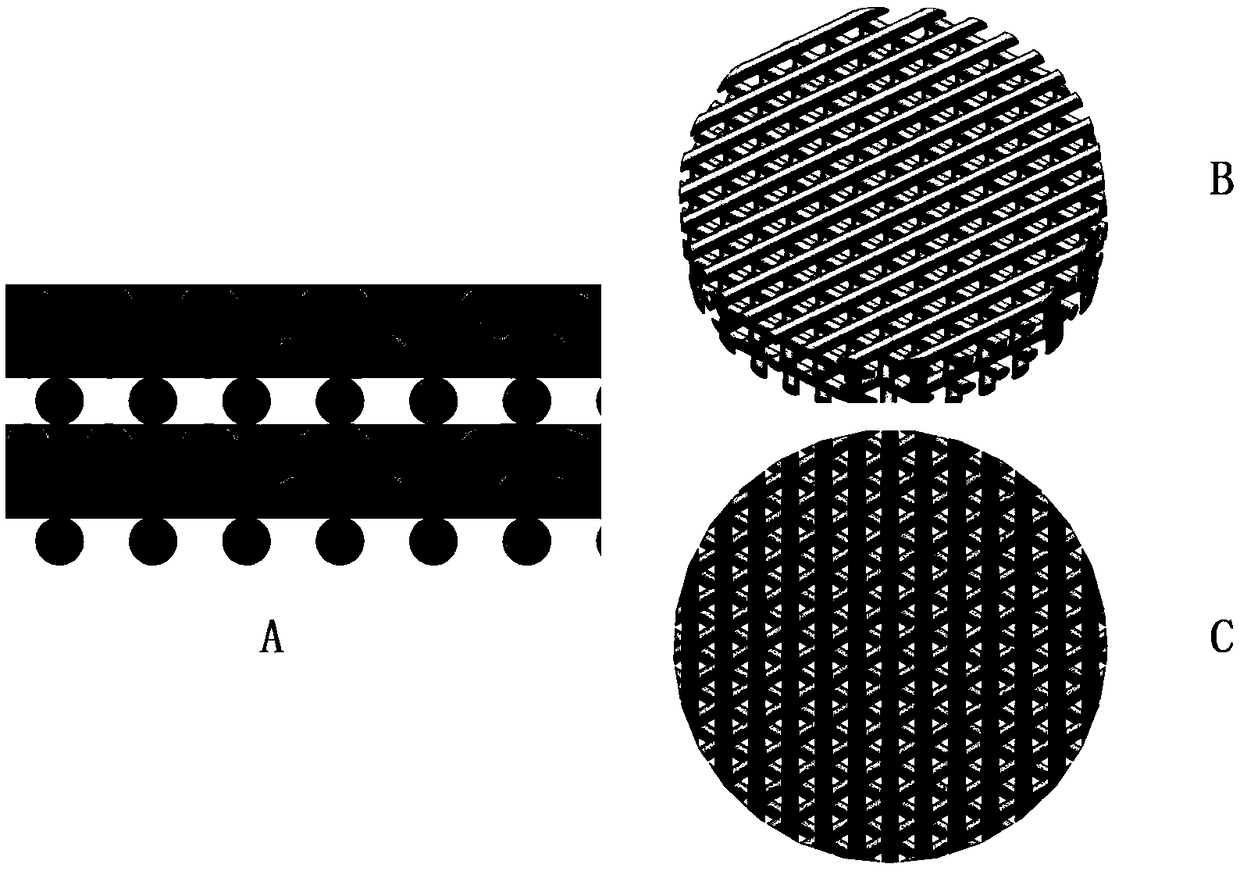
[0047] Using Ti 6Al4V powder as a raw material, a 3D printed Ti scaffold with a 64% porosity fiber grid structure formed by laser sintering technology ( figure 2 , 3 ). Its shape can be 3D printed and personalized according to the shape of the bone defect in the area to be repaired. It can also be manufactured as a scaffold material with a fixed shape through SLM technology, and it can be filled according to the actual bone defect requirements during the operation.

[0048] 2. Preparation of 3D printed Ti-PDA scaffold

[0049] After the stent structure is constructed by 3D printing, the surface is modified using PDA. The specific steps include:

[0050] 1) preparation concentration is the hydrochloric acid (HCL) solution of 0.5mol / L;

[0051] 2) Weigh 0.61g Tris and add it into 500ml water to dissolve, after stirring, add the prepared 0.5mol / L HCL solution dropwise to adjust to pH=8.5, and prepare Tris-HCL solution for la...


PUM

PropertyMeasurementUnit
concentrationaaaaaaaaaa

Abstract

The invention discloses a 3D printed Ti-PDA-PLGA microsphere bone defect repair stent. A 3D printed Ti stent is prepared by a laser sintering technology. Then, under certain conditions, dopamine is self-polymerized on the fiber surface of the 3D printed Ti stent to form a PDA coating, thereby preparing a 3D printed Ti-PDA stent; then PLGA microspheres carrying VEGF are prepared by a double emulsion-solvent evaporation method, and finally, BMP-2 and PLGA microspheres carrying VEGF are adsorbed and immobilized on the surface of the stent by an adsorption method, and finally the 3D printed Ti-PDA-PLGA microsphere bone defect repair stent is formed. The bone defect repair tissue engineering stent disclosed by the invention has the advantages of reliable mechanical property, high biological activity and safety, convenient implantation, small trauma and low cost, and can be used for the repair treatment of bone defect after bone traumas, bone tumors and bone infection.

Description

technical field [0001] The invention belongs to the field of bone tissue repair and reconstruction, and relates to a 3D printed Ti-PDA-PLGA microsphere bone defect repair bracket. Background technique [0002] The repair and reconstruction of bone defects is one of the clinical problems in orthopedics. It is well known that autogenous bone is considered the gold standard for bone grafting, but its source is limited and may lead to complications such as necrosis of the supply site, postoperative chronic pain, allergic reaction, and infection. While allograft bone and xenograft bone are widely used because they come from a wide range of sources and do not require additional surgical operations, but there are also complications such as slow integration and remodeling, immune rejection, and disease transmission, especially reducing the osteoinductive and osteoconductive. [0003] At present, scaffold materials include bioactive calcium phosphate ceramics, calcium phosphate cem...

Claims


Application Information

Patent Timeline
Patent Type & Authority Applications(China)
IPC IPC(8): A61L27/06A61L27/56A61L27/50A61L27/54A61L27/34
CPCA61L27/06A61L27/34A61L27/50A61L27/54A61L27/56A61L2300/252A61L2300/412A61L2300/414A61L2300/45A61L2300/602A61L2300/622A61L2430/02B22F3/11B22F3/26B22F5/00B33Y10/00B33Y80/00C08L79/02
Inventor 姚庆强王黎明徐燕胡军
Owner 南京冬尚生物科技有限公司
Why Eureka
Try Eureka