Preparation of multilayer cartilage complex containing cytokine microspheres through electrospinning 3D printing

A technology of 3D printing and cytokines, which is applied in prostheses, microcapsules, medical science, etc., can solve the problems that normal cartilage cannot provide friction resistance and support, and is easy to be damaged again, so as to facilitate repair, promote cell proliferation and differentiation, The effect of high structural precision

Inactive Publication Date: 2019-04-19
SHANGHAI NINTH PEOPLES HOSPITAL AFFILIATED TO SHANGHAI JIAO TONG UNIV SCHOOL OF MEDICINE
View PDF8 Cites 7 Cited by
  • Summary
  • Abstract
  • Description
  • Claims
  • Application Information

AI Technical Summary

Problems solved by technology

However, in this method, the generated cartilage does not have the layered structure of normal cartilage. Therefore, it cannot provide the friction resistance and support function of normal cartilage, and it is easy to be dama

Method used


Image

  • Preparation of multilayer cartilage complex containing cytokine microspheres through electrospinning 3D printing
  • Preparation of multilayer cartilage complex containing cytokine microspheres through electrospinning 3D printing
  • Preparation of multilayer cartilage complex containing cytokine microspheres through electrospinning 3D printing

Examples


preparation example Construction

[0028] S2, the preparation of three-phase integrated support;

[0029] S3. Planting of cells: An appropriate amount of bone marrow mesenchymal stem cells is planted in each scaffold area.

[0030] Specifically, in the cultivation of the seed cells in the step S1, the bone marrow mesenchymal stem cells are cultured in a DMEM medium containing 10% fetal bovine serum in an incubator.

[0031] Specifically, the temperature of the incubator is 37° C., and there is 5% CO2 in the incubator.

[0032] Specifically, the preparation of the three-phase integrated stent in the step S2 includes the following steps:

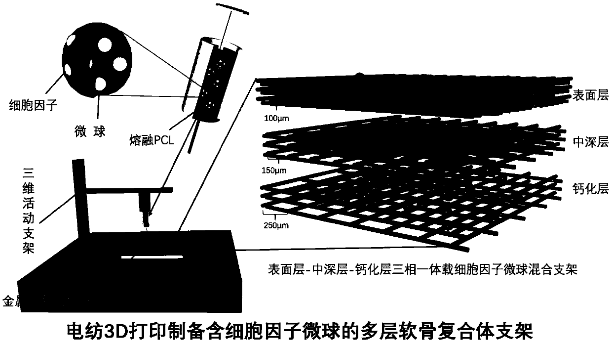
[0033] S21. Model establishment: use 3D printing software to establish a printing model, which is in the shape of a multi-layer cylinder, and the gaps between the structural paths of each layer are adjusted and saved according to needs;

[0034] S22. Preparation of materials: Measure appropriate amounts of biopolymer materials A1, A2, and A3, and microspheres B1, B2, and B3. ...

Embodiment 1

[0041] (1) Use 3D printing software to build a printing model, which is multi-cylindrical, with different path gaps in the surface area, middle-deep area, and calcification layer area, and save it;

[0042] (2) Take an appropriate amount of polymer materials polycaprolactone and polydopamine microspheres, the polydopamine microspheres are respectively loaded with appropriate amount of TGFβ1, BMP7 and IGF cytokines and HA (hydroxyapatite) particles, and an appropriate amount of polyethylene glycol Alcohol, mixed with water to prepare a dispersion liquid, and the microspheres loaded with cytokines are uniformly dispersed in the dispersion liquid;

[0043](3) Equipment preparation: adjust the process parameters of the electrospinning 3D printing equipment: the diameter of the printing wire is 10-50 μm, the printing temperature is 65°C, the air pressure of the barrel is 600-1000KPa during the printing process, and the voltage of the negative high voltage module is - 2.5~-50kV, the...

Embodiment 2

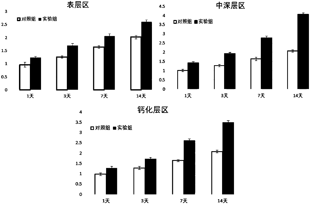
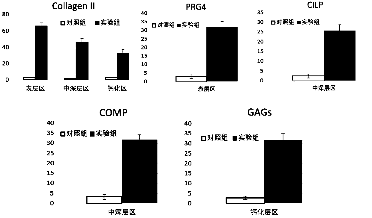
[0045] The cartilage layered scaffold obtained in the above-mentioned Example 1 was characterized for biocompatibility, and the three-phase integrated scaffold mixed with cytokine-loaded microspheres obtained in step (3) was planted with bone marrow stem cells and cultured in an incubator. The scaffolds obtained by three-dimensional printing of caprolactone were planted with bone marrow stem cells as the control group, and compared with each area of ​​the three-phase scaffolds. After 1, 3, 5, and 7 days of culture, the cell viability in the surface area scaffolds was detected by MTT method. The results are shown in Fig. As shown in 2, it shows that the cells on the scaffold obtained by the electrospinning 3D printing technology with cytokine microspheres have good proliferation behavior, the cell viability is better, and the proliferation behavior is more significant than that of the control group.


PUM

PropertyMeasurementUnit
Diameteraaaaaaaaaa

Abstract

The invention belongs to the technical field of cartilage complexes, in particular to preparation of multilayer cartilage complex containing cytokine microspheres through electrospinning 3D printing,which comprises the following steps of: S1, culturing seed cells; S2, preparing a three-phase integrated scaffold; S3, planting cells. According to the method, an electrostatic spinning 3D printing technology is adopted, the fiber diameter and the printing path can be accurately controlled, the diameter of a monofilament can be as fine as 10 micrometers, corresponding loaded cytokine microspheresare mixed in a printing material to be loaded on a printed bracket, by controlling the types and contents of the cytokines, the corresponding seed cells can be induced to differentiate to the corresponding cells on the scaffold layer by layer so as to finally form a component hierarchical structure similar to a normal cartilage tissue, the complex has a layered structure similar to the normal articular cartilage tissue, and has high structural precision; and the loaded cytokine is favorable for promoting cell proliferation and differentiation, and is favorable for repairing cartilage damage.

Description

technical field [0001] The invention belongs to the technical field of cartilage complexes, and in particular relates to electrospinning 3D printing to prepare multilayered cartilage complexes containing cytokine microspheres. Background technique [0002] The damage and degeneration of articular cartilage is a very common form of disease, and it is also one of the main problems faced in orthopedic clinical work. With the aging of the population in our country, and due to the lack of blood vessels and poor self-repair ability of articular cartilage, the incidence of various diseases caused by articular cartilage damage and degeneration is also increasing year by year. Therefore, how to intervene or induce the repair of cartilage damage has become the focus of current research. [0003] For cartilage damage, the current treatment methods are mainly microfracture or cartilage transplantation, but the cartilage formed by the former has poor function, while cartilage transplant...

Claims


Application Information

Patent Timeline
IPC IPC(8): A61L27/12A61L27/16A61L27/18A61L27/20A61L27/22A61L27/38A61L27/54B33Y70/00B33Y10/00
CPCA61L27/12A61L27/16A61L27/18A61L27/20A61L27/222A61L27/227A61L27/3834A61L27/54A61L2300/252A61L2300/412A61L2300/622A61L2430/06B33Y10/00B33Y70/00
Inventor 乔之光韩煜戴尅戎孙彬彬连梅菲唐佳昕
Owner SHANGHAI NINTH PEOPLES HOSPITAL AFFILIATED TO SHANGHAI JIAO TONG UNIV SCHOOL OF MEDICINE
Why Eureka
Try Eureka