Method for detecting cell capable of expressing carcinoembryonic antigen, and application thereof
A carcinoembryonic antigen, cell technology, applied in the field of immune detection, to achieve the effect of low-cost detection and sensitive detection
- Summary
- Abstract
- Description
- Claims
- Application Information
AI Technical Summary
Problems solved by technology
Method used
Image

Examples
Embodiment 1
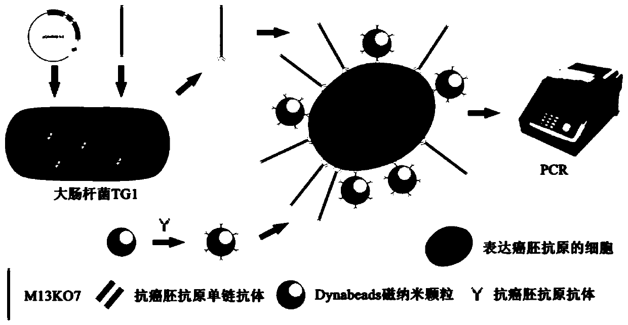
[0059] Example 1 Preparation of anti-carcinoembryonic antigen single-chain antibody fusion virus probe
[0060] (1) Construction of anti-carcinoembryonic antigen single-chain antibody phage library as a probe
[0061] 1. The heavy chain variable region (GenBank: CS681102.1) and the light chain variable region (GenBank: CS681104.1) of the anti-carcinoembryonic antigen single-chain antibody sequence are linked by (Gly4Ser)3, provided by Nanjing Detai Biotechnology Co., Ltd. It was synthesized and cloned between the Sfi I and Not I cloning sites of the pCANTAB 5E vector, and the recombinant plasmid was named pCANTAB 5E-ACEAscFv.
[0062] 2. Transform the recombinant plasmid pCANTAB 5E-ACEAscFv into Escherichia coli TG1 competent cells, spread on an ampicillin-resistant plate, and culture overnight at 37°C.
[0063] 3. Select the positive clones to inoculate into 4mL 2xYT (Tryptone 16g, Yeast Extract 10g, Nacl5g, deionized water to 1L, pH=7.0) on the next day, and culture it on t...
Embodiment 2
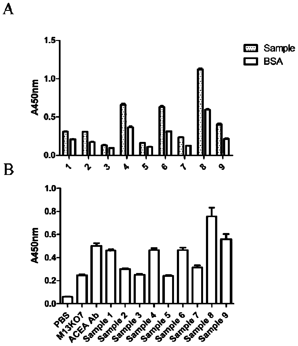
[0073] Example 2 Identification of single-chain antibody fusion virus probes against carcinoembryonic antigen
[0074] (1) Indirect ELISA identification of anti-carcinoembryonic antigen single-chain antibody library specificity
[0075] 1. Use antigen coating solution (Na 2 CO 3 0.159g,NaHCO 3 0.293g, dilute to 100mL, pH=9.6) Dilute carcinoembryonic antigen protein to 2μg / mL, add 100μL per well into a 96-well plate, and coat overnight at 4°C.
[0076] 2. Discard excess CEA protein dilution in the 96-well plate the next day, wash 3 times with 1x PBST (1xPBS containing 0.05% Tween 80, v / v), and dry.
[0077] 3. Add 200 μL of 1% BSA (v / v), block at 37°C for 2 hours, wash with 1xPBST three times, and dry.
[0078] 4. Add 100 μL of 100-fold diluted M13KO7-ACEA scFv, and incubate at 37°C for 1 hour.
[0079] 5. After washing 6 times with 1xPBST and drying, add 100 μL of anti-M13 phage capsid protein g8p antibody diluted 100 times in 1xPBS, and incubate at 37°C for 1 hour.
[...
Embodiment 3
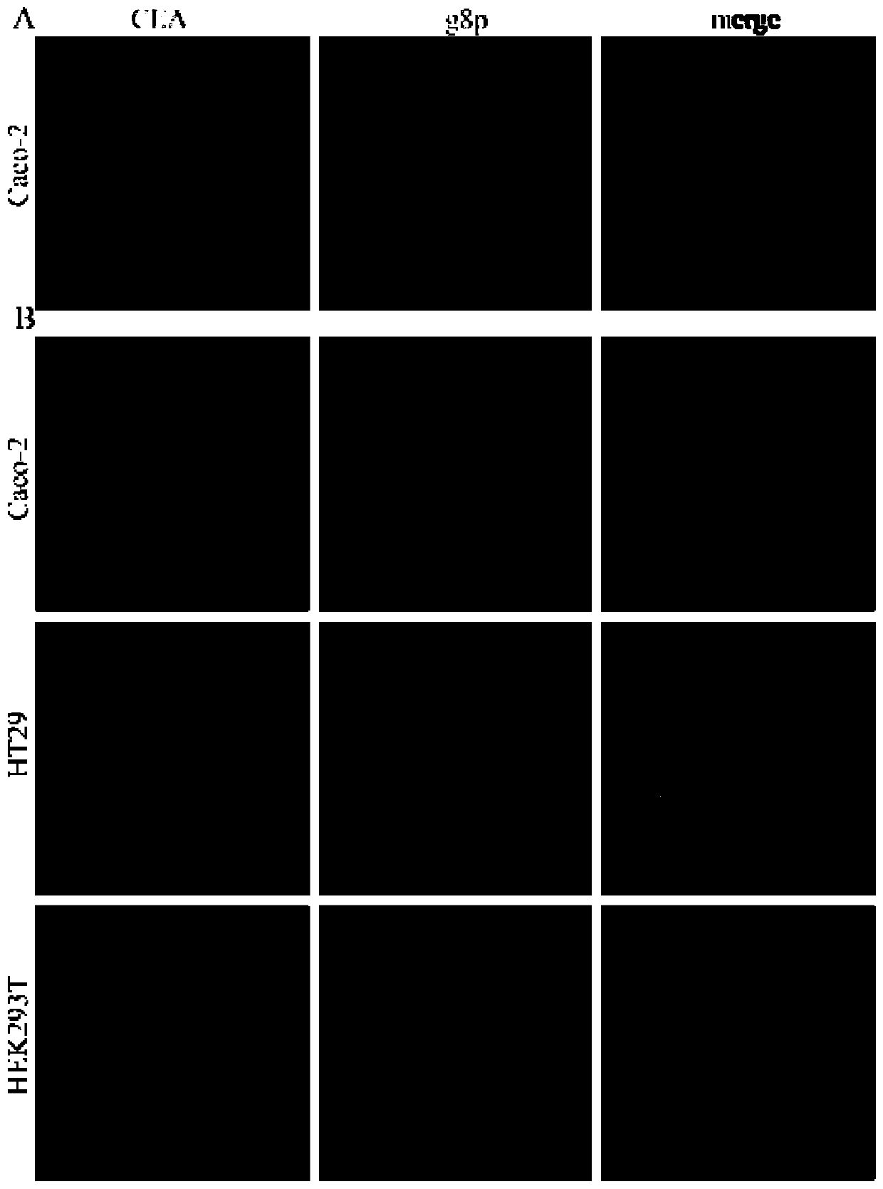
[0098] Example 3 Application of anti-carcinoembryonic antigen single-chain antibody fusion virus probe
[0099] (1) M13KO7-ACEAscFv infected Caco-2 cells with different MOI
[0100] 1. Inoculate Caco-2 cells into a 6-well plate at 37°C, 5% CO 2 Cells were cultured in an incubator, and when the cells grew to 70-80%, M13KO7-ACEA scFv was inoculated at MOI of 0.01, 0.05, 0.1, 0.5, 1, 2 and 5, M13KO7 was inoculated at MOI of 1, and infected for 2 hours.
[0101] 2. Discard the supernatant, wash the 6-well plate three times with pre-cooled 1xPBS, extract the whole genome of the cells by TRIzol method, and reverse transcribe.
[0102] 3. Set up the M13KO7-ACEAscFv virus group and the β-Actin internal reference group for real-time fluorescent quantitative PCR.
[0103] The reaction system and conditions of real-time fluorescence quantitative PCR are as follows:
[0104] 20μL reaction system:
[0105]
[0106] The reaction conditions are:
[0107]
[0108] (2) M13KO7-ACEAsc...
PUM
 Abstract
Description
Claims
Application Information
 - R&D
- Intellectual Property
- Life Sciences
- Materials
- Tech Scout
- Unparalleled Data Quality
- Higher Quality Content
- 60% Fewer Hallucinations
Browse by: Latest US Patents, China's latest patents, Technical Efficacy Thesaurus, Application Domain, Technology Topic, Popular Technical Reports.
© 2025 PatSnap. All rights reserved.Legal|Privacy policy|Modern Slavery Act Transparency Statement|Sitemap|About US| Contact US: help@patsnap.com