Composite collagen membrane for bone grafting in alveolar bone defect area and preparation method of composite collagen membrane

A composite glue and alveolar bone technology, applied in the field of medical devices, can solve the problem of unclear fusion of osteoblasts, and achieve the effects of easy industrial production, low antigenicity and good adhesion

Pending Publication Date: 2019-12-13
CHANGZHOU INST OF MATERIA MEDICA +1
View PDF11 Cites 7 Cited by
  • Summary
  • Abstract
  • Description
  • Claims
  • Application Information

AI Technical Summary

Problems solved by technology

The absorbable biofilm disclosed in this patent is a three-layer composite structure. The outer dense layer is used to prevent the invasion of non-osteoblasts, but it is unclear whether the inner side is conducive to the fusion of osteoblasts.

Method used


Image

  • Composite collagen membrane for bone grafting in alveolar bone defect area and preparation method of composite collagen membrane
  • Composite collagen membrane for bone grafting in alveolar bone defect area and preparation method of composite collagen membrane
  • Composite collagen membrane for bone grafting in alveolar bone defect area and preparation method of composite collagen membrane

Examples


Embodiment 1

[0047] (Example 1, Composite Collagen Membrane for Bone Grafting in Alveolar Bone Defect Area)

[0048] The composite collagen film of this embodiment is a double-layer structure, consisting of a dense collagen fiber film layer and a collagen sponge layer; the composition of the collagen fiber film layer is pure bovine collagen fibers, and the composition of the collagen sponge layer is composed of bovine collagen fibers and hyaluronic acid Composition, wherein the mass percentage of hyaluronic acid is 1% to 10% (3% in this embodiment).

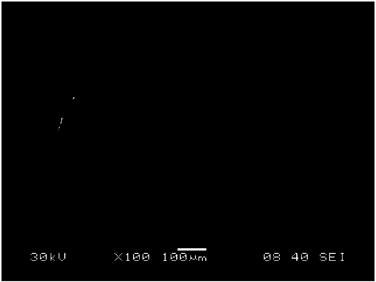
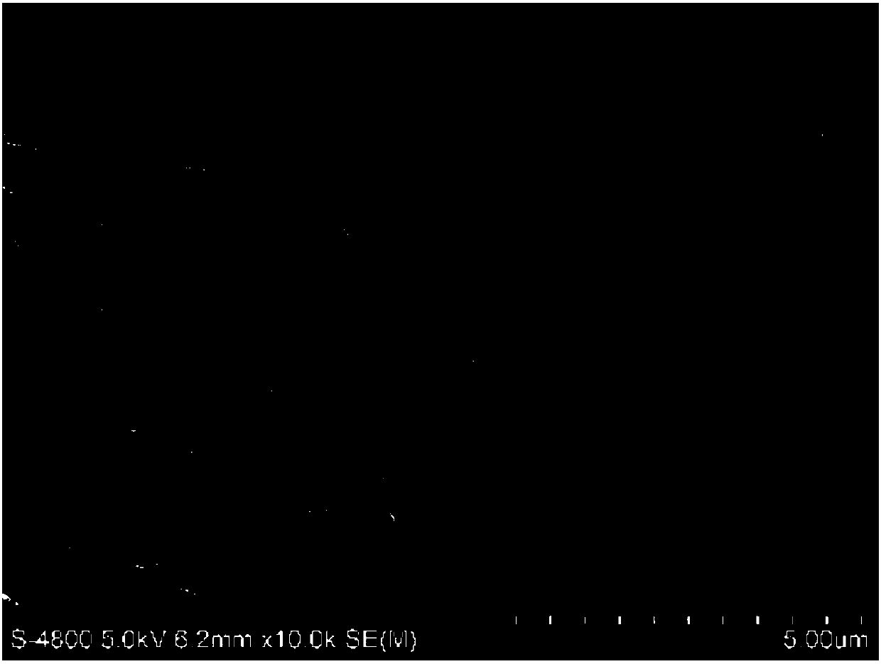
[0049] The scanning electron micrograph of the collagen sponge layer is shown in figure 1 ; The scanning electron micrograph of the collagen fiber membrane layer is shown in figure 2 and image 3 .

[0050] The thickness of the composite collagen membrane is 0.8mm-2mm, and the ratio of the thickness of the collagen fiber membrane layer to the thickness of the collagen sponge layer is 1:2-4. In this embodiment, the thickness of the collag...

Embodiment 2

[0053] (Example 2, preparation method of composite collagen membrane for bone grafting in alveolar bone defect area)

[0054] This embodiment prepares the composite collagen membrane described in Example 1. The preparation process first prepares a collagen fiber membrane, and then forms a collagen sponge layer above the collagen fiber membrane, which specifically includes the following steps:

[0055] ① Preparation of raw materials. The cowhide (without mad cow disease) from a qualified slaughterhouse is washed, washed and dehaired, and then the lower layer of the cowhide with flesh and the upper layer of the epidermis are removed using a piece of hide to obtain the collagen fiber layer. Soak the fiber layer in 0.5% to 2% sodium carbonate solution, turn it over and over again, and then dry it. The collagen fiber layer is added with acetone to degrease and air-dry several times to obtain raw materials.

[0056] ②Removal of impurities and homogenization. Break the dehaired an...

Embodiment 3

[0072] (Example 3, preparation method of composite collagen membrane for bone grafting in alveolar bone defect area)

[0073] All the other preparation methods of the composite collagen film of the present embodiment are the same as in Example 2, except that:

[0074] In step ④, the collagen fiber solution obtained in step ③ is taken, and dried in a vacuum drying oven at 35±2°C for 8-15 hours to obtain a dried sheet, which is a dense collagen fiber membrane.


PUM

PropertyMeasurementUnit
thicknessaaaaaaaaaa
thicknessaaaaaaaaaa
thicknessaaaaaaaaaa

Abstract

The invention discloses a composite collagen membrane for bone grafting in an alveolar bone defect area and a preparation method of the composite collagen membrane, the composite collagen membrane isof a double-layer structure and is composed of a compact collagen fiber membrane layer and a collagen sponge layer; wherein the collagen fiber membrane layer is made of bovine collagen fibers, and thecollagen sponge layer comprises bovine collagen fibers and hyaluronic acid. The compact layer can prevent non-osteoblasts from growing towards a bone grafting area, but allows fibroblasts to be attached along collagenous fibers; and a three-dimensional frame structure of the collagen sponge layer serving as the inner side is beneficial to osteoblast fusion and bone regeneration supporting. The composite collagen membrane has low antigenicity and excellent biocompatibility and has enough barrier time after being used for a human body, and the membrane can keep a complete structure in a wet state and achieve the effect of being attached to the bone wall; after the composite collagen membrane is used for about 1-2 months, the composite collagen membrane starts to be automatically degraded, and is finally decomposed into components which are non-toxic and harmless to a human body and are absorbed by tissues.

Description

technical field [0001] The invention relates to a medical device, in particular to a composite collagen film for bone grafting in alveolar bone defect areas and a preparation method thereof. Background technique [0002] Dental implant is a commonly used method of oral restoration. The survival of implant is closely related to its good initial stability, and the stability depends on the existence of sufficient alveolar bone in the implant area. After the natural tooth is lost, the remaining alveolar ridge shows irreversible and continuous absorption, which leads to a decrease in the height and density of the alveolar bone, resulting in insufficient bone mass in the implant area, which affects the stability of the implant. Fracture, perforation and other complications. One of the most important remedial methods for bone defects in the implant area is to guide bone regeneration in the implant area. [0003] Guided bone regeneration technology refers to the use of absorbable ...

Claims


Application Information

Patent Timeline
Patent Type & Authority Applications(China)
IPC IPC(8): A61L27/40A61L27/24A61L27/20A61L27/50A61L27/56A61L27/58
CPCA61L27/24A61L27/20A61L27/56A61L27/50A61L27/58A61L2430/12
Inventor 李德华管汉亮管湘宁何浩明
Owner CHANGZHOU INST OF MATERIA MEDICA
Why Eureka
Try Eureka