Injectable medical wound dressing with antibacterial function
A wound and functional technology, applied in medical science, pharmaceutical formulations, bandages, etc., can solve problems such as limited antibacterial ability, poor effect of promoting wound healing, and severe infection that is difficult to heal, so as to promote repair and good self-healing performance, effect of good coverage
- Summary
- Abstract
- Description
- Claims
- Application Information
AI Technical Summary
Problems solved by technology
Method used
Image
Examples
Embodiment 1
[0051] The preparation of embodiment 1 raw material
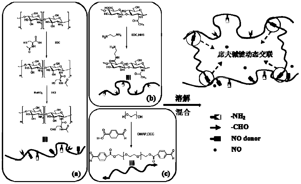
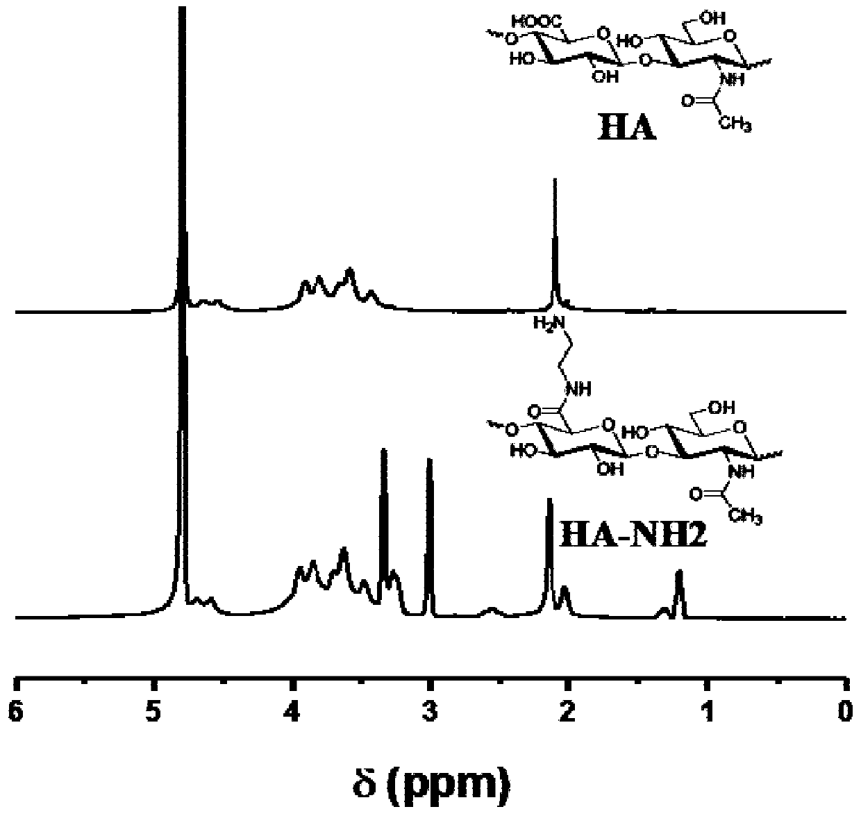
[0052] figure 1 a is amino hyaluronic acid (HA-NH 2 ), the specific synthesis route is as follows: Weigh 200 mg of low molecular weight sodium hyaluronate HA-TLM20-40 into a 250 mL round bottom flask, add 100 mL of deionized water and stir until completely dissolved at room temperature, and use 0.5 M The pH of the solution was adjusted to 5.5 with hydrochloric acid, and then 194.8 mg of EDC and 117.0 mg of NHS were added to activate the carboxyl group at room temperature for 30 min, and then 152.7 mg of ethylenediamine was added to react overnight at room temperature. After the reaction is completed, put the above solution into a dialysis bag with a molecular weight cut-off of 3500Da for dialysis purification, and finally pre-freeze and freeze-dry at -20°C to obtain HA-NH 2 , its H NMR spectrum is shown in figure 2 , where the new peaks at 2.03ppm and 3.01-3.34ppm are the peaks of hydrogen on the grafted ethylenediamine...
Embodiment 2
[0057] 1) HA-NH 2 / Preparation of CS-SNO solution
[0058] Add polymer HA-NH per 100 μL ultrapure water 2 5mg and CS-SNO 10.1mg, stir well with a magnetic stirrer to form a homogeneous solution at room temperature.
[0059] 2) Preparation of DF-PEG 2000 solution
[0060] Add polymer DF-PEG 2000 30 mg per 100 μL of ultrapure water, use a magnetic stirrer to stir well, and form a homogeneous solution at room temperature.
[0061] 3) Preparation of composite gel
[0062] Calculated by volume, take 1 part of HA-NH2 / CS-SNO solution and 1 part of DF-PEG 2000 solution, mix evenly under the magnetic stirrer, adjust the pH to neutral with 3M NaOH, and let it stand to get compound gel.
Embodiment 3
[0064] 1) HA-NH 2 / Preparation of CS-SNO solution
[0065] Add polymer HA-NH per 100 μL ultrapure water 2 5mg and CS-SNO 10.1mg, stir well with a magnetic stirrer to form a homogeneous solution at room temperature.
[0066] 2) Preparation of DF-PEG 2000 solution
[0067] Add polymer DF-PEG 2000 40 mg per 100 μL of ultrapure water, use a magnetic stirrer to stir well, and form a homogeneous solution at room temperature.
[0068] 3) Preparation of composite gel
[0069] By volume, take HA-NH 2 1 part of CS-SNO solution and 1 part of DF-PEG 2000 solution were mixed evenly under the magnetic stirrer, and the pH was adjusted to neutral with 3M NaOH, and the composite gel could be obtained after standing still.
[0070] Figure 4 The injectable schematic diagram of the medical wound dressing sample prepared in this example shows that the dressing of the present invention can be injected so that it can better adhere to wounds of different shapes.
[0071] Figure 5 It is a ...
PUM
 Abstract
Description
Claims
Application Information
 - R&D
- Intellectual Property
- Life Sciences
- Materials
- Tech Scout
- Unparalleled Data Quality
- Higher Quality Content
- 60% Fewer Hallucinations
Browse by: Latest US Patents, China's latest patents, Technical Efficacy Thesaurus, Application Domain, Technology Topic, Popular Technical Reports.
© 2025 PatSnap. All rights reserved.Legal|Privacy policy|Modern Slavery Act Transparency Statement|Sitemap|About US| Contact US: help@patsnap.com


