Preparation method of specific peptide fragment mass spectrometry sample

A mass spectrometry, species-specific technology, applied in the field of biochemistry, can solve the problems of ignoring the detection and analysis limitations of mass spectrometry technology, and achieve the effect of low affinity

Pending Publication Date: 2020-02-28
ANYANG NORMAL UNIV
View PDF11 Cites 3 Cited by
  • Summary
  • Abstract
  • Description
  • Claims
  • Application Information

AI Technical Summary

Problems solved by technology

[0006] The above reports all focus on the fine discovery of proteins and how to biotinyla...

Method used


Image

  • Preparation method of specific peptide fragment mass spectrometry sample
  • Preparation method of specific peptide fragment mass spectrometry sample
  • Preparation method of specific peptide fragment mass spectrometry sample

Examples


Embodiment 1

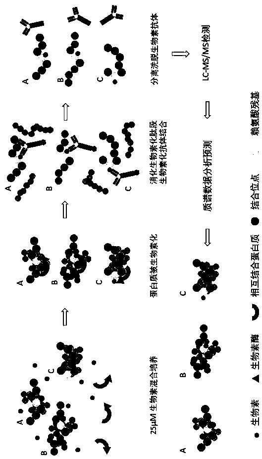
[0023] Embodiment 1: Preparation method of HEK293T cell-specific peptide mass spectrometry analysis sample:

[0024] A: Take the HEK293T cells to be analyzed, collect 5 million cells in a biotin culture environment, and dissolve them in 2 ml of cell lysis buffer. The composition of the cell lysis buffer is 50 mM triethylammonium bicarbonate, 8 M urea, 10nm protease inhibitor, lysed by circulating ultrasound in an ice bath, quantified the concentration of the protein solution, and then digested the peptide with trypsin;

[0025] B: Trypsin method to digest peptides, a total of 15 mg protein lysis solution, add dithiothreitol at a final concentration of 5 mM, incubate at room temperature for 1 hour, then add iodoacetamide at a final concentration of 10 mM, and incubate at room temperature in the dark For 20 minutes, finally add 3 times the total volume of 50 mM triethylammonium bicarbonate and 150 µg trypsin, and incubate at 37°C with shaking for more than 12 hours; the digested...

Embodiment 2

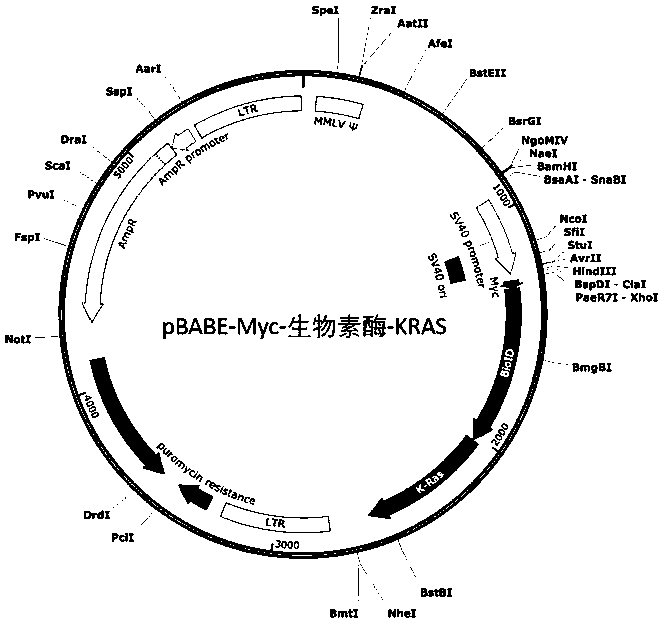
[0033] Example 2: HEK293T cell example of Myc-biotididase-KRAS fusion protein

[0034] (1) Preparation of HEK293T cells expressing Myc-biotidase-KRAS fusion protein

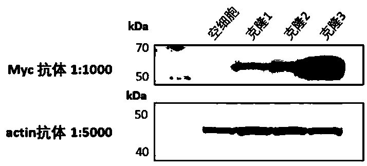
[0035] The gene-synthesized fusion gene fragment containing Myc-tag biotinidase (5' end) and KRAS (3' end) was inserted into the pBABE plasmid behind the SV40 promoter through homologous recombination ( figure 2). The fusion gene fragment plasmid was transiently transfected into HEK293T cells using lipofectamine3000 and packaged into retrovirus. HEK293T cells were infected with virus that was filtered through a 0.22 μm filter to remove cell debris for 24 hours, and pressure-selected with 1 μM puromycin for 1 week, and single clones were selected and transferred to 96-well plates for further culture for 2 weeks. image 3 As shown, the specific Myc monoclonal antibody was used to identify the Myc-biotididase-KRAS target band (55 kDa in size) in Western blotting, and cell clones highly expressing the fusion prote...


PUM


Abstract

A preparation method of a specific peptide fragment mass spectrometry sample comprises the following steps: A, taking sample cells to be analyzed, collecting more than 2 million cells in a biotin culture environment, dissolving the cells in a cell lysis buffer solution, carrying out ultrasonic lysis, quantifying the concentration of a protein solution, and carrying out trypsin digestion of a peptide fragment; B, digesting the peptide fragment by a trypsin method; carrying out freeze-drying at-80 DEG C after digestion; C, preparing a biotin antibody combined with Protein G beads; D: using 1 mlof capture buffer solution to dissolve the obtained freeze-dried peptide fragments; E, combining antibodies and peptide fragments in the step C and the step D, incubating at 4 DEG C for 2 hours, and eluting with an elution buffer solution for 4 times; and F, loading 200 microliters of microfiltration column prepared by a gun head into the total supernate through a 3M C18 solid-phase extraction membrane, eluting to remove salt, dissolving peptide fragments, and carrying out mass spectrometric detection. According to the method, the natural biotinylated protein in the cell can be accurately checked.

Description

technical field [0001] The invention relates to protein analysis research, in particular to a method for preparing a sample for mass spectrometry analysis of specific peptides, and belongs to the technical field of biochemistry. Background technique [0002] Identifying biotinylated proteins is a hot issue in the biological industry, which is related to the exploration of protein post-translational functions and mutual binding proteins. The traditional biotinylated protein avidin method generally uses trypsin digestion of the protein on Protein G beads after avidin is bound to the biotinylated protein. But due to the covalent binding between avidin and biotin the affinity constant (K) is 10 15 mol / L, which is at least 10,000 times higher than the binding affinity of the antibody to the antigen. When the biotin peptide is subsequently eluted, it is difficult to elute and release the bound biotinylated lysine residue site, thus causing the recognized peptide The segment lack...

Claims


Application Information

Patent Timeline
IPC IPC(8): G01N33/68G01N33/531C12P21/06
CPCG01N33/6848G01N33/531C12P21/06
Inventor 徐耀瑜马军岩司伟杰吴汉夔
Owner ANYANG NORMAL UNIV
Why Eureka
Try Eureka