Fluorescent probe for rapidly displaying normal state and near-zero state of potential of cell membrane and application thereof

A fluorescent probe, normal state technology, applied in the field of fluorescent probes, to achieve the effect of eliminating tedious steps, high specificity and high selectivity

Active Publication Date: 2020-05-05
SHANDONG UNIV
View PDF3 Cites 2 Cited by
  • Summary
  • Abstract
  • Description
  • Claims
  • Application Information

AI Technical Summary

Problems solved by technology

[0007] In view of the deficiencies of the current methods for measuring cell membrane potential, the problem to be solved by the present invention is to provide a flu

Method used


Image

  • Fluorescent probe for rapidly displaying normal state and near-zero state of potential of cell membrane and application thereof
  • Fluorescent probe for rapidly displaying normal state and near-zero state of potential of cell membrane and application thereof
  • Fluorescent probe for rapidly displaying normal state and near-zero state of potential of cell membrane and application thereof

Examples


Embodiment 1

[0029] Example 1 Preparation of the fluorescent probe (CQ12) that quickly displays the normal state and near-zero state of cell membrane potential according to the present invention

[0030] Add 0.399g of N-dodecylcarbazole-3-formaldehyde, 5ml of ethanol, and protect with nitrogen in a dry three-necked flask. The temperature is increased to 60°C and all dissolved after heating for 30min. Add the compound 1-(2-hydroxyethyl ) 0.315g of 4-methylquinoline iodide salt, add 5 drops of piperidine, heat to reflux overnight, stop heating, evaporate the solvent after cooling, recrystallize with ethanol, and dry to obtain 0.2g of red powder, yield 30% is CQ12.

[0031] The reaction formula for preparing the compound CQ12 is as follows:

[0032]

[0033] 1 H NMR (400MHz, DMSO-d6), δ (ppm): 9.18 (d, J = 8Hz, 1H), 9.15 (d, J = 8Hz, 1H), 8.89 (d, J = 1.6Hz, 1H), 8.56 (d,J=8.6Hz,1H), 8.52(d,J=6.4Hz,1H), 8.42(q,J=9.2Hz,2H), 8.25(t,J=8.8Hz,2H), 8.14(d ,J=8.8Hz,1H),8.07(t,J=7.6Hz,1H),7.77(d,J=8.4Hz,...

Embodiment 2

[0034] Example 2 Cultivation of cancer cells (SiHa and HeLa) and normal cells (Fibroblast)

[0035] At 37℃, 5% CO 2 In a saturated humidity incubator, the SiHa, HeLa and Fibroblast cell lines were grown adherently in H-DMEM culture medium containing 10% fetal bovine serum (containing 1% double antibody). After the cells have grown to the logarithmic phase, SiHa, HeLa, and Fibroblast cells were spliced ​​and cultured. The specific method is: ①Soak the cover glass in absolute ethanol for 30 minutes, put it in a disposable 35mm petri dish after drying with an alcohol lamp; ②Wash the cells that grow to the logarithmic phase in the cell dish three times with PBS. After digesting with 1mL 0.25% trypsin for 3-5 minutes, carefully pour out the enzyme solution, add a small amount of fresh culture solution, pipette the cells to make a uniform cell suspension, after the cell count, use the cell culture solution to adjust the final cell concentration 1×10 5 Pieces / ml, inoculate into a petri...

Embodiment 3

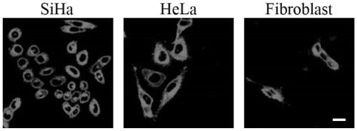
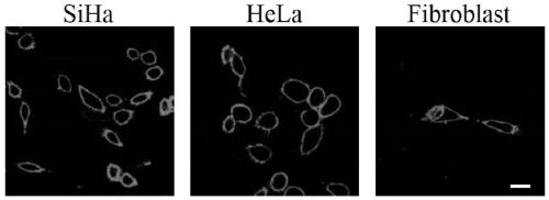
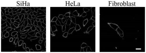
[0036] Example 3 Fluorescent probe CQ12 stains live SiHa, HeLa and Fibroblast cells

[0037] The cell slides prepared in Example 2 were washed three times with PBS, and live SiHa, HeLa and Fibroblast cells were stained with 2μM CQ12, incubated at room temperature for 20 minutes, then aspirated the culture medium, and washed three times with PBS to remove unbound cells The cell growth side is covered on a glass slide, and the cell coloring part, fluorescence distribution and brightness change are observed under the excitation of 473nm laser with a laser scanning confocal fluorescence microscope. It is found that the fluorescent probe CQ12 can pass through The cell membrane enters the cell, and the cell membrane and cytoplasm are stained, emitting bright red fluorescence.

[0038] See the result figure 1 .

[0039] The results suggest that the membrane potential state of living cells is normal. When the cell is in the normal membrane potential state, the fluorescent probe CQ12 can eas...


PUM


Abstract

The invention discloses a fluorescent probe for rapidly displaying a normal state and a near-zero state of the potential of a cell membrane. The fluorescent probe is a compound of 4-[2-(9-dodecyl-9H-carbazole-3) vinyl]-1-(2-hydroxyethyl)-quinoline iodate, which is called CQ12 for short. The invention also discloses an application of the fluorescent probe in detection of a normal state and a near-zero state of the potential of a cell membrane. When the potential of a cell membrane is in a normal state, the probe enables the cell membrane and cytoplasm to be dyed; and when the potential of the cell membrane is in a near-zero state, only the cell membrane is dyed by the probe. Moreover, the probe is not interfered by mitochondrial membrane potential when being used for measuring the potentialof the cell membrane, and shows higher specificity. Compared with a currently used cell membrane potential probe product, the visual and rapid distinguishing mode of the probe has the advantages thatcalibration and standard curve drawing are not needed in the using process, and time and labor are saved. The probe has wide application prospects in the aspects of biological research and medical diagnosis.

Description

Technical field [0001] The invention relates to a fluorescent probe for measuring cell membrane potential and its application, in particular to a fluorescent probe for rapidly displaying the normal state and near zero state of the cell membrane potential by using fluorescent staining image changes and its application. Background technique [0002] Cell membrane potential is the potential difference between the inside and outside of the cell membrane when the cell is resting. It is an important indicator reflecting cell activity and cell metabolism. The normal membrane potential of most cells is negative inside and positive outside, in the range of -50~-100mV. Inside. However, in some types of cells, the cell membrane potential will decrease significantly. For example, U.V. Lassen found in 1967 that the cell membrane potential of human red blood cells was less than -14mV; S. Hodson reported in 1989 that the cell membrane potential of rabbit corneal endothelial cells was around 0m...

Claims


Application Information

Patent Timeline
IPC IPC(8): C07D401/06C09K11/06G01N21/64
CPCC07D401/06C09K11/06G01N21/6428C09K2211/1029
Inventor 何秀全张华淼于晓强刘志强
Owner SHANDONG UNIV
Why Eureka
Try Eureka