Preparation method of mycoplasma synoviae monoclonal antibody

A technology of Mycoplasma synovialis and monoclonal antibody, which is applied in the directions of microorganism-based methods, biochemical equipment and methods, chemical instruments and methods, etc., can solve the problem of inability to produce an immune response to the whole Mycoplasma bacteria.

Pending Publication Date: 2020-10-09
中元牧康(武汉)检测技术服务有限公司
View PDF0 Cites 2 Cited by
  • Summary
  • Abstract
  • Description
  • Claims
  • Application Information

AI Technical Summary

Problems solved by technology

The development of monoclonal antibodies with high titer and strong specificity is the demand of the veterinary diagnostic industry. In Mycoplasma synovialum, there are nearly 100 pseudo-proteins on the membrane surface. Most of the monoclonal antibodies against Mycoplasma hydras use proteins with known sequences on the membrane surface as antigens, and they only target a specific protein, and cannot react against the whole bacteri

Method used


Image

  • Preparation method of mycoplasma synoviae monoclonal antibody
  • Preparation method of mycoplasma synoviae monoclonal antibody

Examples


Embodiment 1

[0026] Embodiment 1: the preparation of antigen

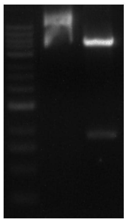
[0027] (1) Insert the P20 gene and P13 gene into the expression vector PET-32a to obtain the recombinant expression vector, transform the Escherichia coli competent cell DH5α with the recombinant expression vector, and screen out the positive clone recombinant plasmid after enzyme digestion, PCR identification and sequencing PET-32a-P20-P13; after BamHI / / XhoI digestion and electrophoresis detection of recombinant plasmid PET-32a-P20-P13 figure 1 As shown, the P20 gene sequence is shown in the sequence listing SEQ ID NO: 1, and the P13 gene is shown in the sequence listing SEQ ID NO: 2; the gene sequence of the PET-32a-P20-P13 recombinant protein is shown in the sequence listing SEQ ID NO: 2. ID NO:3 shown.

[0028] The amino acid sequence of the protein encoded by the P20 gene is shown in the sequence listing SEQ ID NO: 4, and the amino acid sequence of the protein encoded by the P13 gene is shown in the sequence listing SEQ I...

Embodiment 2

[0031] Example 2: Mouse immunization, cell fusion and subcloning

[0032] 1) For the first immunization, Freund's complete adjuvant and an equal amount of recombinant protein were used to emulsify, and after emulsification, 5 6-8 week-old Balb / c female mice were subcutaneously inoculated, and each mouse was inoculated with a dose of 200ug of recombinant protein. After that, immunize once every two weeks. After the second immunization, the adjuvant was replaced by Freund's incomplete adjuvant. After three immunizations, blood was collected from the tail vein at intervals of one week to detect antibodies, and the inactivated whole bacteria of Mycoplasma synovium was used as the coating antigen. Perform antibody testing.

[0033] 2) The mouse with the highest antibody titer was selected as the mouse for cell fusion, and the recombinant protein was inoculated intraperitoneally before the fusion for booster immunization once.

[0034] 3) Preparation of myeloma cells: Select SP2 / 0 ...

Embodiment 3

[0039] Example 3: Preparation and verification of antibodies

[0040] 1) Antibody production: The five cell lines obtained above were successively scaled up and cultured to gradually realize large-scale production of antibodies. The process is as follows, first recover in a 24-well cell plate, add feeder cells to promote the rapid growth of the hybridoma cell line during recovery, transfer the cells to a T25 cell culture flask for culture when the growth reaches 75% of the bottom of the well, and add feeder cells at the same time . When the T25 cell culture flask grew to 75% of the bottom area of ​​the flask, the hybridoma cells were transferred to the T75 cell culture flask, then to the T225 cell culture flask, and then transferred to a 10-liter spinner bottle for culture. The titer of the antibody was detected by retaining samples at each stage to determine the genetic stability of the cells. The results of the titer determination are shown in Table 1:


PUM


Abstract

The invention discloses a preparation method of a mycoplasma synoviae monoclonal antibody. The conservative analysis is carried out on two imaginary membrane proteins with the mycoplasma membrane surface protein sizes of 20Kda and 13Kda; then recombinant plasmids synthesizing, protein inducible expression, and affinity chromatography purification are carried out; the purified recombinant protein is used as immunogen to immunize BALB/c mice, cell fusion is carried out after antibody detection is qualified, a single monoclonal antibody cell strain is obtained after subcloning of fusion cells, and the prepared monoclonal antibody can well and specifically recognize mycoplasma synoviae antigen.

Description

technical field [0001] The invention belongs to the technical field of monoclonal antibody preparation, in particular to a method for preparing a synovial fluid mycoplasma monoclonal antibody. Background technique [0002] Mycoplasma is a class of smallest prokaryotic microorganisms that can grow and reproduce in non-living media between bacteria and viruses. Mycoplasma synoviae is considered to be a pathogen that can specifically cause synovium and joint capsule diseases in chicken joints. Mycoplasma synoviae has no cell wall structure, and its morphology is pleomorphic. Its genome and proteome are very small. Mycoplasma synovia mainly interacts with the host through membrane surface proteins to complete the process of adhesion and colonization. The immune response with the host is also mainly based on membrane surface proteins. In recent years, the incidence of Mycoplasmasynoviae (MS) in chickens has been increasing, which can cause systemic damage to poultry and cause...

Claims


Application Information

Patent Timeline
IPC IPC(8): C07K16/12C07K19/00C12N15/62C12N15/70C12N5/20C12R1/91
CPCC07K16/1253C07K14/30C12N15/70C07K2317/35Y02A50/30
Inventor 李冉郭学波李淑云张懿付博战爽何诗雨祁喆李海霞陈晨潘珊
Owner 中元牧康(武汉)检测技术服务有限公司
Why Eureka
Try Eureka