
A Visual and Quantitative Method for Labeling Aggregated Functional Proteins in Cells

A medium-aggregation-type, cell-model technology, applied in the field of biological probes and protein detection, can solve the problems of aggregation quenching effect, fluorescence quenching of fluorescent molecules, etc., achieve simple operation, good biocompatibility, and avoid aggregation quenching effect of effect

Active Publication Date: 2022-02-25
INST OF HIGH ENERGY PHYSICS CHINESE ACAD OF SCI
View PDF0 Cites 0 Cited by
  • Summary
  • Abstract
  • Description
  • Claims
  • Application Information

AI Technical Summary

Problems solved by technology

[0007] The present invention aims at using antibodies to identify protein sites in the current protein detection process to provide protein signals through fluorescence amplification. Most of the antibodies are connected to traditional organic fluorescent molecules such as FITC and TRITC. Long-term exposure is prone to fluorescence quenching and fluorescence in the protein aggregation area. For the problem of molecular aggregation quenching effect, a method for visually and quantitatively labeling aggregated functional proteins in cells was proposed, and the aggregation-induced luminescent probe AIE-cRGD was used to stain integrin, which solved the aggregation quenching effect of antibody recognition , which facilitates the specific detection of aggregated activated proteins

Method used


Image

  • A Visual and Quantitative Method for Labeling Aggregated Functional Proteins in Cells
  • A Visual and Quantitative Method for Labeling Aggregated Functional Proteins in Cells
  • A Visual and Quantitative Method for Labeling Aggregated Functional Proteins in Cells

Examples


Embodiment 1

[0030] Example 1: Construction and characterization of aggregation-induced luminescent probe AIE-cRGD

[0031] The aggregation-induced luminescent probe AIE-cRGD contains aggregation luminescent molecules and peptides targeting integrin, 1,2-bis(4-carboxyphenyl)-1,2-styrene and cRGD are mixed according to the same molar ratio, and then Add the catalyst EDC-NHS for catalysis, and react overnight to obtain the aggregation-induced luminescent probe AIE-cRGD;

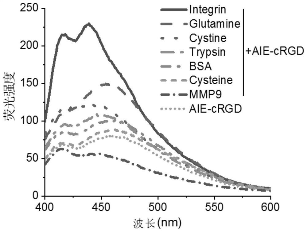
[0032]Dimethyl sulfoxide DMSO and water were formulated into a test system at a ratio of 1:100, and the ultraviolet absorption and fluorescence spectra of the aggregation-induced luminescence probe AIE-cRGD with different concentrations were measured in the test system, and the ultraviolet absorption spectra showed that different concentrations of AIE-cRGD The aggregation-induced luminescence probe AIE-cRGD has an absorption peak at 230nm, and the fluorescence spectrum shows that the aggregation-induced luminescence probe A...

Embodiment 2

[0033] Example 2: Concentration of aggregation-induced luminescence probe AIE-cRGD

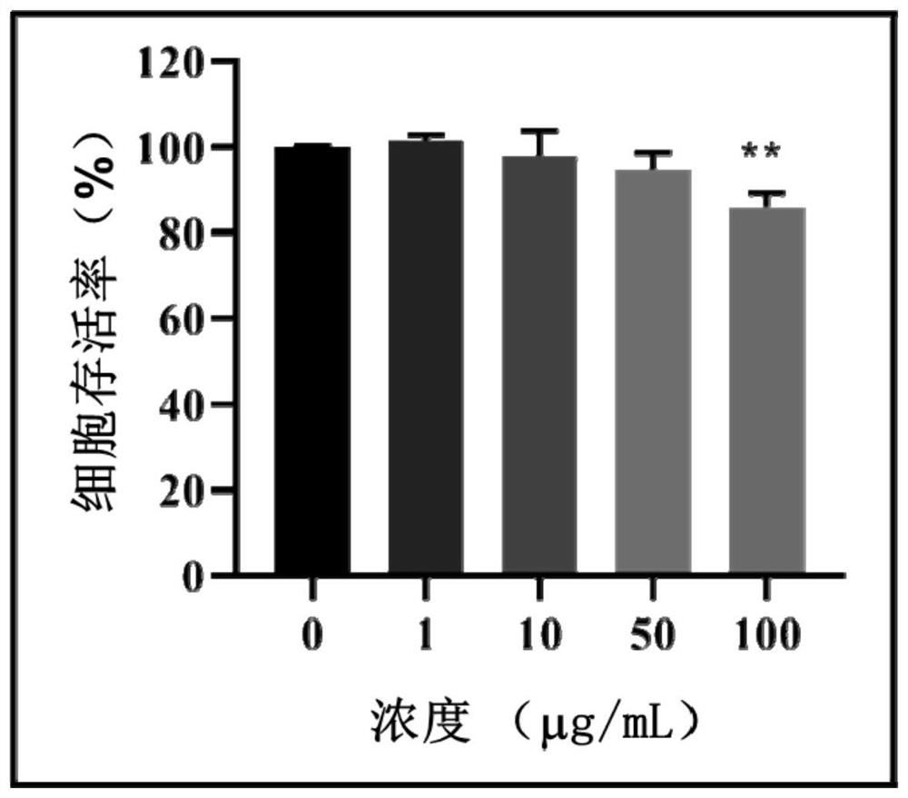
[0034] Taking breast tumor cell MCF-7 as an example, determine the concentration of AIE-cRGD; set the concentration of AIE-cRGD to 1 μg / mL, 10 μg / mL, 50 μg / mL and 100 μg / mL respectively. mL, and then MCF-7 cells were placed in various concentrations of aggregation-induced luminescence probe AIE-cRGD solutions, and the cell counting reagent CCK-8 was used to detect the concentration of MCF-7 cells in different concentrations of aggregation-induced luminescence probe AIE-cRGD solutions. By analyzing the effect of the concentration of aggregation-induced luminescence probe AIE-cRGD on cell viability, it was found that when the concentration of aggregation-induced luminescence probe AIE-cRGD was 1 μg / mL, 10 μg / mL and 50 μg / mL, the cell viability did not occur significantly changes, and when the aggregation-induced luminescent probe AIE-cRGD concentration was 100 μg / mL, the cell viability decreased...

Embodiment 3

[0035] Example 3: Detection of the targeting protein of the aggregation-induced luminescent probe AIE-cRGD

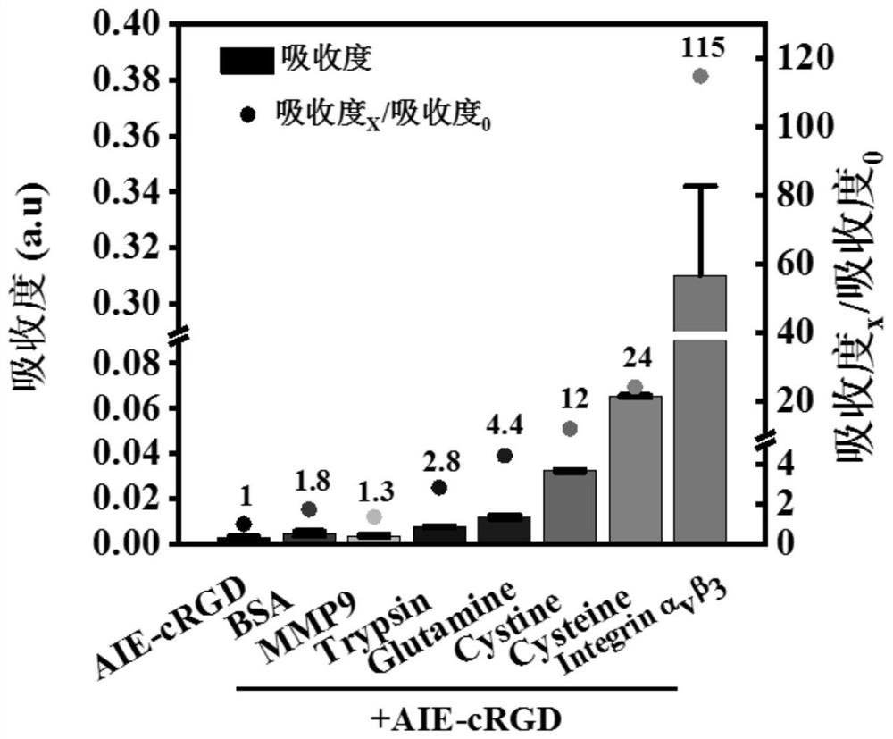
[0036] Dilute bovine serum albumin BSA, trypsin, glutamine Glutamine, cysteine ​​Cysteine, cystine Cystine, matrix metalloproteinase MMP9 and integrin αvβ3 to 100 μg / mL respectively, and aggregate-induced luminescent probe AIE - Dilute the concentration of cRGD to 100 μg / mL, then add each protein solution and aggregation-induced luminescent probe AIE-cRGD solution into 300 μL phosphate buffered saline solution according to the volume ratio of 1:1 and mix, and measure 230nm for each protein solution Changes in the ultraviolet absorption intensity of the aggregation-induced luminescent probe AIE-cRGD at the place, and analyze the response of the aggregation-induced luminescent probe AIE-cRGD to each protein, such as figure 2 and image 3 As shown, the absorbance of the AIE-cRGD solution at 230nm is 0.0027, and the absorbance of the AIE-cRGD solution at 230nm is 0.31 aft...


PUM


Abstract

The invention relates to the technical field of biological probes and protein detection, and provides a method for visually and quantitatively labeling aggregated functional proteins in cells, by constructing and characterizing the aggregation-induced luminescent probe AIE-cRGD, and measuring the absorption peak of the probe AIE-cRGD and the fluorescence emission peak and the applicable concentration, the detection confirmed that the target protein of the probe was integrin αvβ3, the suspension survival cell model was established based on the poly-HEMA plating method, and the cells in the suspension survival cell model were induced to grow in an environment of detachment, Simulate the anti-apoptotic state of cells, use probes to label integrins in cells and perform imaging. When integrins are internalized into endosomes, determine the distribution of integrins in detached cells and detect phosphorylation Colocalization of focal adhesion kinase p‑FAK with activated integrins on endosomes determines cell resistance to apoptosis The invention can be directly used for the staining of living cells, lays a foundation for the visualization of physiological functions, and is beneficial to high-quality imaging and high-sensitivity online sensing monitoring.

Description

technical field [0001] The invention relates to the technical field of biological probes and protein detection, in particular to a method for visually and quantitatively marking aggregated functional proteins in cells. Background technique [0002] Integrins are widely distributed on the cell surface and consist of two subunits, α and β, as transmembrane heterodimers. They are a type of transmembrane receptor glycoproteins, and their ligands are mainly extracellular matrix (ECM) proteins, such as vitreous Binding protein, laminin, etc. Most integrins have longer extracellular domains, transmembrane regions and shorter cytoplasmic domains, and their different domains have different functions. For the extracellular domain, it can connect with the extracellular matrix by recognizing the specific sequence of the extracellular matrix protein, that is, arginine-glycine-aspartic acid (RGD sequence), and mediate between cells and extracellular matrix, cells and Adhesion signals be...

Claims


Application Information

Patent Timeline
Patent Type & Authority Patents(China)
IPC IPC(8): G01N21/64
CPCG01N21/6458G01N21/6428
Inventor 邢更妹常亚男陈奎李娟
Owner INST OF HIGH ENERGY PHYSICS CHINESE ACAD OF SCI
Learn More