
Adeno-associated virus capable of being used for bimodal imaging and application of adeno-associated virus

A technology of recombinant adenovirus and virus, applied in the direction of application, virus, virus/phage, etc., can solve the problems of no virus vector carrying AQP1 gene, low detection sensitivity, etc., achieve wide host range, low immunogenicity, stable and long-lasting expression Effect

Pending Publication Date: 2021-06-25
INNOVATION ACAD FOR PRECISION MEASUREMENT SCI & TECH CAS
View PDF3 Cites 5 Cited by
  • Summary
  • Abstract
  • Description
  • Claims
  • Application Information

AI Technical Summary

Problems solved by technology

The limitation of the second type of reporter gene is that the detection sensitivity is low, and a large amount of expression (2.5μM) is required to produce an observable contrast effect
[0005] At present, there is no report on the direct use of viral vectors to carry the AQP1 gene for MRI imaging in living animals

Method used


Image

  • Adeno-associated virus capable of being used for bimodal imaging and application of adeno-associated virus
  • Adeno-associated virus capable of being used for bimodal imaging and application of adeno-associated virus
  • Adeno-associated virus capable of being used for bimodal imaging and application of adeno-associated virus

Examples


Embodiment 1

[0023] Example 1: Construction of plasmid pAAV-CAG-AQP1-2A-EGFP-WPRE-pA carrying AQP1 and EGFP genes and preparation of recombinant adeno-associated virus rAAV-CAG-AQP1-2A-EGFP-WPRE-pA

[0024]In order to increase the expression level of the target gene, we selected the strong eukaryotic promoter CAG promoter. In order to make the fusion expression of AQP1 gene and green fluorescent protein (EGFP) reporter gene, we selected 2A self-cleaving polypeptide coding sequence as the connecting sequence of AQP1 gene and EGFP gene. In order to improve the post-transcriptional stability of the target gene, we added WPRE post-transcriptional regulatory elements and polyA sequences at the 3' end of the target gene. We used conventional molecular cloning methods to insert the CAG promoter (shown in SEQ ID NO.1), the AQP1 gene (shown in SEQ ID NO. ), 2A junction sequence (shown in SEQ ID NO.3), EGFP gene (shown in SEQ ID NO.4), WPRE post-transcriptional regulatory element (shown in SEQ ID N...

Embodiment 2

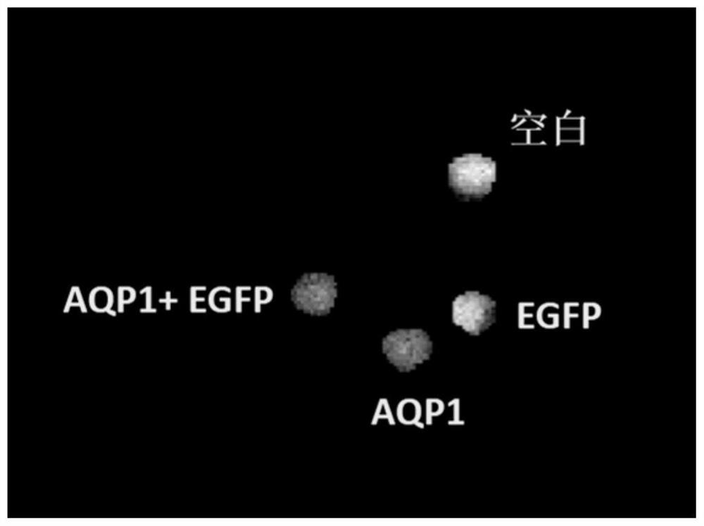
[0027] Example 2: MRI imaging observation of BHK cells transfected with pAAV-CAG-AQP1-2A-EGFP-WPRE-pA plasmid

[0028] In order to verify the MRI imaging effect of AQP1 mediated by the pAAV-CAG-AQP1-2A-EGFP-WPRE-pA plasmid, the pAAV-CAG-AQP1-2A-EGFP-WPRE-pA plasmid (AQP1-EGFP) was transfected with Lipfectamine 2000 BHK cells were transfected, and simultaneously transfected with pAAV-CAG-AQP1-WPRE-pA plasmid (AQP1) expressing only AQP1 protein and pAAV-CAG-EGFP-WPRE-pA plasmid (AQP1-EGFP) expressing only EGFP protein as a control. figure 2 DWI-MRI imaging results of cell pellets after transfection of BHK cells with different plasmids are shown. The cells transfected with pAAV-CAG-AQP1-2A-EGFP-WPRE-pA plasmid (AQP1-EGFP) showed the darkest DWI signal intensity. The results indicated that the simultaneous expression of AQP1 protein and EGFP protein could produce a stronger DWI-MRI contrast effect compared to the expression of AQP1 protein alone.

Embodiment 3

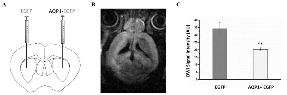
[0029] Example 3: MRI imaging observation of mouse brain injected with rAAV2-CAG-AQP1-2A-EGFP-WPRE-pA and control virus rAAV2-Ef1α-EGFP-WPRE-pA

[0030] The rAAV2-CAG-AQP1-2A-EGFP-WPRE-pA virus was injected into the right dorsal striatum brain area of ​​the mouse brain (CPu, coordinates: AP-0.5mm; ML-2mm; DV- 3.3mm), the titer is 6×10 11 vg / ml, the injection volume was 2 μL. The control virus rAAV2-Ef1α-EGFP-WPRE-pA was injected in the left dorsal striatum brain area (coordinates: AP-0.5mm; ML 2mm; DV-3.3mm), and its titer was 2.4×10 12 vg / ml, the injection volume is 0.5 μL. Schematic diagram of virus injection as image 3 As shown in A.

[0031] After 3 weeks, use a 7.0T magnetic resonance imaging machine to observe the animal brain with MRI in vivo. The diffusion-weighted imaging sequence used is the SE-DWI sequence based on STEAM, and the parameters are: TR=3000ms, TE=24ms, and the average number of times is 2, and the total duration of the sequence is 51min12s. The d...


PUM


Abstract

The invention discloses an adeno-associated virus capable of being used for bimodal imaging and application of the adeno-associated virus. A CAG promoter, an AQP1 gene, a 2A connecting sequence, an EGFP gene, a WPRE after-transcription adjustment element and a polyA sequence are sequentially inserted in the middle of ITR of an AAV vector plasmid. After animal living nerve cells are infected by the prepared recombinant adeno-associated virus, expression of AQP1 and EGFP proteins can be mediated in the nerve cells, so that an expression region has green fluorescence signals, diffusion weighted MRI (Magnetic Resonance Imaging) signals can be changed, and the characteristic of bimodal imaging is achieved. The expression level of the exogenous gene transduced by the virus vector disclosed by the invention can be observed in situ in a living body tissue through noninvasive magnetic resonance imaging, can also be observed in an in-vitro tissue through fluorescence imaging, and can be used for magnetic resonance imaging contrast, neuron marking, neural circuit tracing and the like.

Description

technical field [0001] The invention belongs to the technical field of virus vectors, in particular to an adeno-associated virus carrying a fusion gene of type 1 aquaporin and green fluorescent protein and its application. Background technique [0002] Detection of gene expression in living animals by non-invasive imaging methods is very critical for gene transduction, gene therapy, cell transplantation, cell therapy and other fields. In addition, the method of detecting gene expression in vivo will help basic biological research. During gene transduction or cell transplantation, a reporter gene is often used to mark the expression of a gene of interest or gene of interest. Currently, the most commonly used reporter genes are those based on fluorescent proteins and chemiluminescent proteins. Since it is difficult for light to penetrate deep tissues, it is difficult for optical imaging to obtain gene expression results in opaque deep tissues at the living level. Therefore, i...

Claims


Application Information

Patent Timeline
IPC IPC(8): C12N15/864C12N7/01C12N15/62C12N15/65A61K49/00A61K49/18
CPCC12N15/86C12N7/00C12N15/65A61K49/0002A61K49/0047A61K49/0097A61K49/1896C12N2750/14143C12N2750/14121C12N2830/48
Inventor 郑宁李梅吴阳王杰徐富强
Owner INNOVATION ACAD FOR PRECISION MEASUREMENT SCI & TECH CAS
Learn More