Targeted nano magnetic resonance contrast agent for articular cartilage injury as well as preparation and application of targeted nano magnetic resonance contrast agent
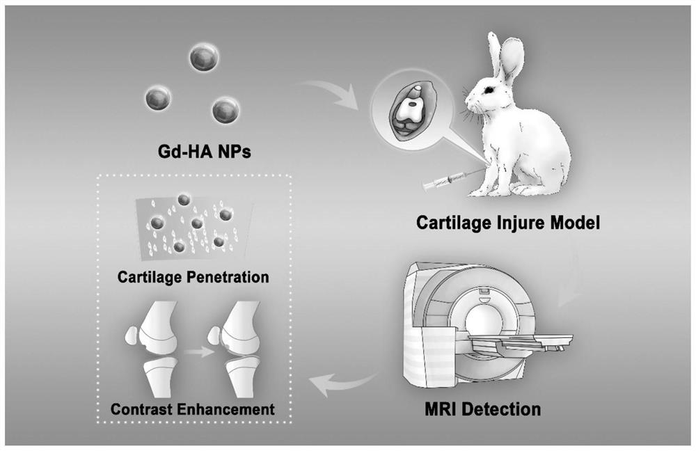
A magnetic resonance contrast agent and articular cartilage technology, applied in the field of molecular imaging, can solve the problems of interfering with the imaging effect of the articular cartilage defect area, the delayed enhancement is not obvious, the relaxation rate of Gd agent is low, etc., so as to prevent joint effusion. The effect of preventing interference, ensuring MRI signal strength, improving efficiency and safety
- Summary
- Abstract
- Description
- Claims
- Application Information
AI Technical Summary
Problems solved by technology
Method used
Image
Examples
Embodiment 1
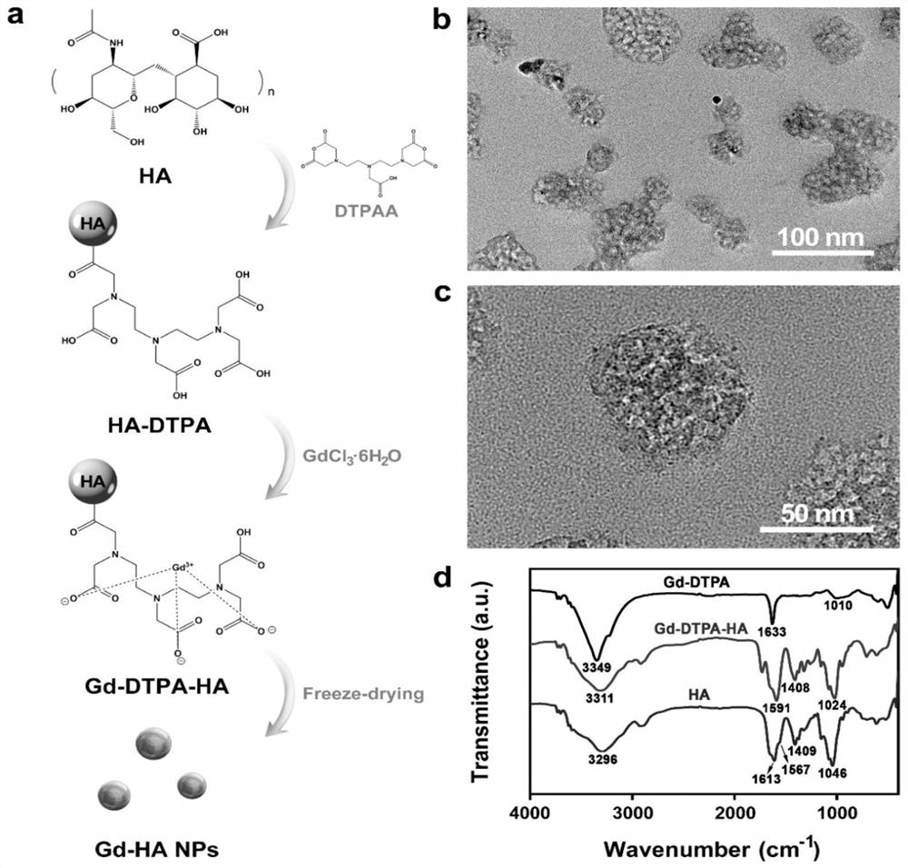
[0045] Example 1: Preparation and physical and chemical characterization of targeted nano-magnetic resonance contrast agent
[0046] a. Take 200mg of hyaluronic acid HA (0.5mM) with a molecular weight of 990,000Da and fully dissolve it in a 50ml test tube, add 20mL of double distilled water, stir at room temperature for 1h until clear and transparent, add 1% (2-3mg) 5 carboxyfluorescein ( 5-FAM SE) was stirred for 1-2h; 90mg of diethylenetriaminepentaacetic dianhydride (0.25mM) was added to 600μL dimethyl sulfoxide to dissolve, and stirred at room temperature for 2-3h; Gadolinium (0.25mM), stirred for 0.5h, put the reaction solution into a dialysis bag with a molecular weight cut-off of 30,000, dialyzed in double distilled water for 24h-48h, pay attention to protect it with tinfoil paper, and finally prepare a solution of 14.26mmol / L A: The mass ratios of diethylenetriaminepentaacetic dianhydride, hyaluronic acid, gadolinium chloride hexahydrate and 5-carboxyfluorescein are 1:...
Embodiment 2
[0050] Example 2: Study on in vitro relaxation rate of targeted nano-magnetic resonance contrast agent
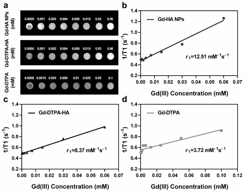
[0051] Take an appropriate amount of nano-magnetic resonance contrast agent solution samples, prepare different concentrations of HA-DTPA-Gd NPs solutions with double-distilled water, Gd(III) concentration 0.0005–0.06mM, and place them in 2ml centrifuge tubes. Then prepare DTPA-Gd with corresponding gadolinium concentration as a control. The tubes are placed sequentially in plastic test tube racks and placed in boxes filled with water. The clinical 3.0T MRsystem (Discovery MR750, GE Medical System, USA) and supporting animal coils were used for T1 Map sequence scanning to evaluate the effect of negative reinforcement. The T1 Map was post-processed with GE Function Tool 4.6 special software.
[0052] Susceptibility such as image 3 As shown in a, compared with the control group, with the gradual increase of gadolinium ion concentration, T1 gradually increased. After meas...
Embodiment 3
[0053] Example 3: Research on the change of signal-to-noise ratio of targeted nano-magnetic resonance contrast agent in vivo.
[0054] Gd-HA-NPs, Gd-DTPA-HA and Gd-DTPA were injected into rabbit knee articular cartilage injury model respectively. After injection, the knee joint was imaged with a Siemens Verio 3T magnetic resonance imaging system equipped with a rabbit knee coil.
[0055] Such as Figure 4 Articular cartilage (red dotted line) and damaged area (blue dotted line) were not clearly visible in the two groups before contrast agent injection in a and b. One hour after the intra-articular injection of Gd-DTPA, the signal intensity of the entire joint cavity increased, but the image of the lesion was only slightly enhanced, and the outline of the cartilage was still unclear. At 2h and 3h after injection, the signal intensities of cartilage and lesion remained very close. One hour after injection, the signal intensity of cartilage and injury sites in Gd-DTPA-HA group...
PUM
| Property | Measurement | Unit |
|---|---|---|
| Diameter | aaaaa | aaaaa |
| Particle size | aaaaa | aaaaa |
Abstract
Description
Claims
Application Information
 - R&D
- Intellectual Property
- Life Sciences
- Materials
- Tech Scout
- Unparalleled Data Quality
- Higher Quality Content
- 60% Fewer Hallucinations
Browse by: Latest US Patents, China's latest patents, Technical Efficacy Thesaurus, Application Domain, Technology Topic, Popular Technical Reports.
© 2025 PatSnap. All rights reserved.Legal|Privacy policy|Modern Slavery Act Transparency Statement|Sitemap|About US| Contact US: help@patsnap.com


