Ultrasonic microvesicle as immuno adjuvant and vaccine carrier

A technology of ultrasonic microbubbles and vaccine carriers, applied in the field of biomedical engineering, can solve the problems that it is difficult to meet the needs of the development of new peptide vaccines and fail to stimulate immune responses, achieve safe repeatability, improve nucleic acid expression levels, and target good effect

Active Publication Date: 2007-03-28
许川山 +2
View PDF0 Cites 5 Cited by
  • Summary
  • Abstract
  • Description
  • Claims
  • Application Information

AI Technical Summary

Problems solved by technology

Secondly, co-immunization of aluminum compounds with many recombinant or synthetic peptide vaccine antigens or nucleic acid (DNA) vaccine antigens failed to

Method used


Image

  • Ultrasonic microvesicle as immuno adjuvant and vaccine carrier
  • Ultrasonic microvesicle as immuno adjuvant and vaccine carrier

Examples


example 1

[0017] Example 1: Preparation of Surfactant-like Microbubbles

[0018] Mix the surfactant Span60; Tween 80 in a ratio of 1:1, and at the same time use NaCl:KCl:Na 2 HPO 4 :KH 2 PO 4According to the mass ratio of 200:30:5:1, add 100ml of deionized water, and adjust the pH value of the mixed solution to 7.36 with NaOH, which is used as the medium; add 100ml of the medium into the mixture of Span60 and Tween80, Heat the mixture obtained above on a magnetic stirrer, and heat the temperature to 120°C for 10 minutes, and mix thoroughly to make it a milky suspension system; , add 0.6ml of perfluoropropane gas at a rate of 0.5ml / s; place the probe of the vibrometer at 0.5-2.0cm below the liquid surface for sonic vibration treatment, and vibrate at 30% of the maximum output power for 3 minutes. The emulsified mixed solution after sonication was divided into 3 layers after standing for 35 minutes, and the middle layer was taken out and diluted with phosphate buffer solution to obtai...

example 2

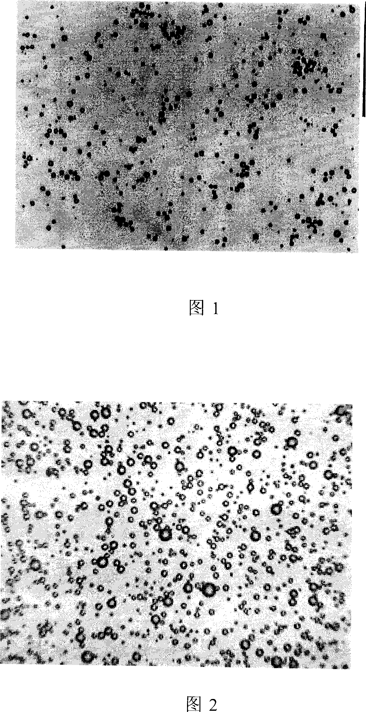
[0019] Example 2: Preparation of Lipid Microvesicles

[0020] Lecithin, cholesterol, polyethylene glycol stearyl ethanolamine were dissolved in chloroform at a mass ratio of 1:3:3, and evaporated in a rotary vacuum to form a film; 0.9% sodium chloride solution, propylene glycol and glycerin (0.9% chlorine Sodium chloride solution: propylene glycol: glycerin = 8:1:1), shake to wash the membrane to form a liposome suspension. Freeze overnight. After thawing, vibrate for 80 s with a vibrometer at 30% of the maximum output power, and at the same time slowly fill in 0.6 ml of perfluoropropane gas below through a three-way tube to form lipid fluorocarbon microbubbles (as shown in Figure 2).

example 3

[0021] Example 3: Preparation of polymer material-polylactic acid / glycolic acid (PLGA) microbubbles

[0022] Add 0.1g of camphor into 20ml of dichloromethane, stir fully to make it completely dissolved; add 1.0g of high molecular material polylactic acid / glycolic acid polymer (PLGA) into the above solution, stir until it is completely dissolved, and then add 5% After 3ml of ammonium chloride is added, vibrate for 40s with a vibrator at 30% of the maximum output power to form a milky white emulsion; add the emulsion to 3% polyvinyl alcohol and homogenize it for 5 minutes, then add 2% isopropanol solution, stirred with a magnetic stirrer at room temperature for 2-5 hours, centrifuged at a high speed for 5 minutes (speed 3000-5000 rpm), repeated 3 times; added 5% mannitol and fully mixed into a milky white solution to obtain PLGA microbubbles (as shown in Figure 3)


PUM

PropertyMeasurementUnit
Particle sizeaaaaaaaaaa

Abstract

The present invention belongs to the field of biomedical engineering, in the more concrete, said invention relates to a new type ultrasonic microvesicle which can be used as immunological adjuvant and vaccine carrier. It is characterized by that the vaccine antigen substance is adhered on the microvesicle surface and/or covered in microvesicle interior so as to synthesize the vaccine antige carried ultrasonic microvesicle. When it is used, the vaccine antigen carried ultrasonic microvesicle can systematically or locally act on target tissue, the ultrasonic wave can be used to break the microvesicle of target tissue area, and orietationally release vaccine antigen substance so as to raise the immunological activity of antigen.

Description

technical field [0001] The invention belongs to the field of biomedical engineering, and more specifically, the invention relates to a novel ultrasonic microbubble used as an immune adjuvant and a vaccine carrier. Background technique [0002] Traditional vaccines using inactivated or attenuated pathogens, cell components (such as capsular polysaccharides) or inactive bacterial toxins can usually induce the body to produce neutralizing antibodies to prevent infection, and immune adjuvants play an important role in this process . The immune adjuvant is a substance that does not produce immunogenicity when administered alone, but induces mucosal and / or systemic immunity when combined with an antigen. The only adjuvants currently used in human vaccines on a large scale are aluminum compounds, the most important of which is Al(OH) 3 . There are two types of vaccines using aluminum compounds in modern times, one is aluminum compound precipitation vaccines; the other is aluminu...

Claims


Application Information

Patent Timeline
IPC IPC(8): A61K39/385A61K38/39A61K47/30
Inventor 许川山王志刚于廷和刘志君
Owner 许川山
Why Eureka
Try Eureka