Magnetic nanoparticles for disease diagnostics

a technology of magnetic nanoparticles and disease diagnosis, applied in the field of magnetic nanoparticles, can solve the problems of limiting the choice of neuronal disease diagnostic agents, affecting the clinical outcome, so as to improve the clinical outcome, improve the clinical effect, and improve the pharmacokinetics.

Active Publication Date: 2019-01-15
NANO & ADVANCED MATERIALS INST
View PDF1 Cites 0 Cited by
  • Summary
  • Abstract
  • Description
  • Claims
  • Application Information

AI Technical Summary

Benefits of technology

[0009]The present disclosure provides magnetic nanoparticles having improved long term stability, improved pharmacokinetics, ability to penetrate the blood brain barrier, and long circulation half life with reduced toxic effects on the patient.

Problems solved by technology

However, these diagnostic agents can be disadvantaged by toxicity of the conjugated peptides or antibodies and their large molecular weight may inhibit the penetration of such targeting agents through biological barriers, such as blood-brain barrier, small intestine, nasal, skin and mouth mucosa.
The blood brain barrier can thus severely limit the choice of diagnostic agents for neuronal diseases.
However, these approaches are complicated and expose patients to radiation, which may increase the risk of continuous disease monitoring.
MRI contrast agents, and in particular iron-based MRI contrast agents, are susceptible agglomeration and exhibit poor in vivo distribution and half-life.

Method used


Image

  • Magnetic nanoparticles for disease diagnostics
  • Magnetic nanoparticles for disease diagnostics
  • Magnetic nanoparticles for disease diagnostics

Examples


example 1

[0095]Synthesis and Characterization of Metal-curcumin Complexes

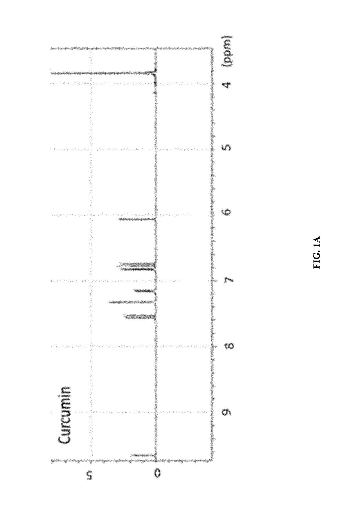
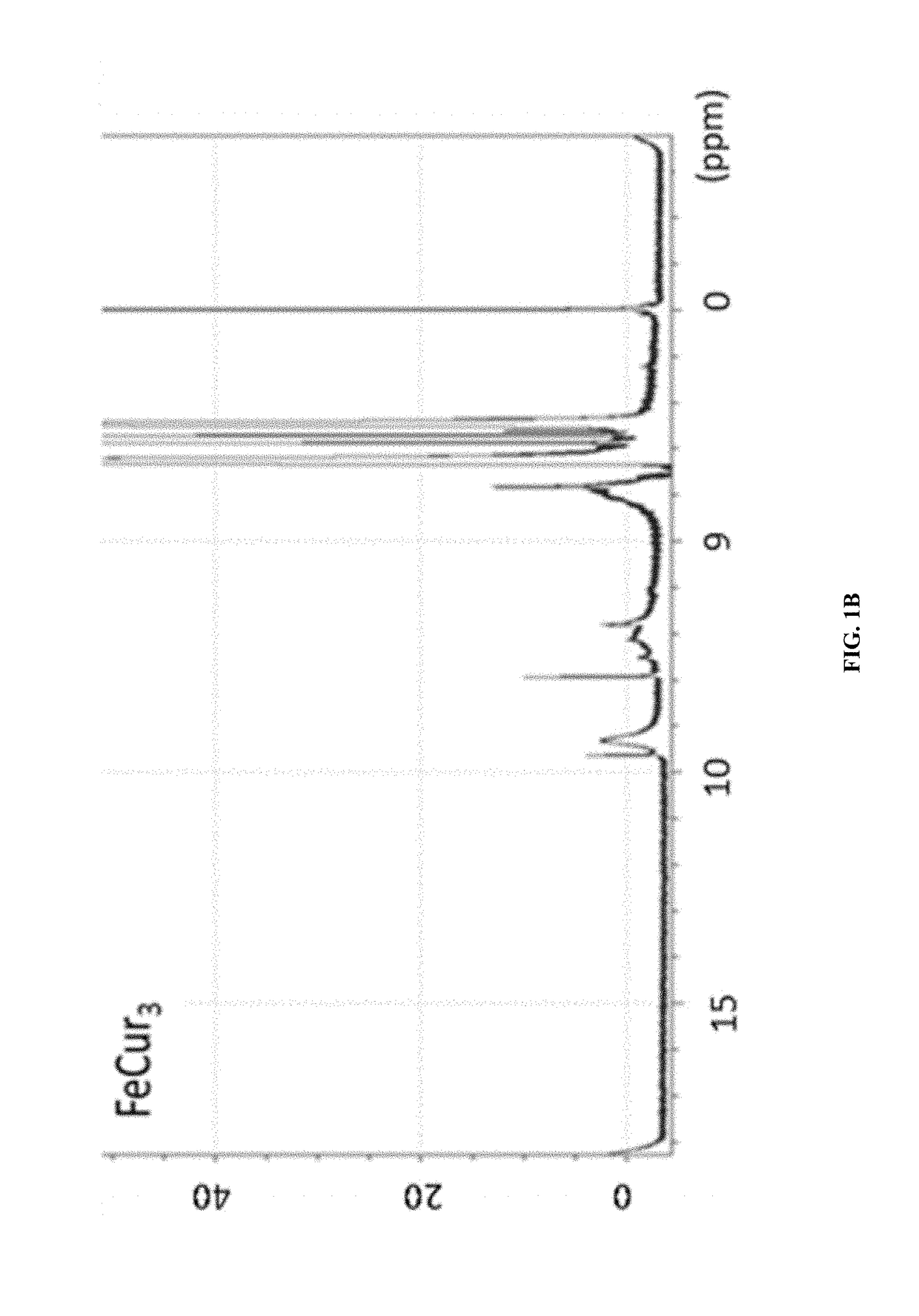
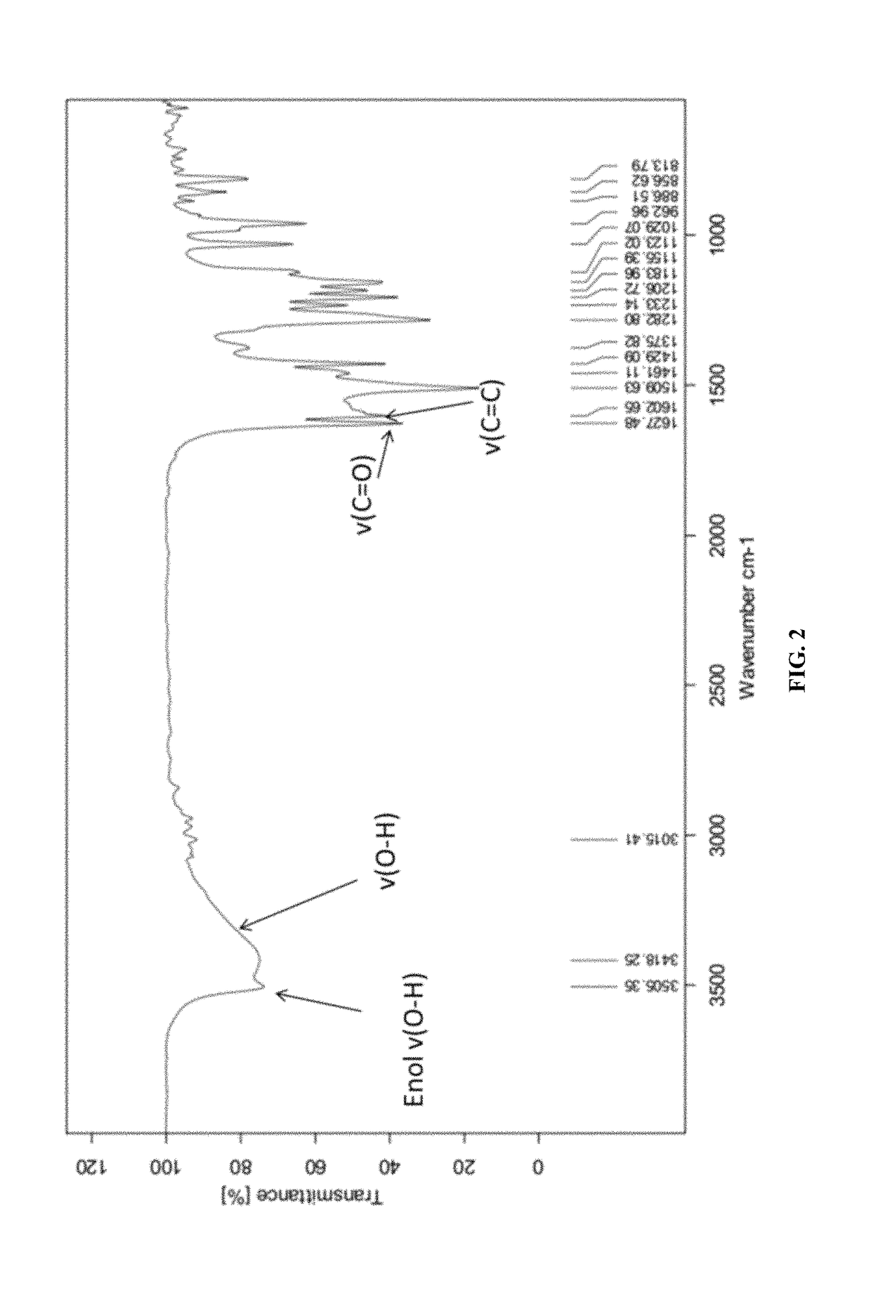
[0096]The synthetic strategy is similar to the reported literature (Sigurdsson et al., 2008; Wadghiri et al., 2003) with modification. Iron-curcumin complex will be described as an example for the synthetic procedure. Iron-curcumin complex was synthesized by dissolving curcumin with minimal amount of dimethylformamide. The iron solution (e.g., Fe(NO3)3 or FeCl3) with one-third mole ratio of curcumin was pre-dissolved in dimethylformamide and added dropwise to the curcumin solution with stirring. The solution was turned to deep red and stirred for overnight under darkness at room temperature. The solution was lyophilized and the powder was washed with Milli-Q water and minimal amount of dichloromethane. FIG. 1 shows the 1H-NMR spectrum of iron-curcumin complex showed broad peaks of curcumin demonstrated the conjugated curcumin to the paramagnetic iron. The lower frequency of the C═O and C═C bond observed in the metal com...

example 2

[0097]Encapsulation of Iron-curcumin Complex by Flash Nanoprecipitation (FNP) Method—MIVM

[0098]The synthesized iron-curcumin complex is barely soluble in water due the hydrophobic nature of curcumin. In order to enhance its aqueous solubility, blood circulation time and prevent non-specific uptake by immune system, the iron-curcumin complex was further encapsulated inside polymeric micelle. PEG(2 k amu)-PLA (10 k amu) co-block polymer and iron-curcumin complex were dissolved in organic phase (e.g., DMF or acetone) while co-stabilizer polyvinyl pyrrolidone (PVP) (30 k amu) was dispersed in aqueous phase. The injection ratio is iron-curcumin complex: coblock polymer: PVP=1:2:4. Both streams were co-injected into Multi-inlet vortex mixer (MIVM) with injection rate at 45 ml / min and 5 ml / min for aqueous phase and organic phase, respectively. The PLA tail of PEG-PLA co-block polymer was in favor to adhere on the iron-curcumin complex surface and formed micelle structure. Also, due to the ...

example 3

[0102]Potential MRI Agent of Iron-curcumin Nanoparticles

[0103]The potential of iron-curcumin nanoparticles to be a magnetic resonance imaging agent was investigated by scanning the MRI signal of iron-curcumin nanoparticles in agarose gel. Agarose gel with different concentration of iron-curcumin nanoparticles were prepared and stacked inside 50 mL centrifugal tube. It was then scanned by the Mill instrument to analyze the MRI signal (FIG. 9). The magnetic moment was also measured by vibrating sample magnetometer (FIG. 10A and FIG. 10B)


PUM

PropertyMeasurementUnit
hydrodynamic diameteraaaaaaaaaa
concentrationsaaaaaaaaaa

Abstract

Provided herein is a nanoparticle comprising a metal core and a polymer shell coating the metal core useful as a magnetic resonance contrast agent.

Description

CROSS REFERENCE TO RELATED APPLICATION[0001]The present application claims benefits from U.S. provisional patent application Ser. No. 62 / 496,757 filed Oct. 28, 2016, and the disclosure of which is incorporated herein by reference in their entirety.FIELD OF THE INVENTION[0002]The present disclosure relates to magnetic nanoparticles useful as magnetic resonance imaging contrast agents, formulations thereof, and methods for producing thereof.BACKGROUND OF THE INVENTION[0003]Conventional in vivo active targeting diagnostic agents commonly rely on a specific peptide or antibody, which is capable of specifically binding the target of interest, linked to a signaling agent. However, these diagnostic agents can be disadvantaged by toxicity of the conjugated peptides or antibodies and their large molecular weight may inhibit the penetration of such targeting agents through biological barriers, such as blood-brain barrier, small intestine, nasal, skin and mouth mucosa. The blood-brain barrier ...

Claims


Application Information

Patent Timeline
Patent Type & Authority Patents(United States)
IPC IPC(8): A61K9/00A61K49/10A61K49/18
CPCA61K49/106A61K49/186A61K49/126A61K49/1854A61K49/1857
Inventor CHENG, KWOK KINKWOK, SAU KUEN CONNIEKO, CHUN HAYTSAI, KEVINNG, CHUN FAI
Owner NANO & ADVANCED MATERIALS INST
Why Eureka
Try Eureka