Dendrimer compositions and their use in treatment of diseases of the eye

a technology of dendrimer and composition, which is applied in the field of dendrimer composition and its use in the treatment of eye diseases, can solve the problems of retinal ischemia, death of neurons initiating further activation of microglia, and decrease in blood flow within the eye, and achieve the effect of suppressing or inhibiting inflammatory and/or angiogenic diseases

Active Publication Date: 2019-08-06
THE JOHN HOPKINS UNIV SCHOOL OF MEDICINE
View PDF55 Cites 1 Cited by
  • Summary
  • Abstract
  • Description
  • Claims
  • Application Information

AI Technical Summary

Benefits of technology

[0014]In accordance with an embodiment, the present invention provides a method for treating an inflammatory and / or angiogenic disease in the eye of a subject comprising administering to the subject systemically, a composition comprising dendrimer nanoparticles, wherein the dendrimer nanoparticles comprise poly(amidoamine) (PAMAM) hydroxyl-terminated dendrimers covalently linked to at least one biologically active agent, in an amount effective to suppress or inhibit the inflammatory and / or angiogenic disease in the eye.
[0016]In accordance with another embodiment, the present invention provides a method for treating an inflammatory and / or angiogenic disease in the eye of a subject comprising periodically administering to the subject intravenously, a composition comprising dendrimer nanoparticles, wherein the dendrimer nanoparticles comprise poly(amidoamine) (PAMAM) hydroxyl-terminated dendrimers covalently linked to a biologically active agent, in an amount effective to suppress or inhibit the inflammatory and / or angiogenic disease in the eye.

Problems solved by technology

Retinal vascular occlusion, be it by high intra-ocular pressure in the I / R model or thrombus in BVO, causes a decrease in blood flow within the eye resulting in retinal ischemia.
This causes death of neurons initiating further activation of microglia.
As the disease progresses, the gradual closure of retinal vessels results in retinal ischemia, giving rise to signs including venous abnormalities (beading, loops), intraretinal microvascular abnormalities, and increasing retinal hemorrhage and exudation.
However, systemic delivery would have many advantages beyond current treatments as there are at present no systemic treatments available for ischemic retinopathies or AMD.
Currently, there are no targeted therapies for AMD or DR.

Method used


Image

  • Dendrimer compositions and their use in treatment of diseases of the eye
  • Dendrimer compositions and their use in treatment of diseases of the eye
  • Dendrimer compositions and their use in treatment of diseases of the eye

Examples


example 1

[0090]Characterization of D-Cy5 conjugates. Ethylenediamine-core poly-(amidoamine) [PAMAM] hydroxyl-terminated generation-4 (G4-OH) were labeled with near IR fluorescent dye Cy5 as we reported previously (Molecular Pharmaceutics. 2013; 10:4560-71; Biomaterials. 2012; 33:979-88). Briefly, G4-OH was partially functionalized by 6-amino caproic acid using FMOC protection / deprotection chemistry resulting in bifunctional dendrimers with ˜5-6 NH2 groups on their surface. The resulting bifunctional dendrimers with reactive amine groups were reacted with N-hydroxysuccinimide monoester Cy5 dye to obtain the D-Cy5 conjugate. The resulting conjugates were purified using dialysis and GPC (gel permeation chromatgraphy) and characterized using 1H NMR (FIG. 4, FIGS. 5A-5C, and FIGS. 6A-6B).

[0091]The HPLC chromatogram of bifunctional dendrimer showed elution time of 14.84 min from the column which differed from the elution time of G4-OH dendrimer (elution time 14.42 min) (FIGS. 9A-9B). This indicate...

example 2

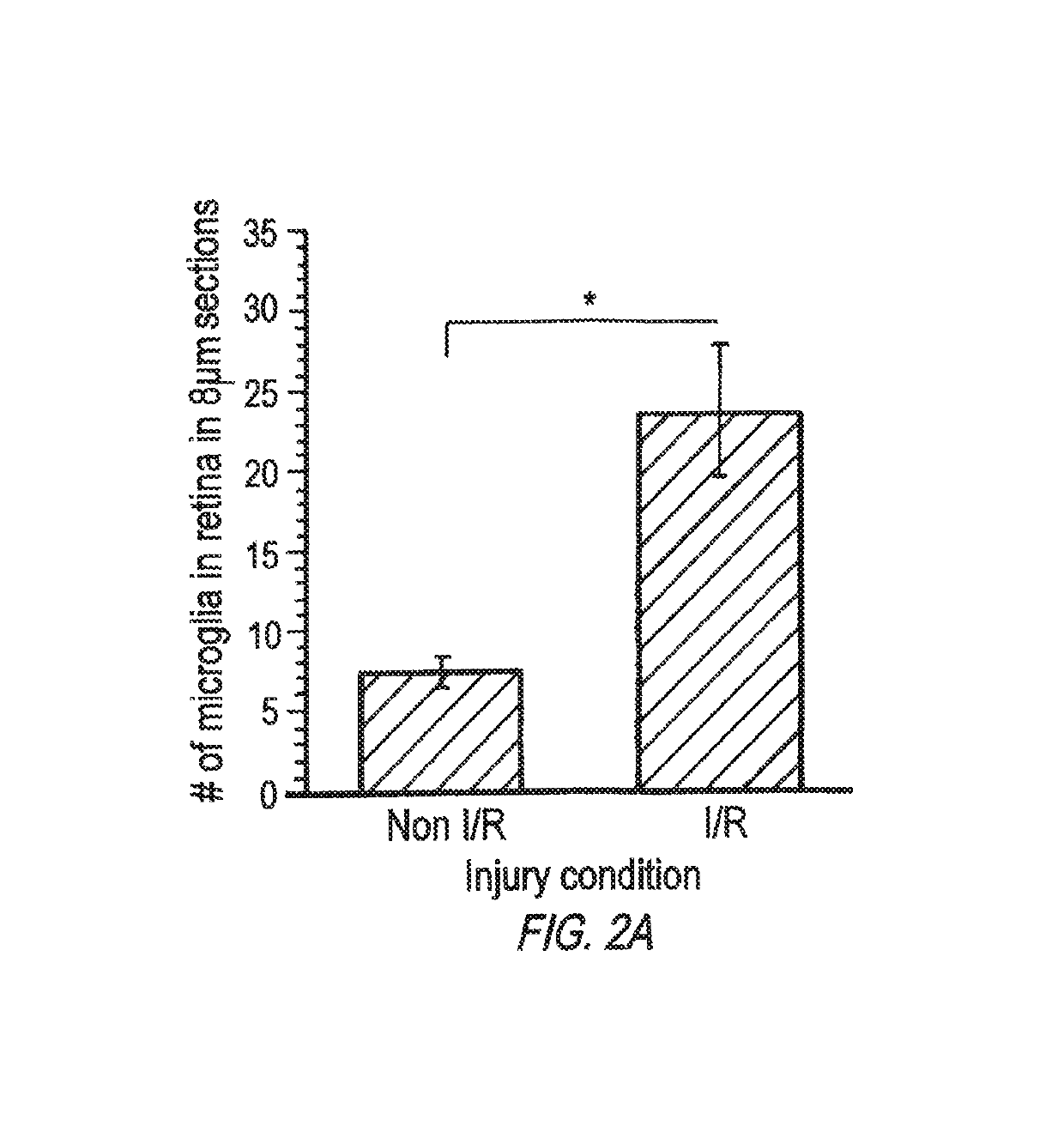
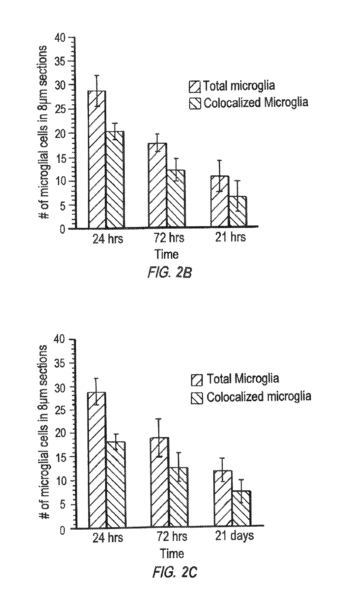
[0092]Ischemia-Reperfusion: Differences in microglial / macrophage population, morphology and retinal structural changes. Iba-1+ resident microglia / macrophages in normal retina were less in number and had ramified morphology with distinctive dendrites. The heterogeneous populations of microglial cells were predominately found in choroid and inner nuclear layer (INL) and very few of them were observed in the outer plexiform layer (OPL). Sections from control, non I / R, eyes 24 hours after intravitreal injection were examined Twenty four hours after injection of D-Cy5, there is no dendrimer retained in retina. Free Cy5 was present throughout inner retina and in the inner plexiform layer (IPL). The retinas had a normal lamination after intravitreal injection. I / R injury led to a structurally damaged retina and marked activation of microglia in the retina and choroid, based on a change from dendritic to round or fusiform morphology. At six days post IR, the retinal microglial / macrophages w...

example 3

[0093]Retinal biodistribution of D-Cy5 upon intravitreal & intravenous administration: Intravitreal Administration. Intravitreal administration of D-Cy5 showed differential biodistribution between normal and I / R retinas. In normal retinas at 24 hours post intravitreal injection of D-Cy5, there was very minimal fluorescence in retina and choroid. There was no fluorescence signal from D-Cy5 after 24 hours suggesting that dendrimers were cleared completely from retina. On the contrary, free Cy5 remained in inner retina at 24 hours post injection. This suggests that D-Cy5 is cleared rapidly from intact retina. In I / R-injured retinas, we observed significant fluorescence signal from D-Cy5 in retinal sections at 24 hours post-injection. Dendrimers (D-Cy5) were observed in Iba-1+ microglia / macrophages in the subretinal space, ONL, INL and in the vicinity of internal limiting membrane (ILM) of retina. We have also observed dendrimer in vitreous and localized in other cells in inner retina a...


PUM

PropertyMeasurementUnit
elution timeaaaaaaaaaa
retention timeaaaaaaaaaa

Abstract

The treatment of many ocular disorders is hampered because of poor penetration of systemically administered drugs into the eye. The tight junctional complexes (zonulae occludens) of the retinal pigment epithelium and retinal capillaries are the site of the blood-ocular barrier. This barrier inhibits penetration of substances, including antibiotics, into the vitreous. Over the last 18 years we have evaluated the nontoxic doses of various drugs. These include antibiotics and antifungals for treatment of bacterial and fungal endophthalmitis, antivirals for treatment of viral retinitis (specifically, when medication with these drugs poses the threat of toxicity to other organs). Intravitreal antineoplastic drugs have been studied to prevent cell proliferation in the vitreous cavity after retinal attachment surgery, which can lead to proliferative vitreoretinopathy (PVR). Furthermore, we evaluated the anti-inflammatory action of dexamethasone and cyclosporine A to reduce intraocular inflammation after intraocular surgery or in uveitis. Because these studies had been performed in the presence of the vitreous, which can slow down the diffusion of the drugs toward the retina, it was necessary to reevaluate the concentration of drugs which could be administered intravitreally in the vitrectomized eye. The nontoxic dose of numerous drugs when added to vitrectomy infusion fluid has also been evaluated. Furthermore, the role of vitrectomy in the treatment of bacterial fungal endophthalmitis has been studied and the role of vitrectomy in this ocular disorder is defined.

Description

REFERENCE TO RELATED APPLICATIONS[0001]This application is a 371 application of International Application No. PCT / US2015 / 028386 filed Apr. 30, 2015, which claims the benefit of U.S. Provisional Patent Application No. 61 / 986,495, filed on Apr. 30, 2014, which is hereby incorporated by reference for all purposes as if fully set forth herein.BACKGROUND OF THE INVENTION[0002]Microglia are the resident macrophages of the brain and retina. They become activated in diseases such as diabetes and retinal degeneration where cells die, causing microglia to phagocytose cellular debris. Activation of retinal microglia occurs in a mouse model of ischemia / reperfusion injury (I / R), as occurs in inflammatory diseases of the eye, including glaucoma, age related macular degeneration (AMD), diabetic retinopathy and branch vein occlusion. Retinal vascular occlusion, be it by high intra-ocular pressure in the I / R model or thrombus in BVO, causes a decrease in blood flow within the eye resulting in retina...

Claims


Application Information

Patent Timeline
Patent Type & Authority Patents(United States)
IPC IPC(8): A61K49/00A61K31/573A61K9/08A61K45/06A61K47/59A61K31/198A61K9/00
CPCA61K31/198A61K9/0019A61K9/0048A61K9/08A61K49/0054A61K45/06A61K47/595A61K49/0032A61K31/573A61P25/02A61P27/02A61P29/00A61P31/04A61P37/06A61P43/00A61P7/00A61P9/00A61P9/10A61P9/12
Inventor RANGARAMANUJAM, KANNANLUTTY, GERARDKAMBHAMPATI, SIVA PRAMODHMISHRA, MANOFBHUTTO, IMRAN
Owner THE JOHN HOPKINS UNIV SCHOOL OF MEDICINE
Why Eureka
Try Eureka