Treatment of aneurysms
a technology for aneurysms and aneurysms, which is applied in the field of treatment of aneurysms, can solve the problems of increasing the risk of rupture, and weakening the tensile strength of the arterial wall, so as to reduce the risk of ruptur
- Summary
- Abstract
- Description
- Claims
- Application Information
AI Technical Summary
Benefits of technology
Problems solved by technology
Method used
Image
Examples
example 1
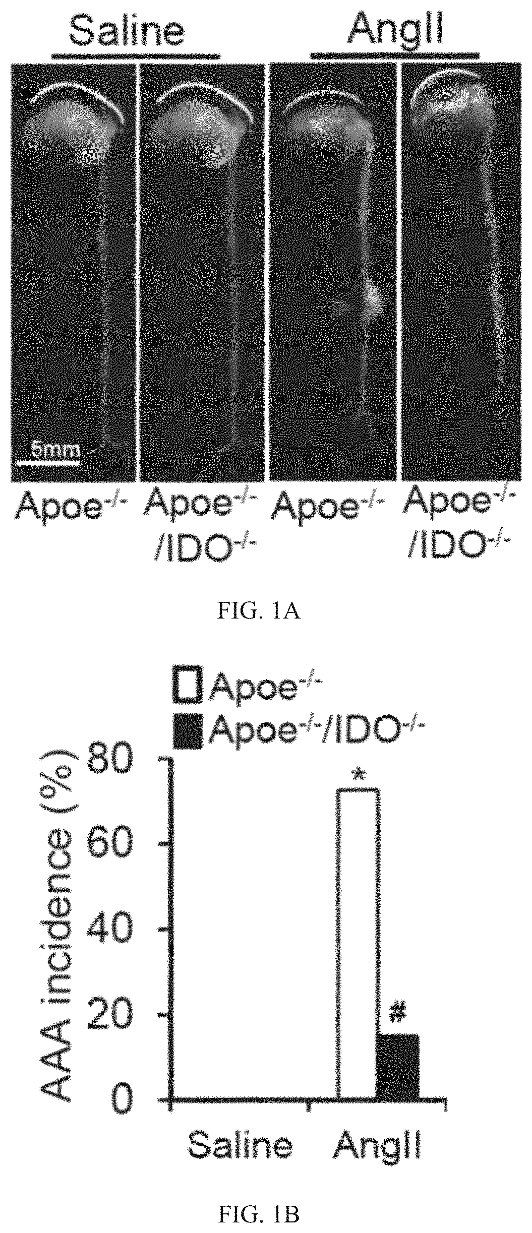
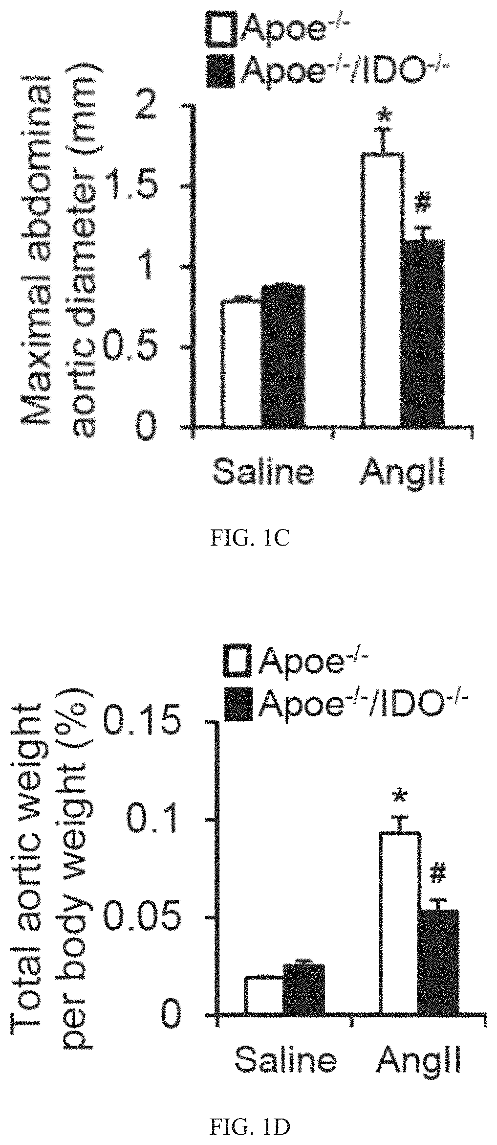
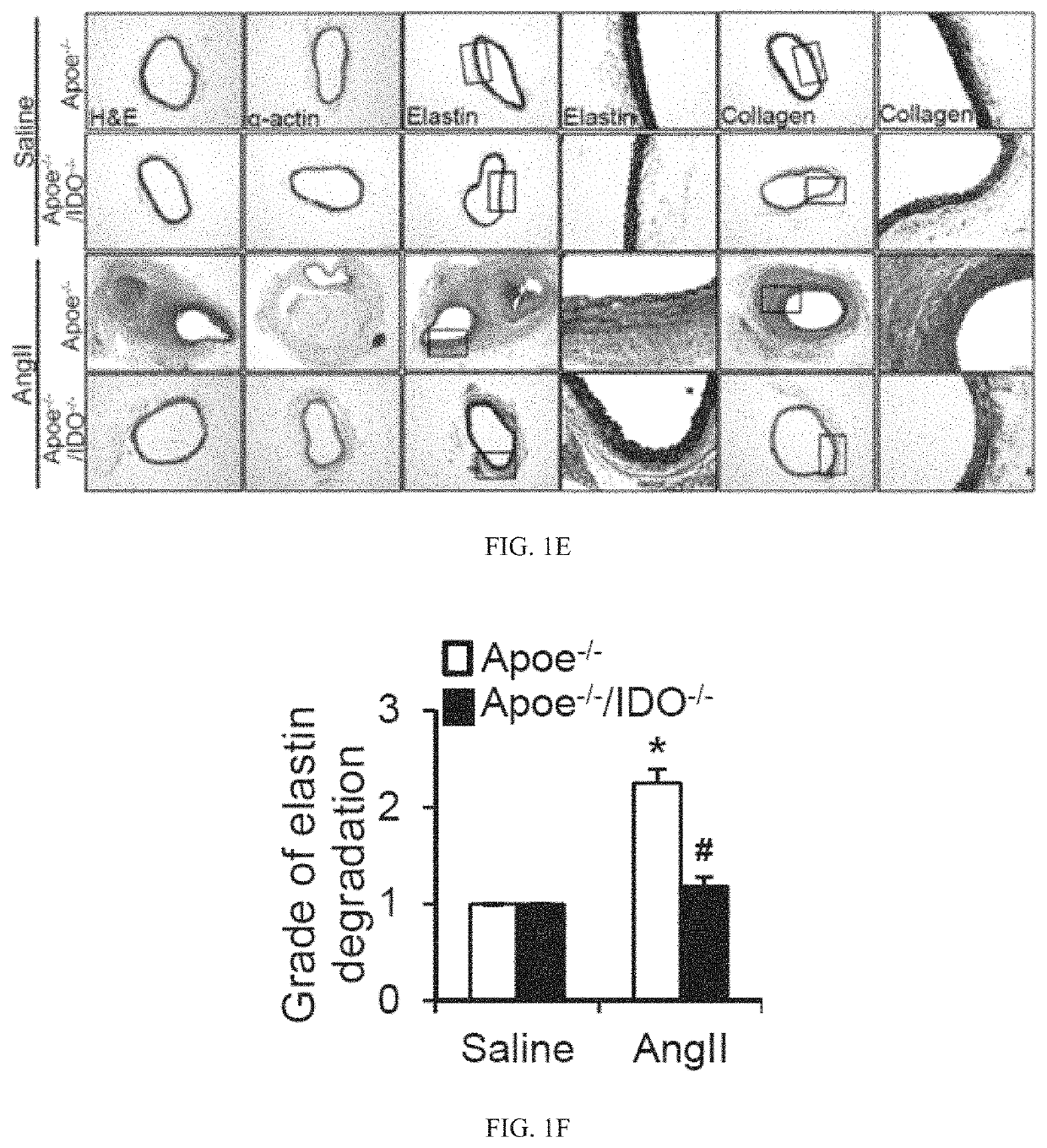
ion Abrogates AngII-Induced AAA Formation
[0351]AngII-induced mouse AAA formation in the atherosclerotic-susceptible strain (ApoE− / −) has become the most widely used model. See Qin Z, et al., Angiotensin II-induced TLR4 mediated abdominal aortic aneurysm in apolipoprotein E knockout mice is dependent on STAT3. J Mol Cell Cardiol. (2015) 87:160-70; Rateri D L, et al., Prolonged infusion of angiotensin II in ApoE (− / −) mice promotes macrophage recruitment with continued expansion of abdominal aortic aneurysm. Am J Pathol. (2011) 179:1542-8; Cassis L A, et al., ANG II infusion promotes abdominal aortic aneurysms independent of increased blood pressure in hypercholesterolemic mice. Am J Physiol Heart Circ Physiol. (2009) 296:H1660-5; Daugherty A and Cassis L A. Mouse models of abdominal aortic aneurysms. Arterioscler Thromb Vasc Biol. (2004) 24:429-34; Owens A P, 3rd, Rateri D L, Howatt D A, Moore K J, Tobias P S, Curtiss L K, Lu H, Cassis L A and Daugherty A. MyD88 deficiency attenuates...
example 2
iency Mitigates MMP2 Upregulation in AAA Mice
[0356]AngII infusion is intensely associated with vascular inflammation, which is considered a key mediator of AngII-induced AAA formation. See Qin Z, et al., Angiotensin II-induced TLR4 mediated abdominal aortic aneurysm in apolipoprotein E knockout mice is dependent on STAT3. J Mol Cell Cardiol. (2015) 87:160-70; Owens A P, et al., MyD88 deficiency attenuates angiotensin II-induced abdominal aortic aneurysm formation independent of signaling through Toll-like receptors 2 and 4. Arterioscler Thromb Vasc Biol. (2011) 31:2813-9. As shown in Table 3, serum concentrations of inflammatory cytokines, including interferon (IFN)-γ, tumor necrosis factor-α, interleukin-6, and cyclophilin A, are elevated in both AngII-infused ApoE− / − and ApoE− / − / IDO-mice. These data indicate that IDO deletion does not alter AngII-induced inflammation. Previous studies demonstrated that IFN-γ mediates AngII-induced Kyn pathway activation in vivo. See Wang Q, et al....
example 3
IDO Deletion Inhibits AAA Formation
[0359]Based on the above-mentioned data, we postulated that the activation of the Kyn pathway in VSMCs promotes AAA formation. To test this hypothesis, reciprocal bone marrow transplants between ApoE− / − and ApoE− / − / IDO− / − mice were performed, in which bone marrow cells were transplanted into irradiated mice. After 6 weeks of engraftment, transplanted mice were treated with AngII (1000 ng / min per kg) for 4 weeks. This led to the formation of AAAs in ApoE− / − mice transplanted with either IDO− / − or IDO+ / + bone marrow cells, with a similar incidence of approximately 70% (FIGS. 3A and 3B). In contrast, an inhibition of AAA formation (20% incidence) was observed in ApoE− / − / IDO− / − mice transplanted with either IDO− / − or IDO+ / + bone marrow cells (FIGS. 3A and 3B).
[0360]No differences in the maximal abdominal aortic diameter (FIG. 3C) and total aortic weight (FIG. 3D) were observed between mice transplanted with IDO− / − bone marrow cells and mice transplante...
PUM
| Property | Measurement | Unit |
|---|---|---|
| size | aaaaa | aaaaa |
| size | aaaaa | aaaaa |
| diameter | aaaaa | aaaaa |
Abstract
Description
Claims
Application Information
 - R&D
- Intellectual Property
- Life Sciences
- Materials
- Tech Scout
- Unparalleled Data Quality
- Higher Quality Content
- 60% Fewer Hallucinations
Browse by: Latest US Patents, China's latest patents, Technical Efficacy Thesaurus, Application Domain, Technology Topic, Popular Technical Reports.
© 2025 PatSnap. All rights reserved.Legal|Privacy policy|Modern Slavery Act Transparency Statement|Sitemap|About US| Contact US: help@patsnap.com


