Methods and kits for the detection of endotoxi

a technology of endotoxin and kits, which is applied in the field of methods and kits for the detection of endotoxin, can solve the problems of difficult rapid diagnosis at an early stage, irreversible shock and death, organ failure, etc., and achieve the effects of increasing respiratory rate, rapid and accurate diagnosis of septicemia, and increasing heart ra

Inactive Publication Date: 2002-12-05
THE TRUSTEES OF THE UNIV OF PENNSYLVANIA
View PDF0 Cites 0 Cited by
  • Summary
  • Abstract
  • Description
  • Claims
  • Application Information

AI Technical Summary

Benefits of technology

[0022] Cell lines such as Chinese hamster ovary (CHO) cells, COS-7 cells or SF9 insect cells can be transfected with cDNA from bovine, sheep, hamster, canine, rat or human A.sub.1 adenosine receptors and / or treated with A.sub.1 adenosine receptor promoters to increase the expression and density of A.sub.1 adenosine receptors used in these assays. In one embodiment, cDNAs can be tagged with hexahistidine and FLAG (H / F) epitope to create an expression vector which can be transfected into cells by means of lipofectin or DEAE / dextran methods to increase the expression of A.sub.1 adenosine receptors in tissues. Membranes of cells containing native or recombinant A.sub.1 adenosine receptors are labeled with a detectable A.sub.1 adenosine receptor agent. The receptors are then solubilized and purified with use of affinity chromatography and SDS polyacrylamide gel electrophoresis. Monoclonal antibodies are then raised against the purified receptors for detection of endotoxin by ELISA.
[0028] Accordingly, methods of the present invention can be used to diagnose septicemia in an animal. By "animal" it is meant to include, but is not limited to, mammals, fish, amphibians, reptiles, birds, marsupials, and most preferably, humans. Thus, in a preferred embodiment, a clinician can measure the levels of endotoxin in a sample from a human and rapidly and accurately diagnose septicemia. In this embodiment, a sample, preferably a plasma sample, is obtained from a human suspected of having or being at risk for septicemia. Signs or symptoms which may lead a clinician to suspect that a human has septicemia include, but are not limited to, fever or hypothermia, chills, rigors, diaphoresis, increased respiratory rate, increased heart rate, altered mental status, confusion, and lowered blood pressure in conjunction with shock. Examples of humans at risk for septicemia include, but are not limited to, those with indwelling catheters such as urinary, intravenous or endotracheal catheters, CSF drainage tubes, joint or wound drainage tubes, chest tubes, or peritoneal catheters for dialysis, and immunocompromised individuals such as those with cancer, leukemia, AIDS or other immunological disorders, diabetes, kidney disease, liver disease, and individuals being treated for cancer by chemotherapy. Endotoxin levels in a sample are then determined in accordance with a method of the present invention.

Problems solved by technology

Following its release, this toxin produces a cascade of complex events which ultimately results in organ failure, irreversible shock and death.
Despite the prevalence and severity of this disease, rapid diagnosis at an early stage is still difficult.
However, by the time such clinical signs and symptoms manifest, irreversible organ and tissue damage may have already occurred.
Further, results from blood cultures require at least 24 hours and do not always provide a definitive diagnosis since only 35% of patients with septicemia have positive blood cultures.
However, this particular method is not quantitative.
However, the LAL test is not sufficiently specific enough for clinical diagnosis of septicemia in human plasma.
Further, substances in human blood having nonspecific amidolytic activities, such as factor Xa, thrombin, and trypsin act directly on the chromogenic substrate (Obayashi J. Lab. Clin. Med. 1984, 104:321-330) and produce false positive results.
However, dilution of the sample results in dilution of the endotoxin level and reduces the sensitivity of detecting low levels of endotoxin in the sample.
Attempts at ultrafiltration to remove high molecular weight interfering substances have also proved unsuccessful because the pore size is too small and endotoxin may be adsorbed on the ultrafiltration membrane (Nawata et al. J. Chromatography 1992, 597:415-424).
However, these ELISAs are also affected by substances in human blood which activate or inhibit LAL as described above and like other LAL assays depend on the availability of the rare horseshoe crabs which are diminishing in populations.

Method used


Image

  • Methods and kits for the detection of endotoxi
  • Methods and kits for the detection of endotoxi
  • Methods and kits for the detection of endotoxi

Examples


example 1

Preparation of A.sub.1 Adenosine Receptors for Binding Experiments

[0036] Frozen sheep brain (Pel Freeze, Rodgers, Ark.) or hamster brain (Harlan Bioproducts, Indianapolis, Ind.) was thawed and homogenized in 10 volumes of cold (4.degree. C.), 0.32 M sucrose. The homogenate was centrifuged at 1,000.times. g for 10 minutes to remove the nuclear fraction. The supernatant was centrifuged at 30,000.times. g for 30 minutes. The resulting pellet was resuspended in 10 ml water and left on ice for 30 minutes to obtain synaptosomal membranes. The suspension was then centrifuged at 48,000.times. g for 10 minutes and the membranes resuspended in 50 mM Tris-HCl (pH 7.4) at a concentration of 8 mg protein / ml, frozen in liquid nitrogen, and stored at -20.degree. C. until binding assay.

[0037] Feline PAECs were grown to confluency in 75 mm.sup.2 culture plates. The confluent cells were washed twice with KSP buffer (10 mM KH.sub.2P0.sub.4, 50 mM sucrose). The cells were scraped and suspended in KSP b...

example 2

Saturation Experiments

[0039] Binding of [.sup.3H] DPCPX or [.sup.3H] CCPA to membranes was carried out in an assay volume of 500 .mu.l Tris-HCl, pH 7.4, MgCl.sub.2 (10 mM) (for membranes from feline PAECs only) containing [.sup.3H] DPCPX or [.sup.3H] CCPA (0.01 to 4 nM). Nonspecific binding of [.sup.3H] DPCPX or [.sup.3H] CCPA was determined in the presence of R-PIA (100 .mu.M) or theophylline (1 mM), respectively. To remove endogenous adenosine, adenosine deaminase was present in all binding assays at a concentration of 0.2 U / ml. Incubations were performed for 2 hours at room temperature for sheep and hamster brain membranes and 4.degree. C. for feline PAEC membranes. Incubations were terminated by rapid removal of the incubation buffer by filtration through Whatman GF / B filters (Whatman Inc., Clifton, N.J.). The membrane bound radioactivity was measured by scintillation counting of the filter paper. To determine Kd, Bmax, and nonspecific binding in sheep and hamster brain and feli...

example 3

Competition Experiments

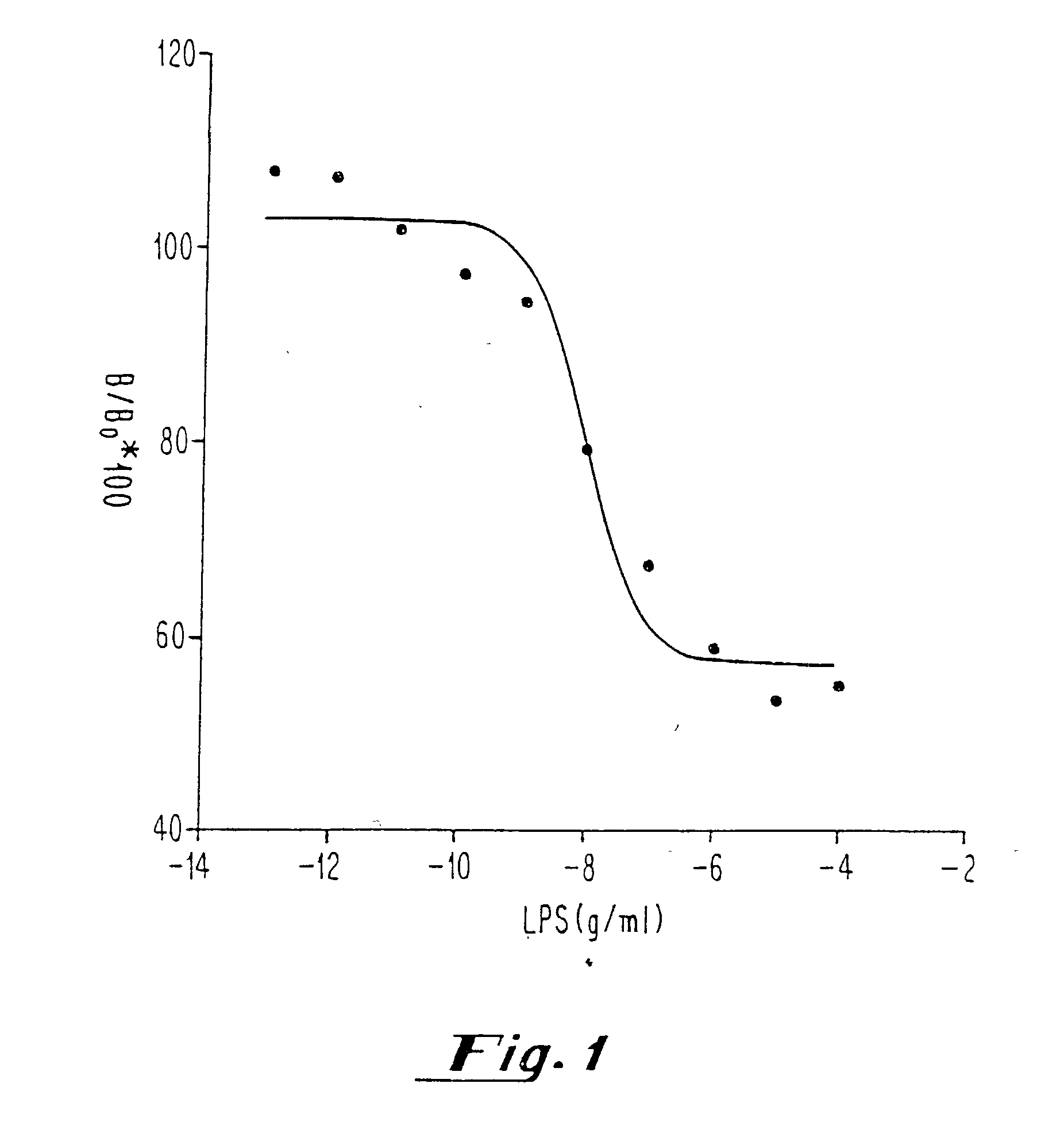
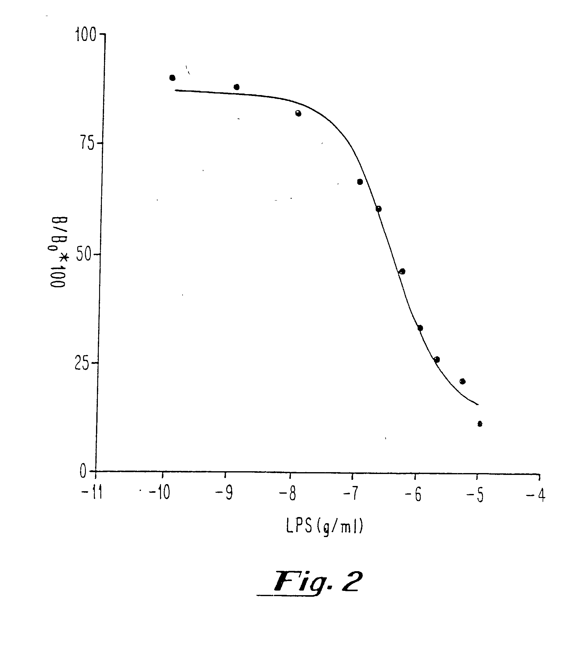
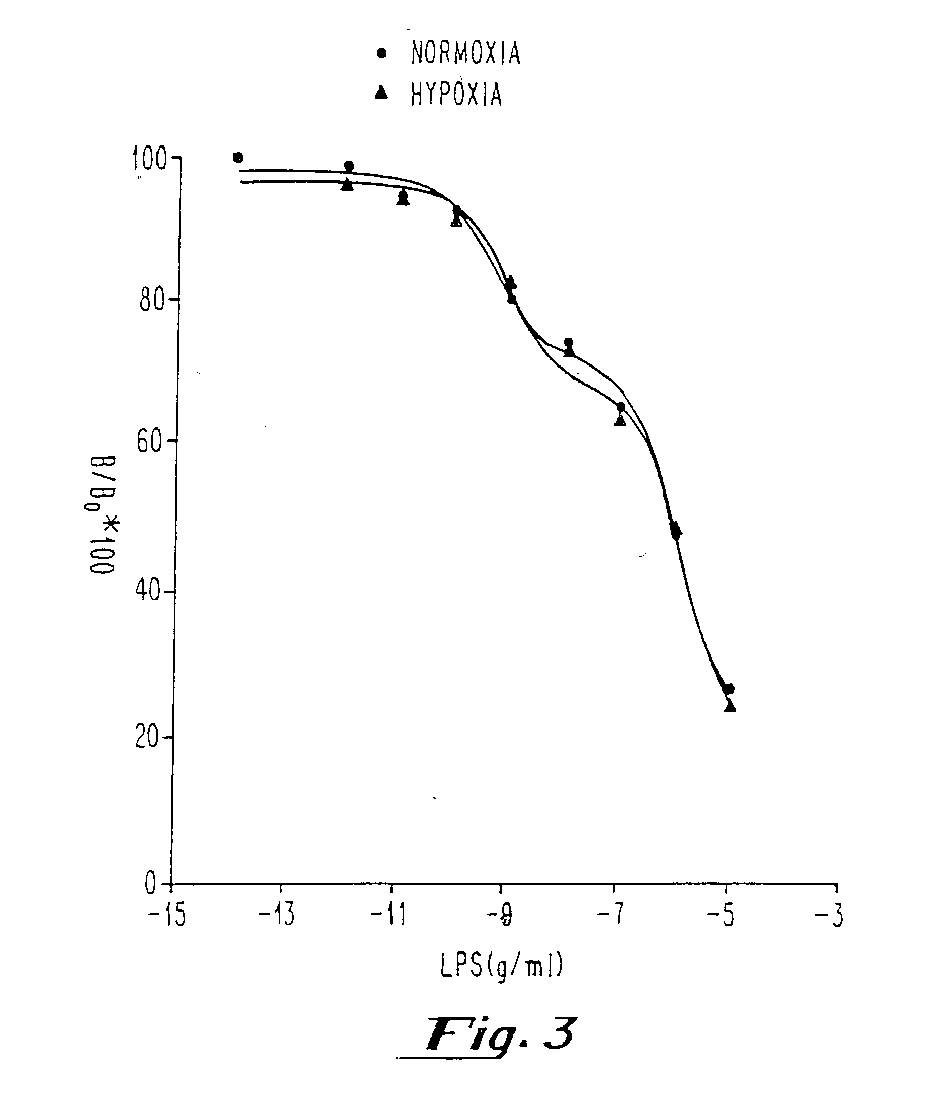
[0040] After determination of the Kd, Bmax and nonspecific binding of [.sup.3H] DPCPX or [.sup.3H] CCPA in membranes prepared from sheep and hamster brains and pulmonary arterial endothelial cells with and without hypoxia, competition experiments with known concentrations of endotoxin [LPS from E. coli 055:B5, E. coli 0111:B4, Serratia marcescens, and Salmonella typhimurium; 0.1 pg / ml-10 .mu.g / ml in saline or plasma) were performed in the presence of [.sup.3H] DPCPX or [.sup.3H] CCPA (0.2-5 nM). Measurements of all samples (saline and plasma) were performed in duplicate.


PUM

PropertyMeasurementUnit
systolic blood pressureaaaaaaaaaa
concentrationsaaaaaaaaaa
pHaaaaaaaaaa

Abstract

Methods for determining levels of endotoxin in a sample are provided. These methods are useful in the diagnosis of septicemia. Kits for the detection of endotoxin are also provided.

Description

[0001] Septicemia is the thirteenth leading cause of death in humans, accounting for up to 100,000 deaths and $5 to $10 billion of health care expenditures annually in the United States, alone (Increase in National Hospital Discharge Survey rates for septicemia--United States, 1979-1987 MMWR 1990, 39:31 -34; Skelter et al. Arch. Microbiol. 1995,164:383-389). It is the most common cause of death in medical and surgical intensive care units, and is associated with a mortality rate of 40 to 90%.[0002] Septicemia is caused by the release of endotoxin, also referred to herein as lipopolysaccharide (LPS), from the outer wall of gram negative bacteria into the blood stream. LPS or endotoxin is released from the outer membrane of the bacteria when they multiply, die, or lyse (Rietschel et al. FASEB J. 1994, 8:217-225). Following its release, this toxin produces a cascade of complex events which ultimately results in organ failure, irreversible shock and death. By binding to cell membrane pr...

Claims


Application Information

Patent Timeline
Patent Type & Authority Applications(United States)
IPC IPC(8): G01N33/53G01N33/50G01N33/566G01N33/569G01N33/577G01N33/579G01N33/68
CPCG01N33/56916G01N33/68G01N33/579
Inventor NEELY, CONSTANCE F.
Owner THE TRUSTEES OF THE UNIV OF PENNSYLVANIA
Why Eureka
Try Eureka