Optical sorting method

a sorting method and optical technology, applied in the field of optical sorting method, can solve the problems of limited scope of the above-mentioned system, limited library size allowed by phage display technology, and inability to direct select activities, etc., to achieve enhanced gene product activity and measure

Inactive Publication Date: 2005-02-17
MEDICAL RESEARCH COUNCIL
View PDF10 Cites 122 Cited by
  • Summary
  • Abstract
  • Description
  • Claims
  • Application Information

AI Technical Summary

Benefits of technology

[0037] Additionally, the genetic elements isolated after a first round of sorting may be subjected to mutagenesis before repeating the sorting by iterative repetition of the steps of the method of the invention as set out above. After each round of mutagenesis, some genetic elements will have been modified in such a way that the activity of the gene products is enhanced (i.e., measurable activity increased by at least 10% relative to wild-type).

Problems solved by technology

Thus, the practical limitation on library size allowed by phage display technology is of the order of 107 to 1011, even taking advantage of λ phage vectors with excisable filamentous phage replicons.
However, the scope of the above systems is limited to the selection of proteins and furthermore does not allow direct selection for activities other than binding, for example catalytic or regulatory activity.
However, selection for “catalytic” or binding activity using SELEX is only possible because the same molecule performs the dual role of carrying the genetic information and being the catalyst or binding molecule (aptamer).
Additionally, proteins may not be selected using the SELEX procedure.
The range of catalysts, substrates and reactions which can be selected is therefore severely limited.
However, none of the methods so far developed have provided molecules of comparable diversity and functional efficacy to those that are found naturally.
Additionally, there are no man-made “evolution” systems which can evolve both nucleic acids and proteins to effect the full range of biochemical and biological activities (for example, binding, catalytic and regulatory activities) and that can combine several processes leading to a desired product or activity.

Method used


Image

  • Optical sorting method
  • Optical sorting method
  • Optical sorting method

Examples


example 1

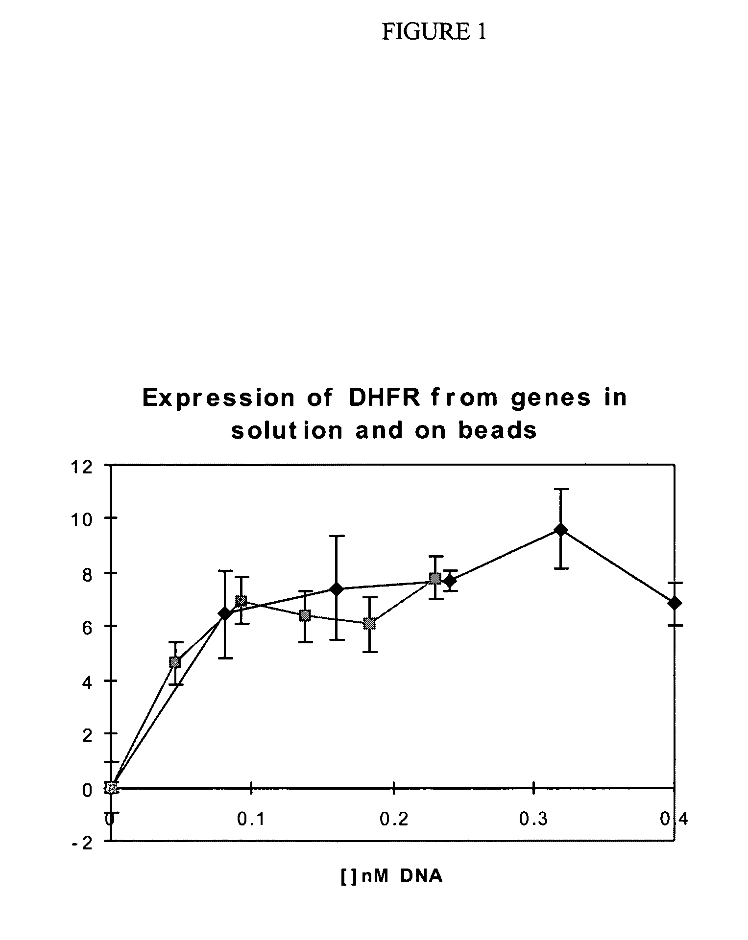
[0226] Enzymes can be expressed from genes in solution and genes attached to paramagnetic microbeads with identical efficiency.

[0227] One format for the selection of genetic elements by using a change in their optical properties is one in which the genetic element comprises a microbead to which the gene is attached. Here it is shown how a gene for an enzyme (E. coli dihydrofolate reductase) can be linked to a paramagnetic bead and is translated in vitro just as efficiently as in solution.

[0228] The E. coli folA gene encoding dihydrofolate reductase (DHFR) is PCR-amplified using oligonucleotides EDHFRFo and EDHFRBa. This DNA is then cloned into the pGEM-4Z vector (Promega) digested with HindIII and KpnI downstream of the lac promoter and the T7 RNA polymerase promoter. The oligonucleotide EDHFRBa appends the efficient phage T7 gene 10 translational start site upstream of the DHFR start codon.

[0229] DNA sequencing identifies a clone which has the correct nucleotide sequence. Bacter...

example 2

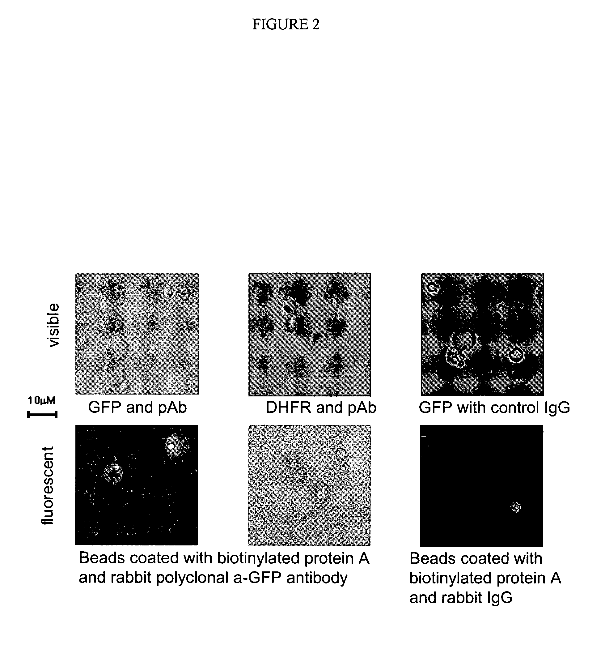
[0236] A fluorescent protein (GFP) can be translated in vitro from genes attached to single microbeads encapsulated in the aqueous compartments of a water-in-oil emulsion and the translated gene-product bound back to the microbeads making them fluorescent.

[0237] One format for the selection of genetic elements is where the genetic element comprises a gene linked to a microbead and the product is coupled back onto the microbead within the microcapsule resulting directly, or indirectly, in a change in the optical properties of the microbead which allows it to be sorted.

[0238] Here it is shown that a fluorescent protein (green fluorescent protein or GFP) can be transcribed and translated in vitro from genes attached to single microbeads encapsulated in the aqueous compartments of a water-in-oil emulsion and the translated gene-product bound back the microbeads making them fluorescent.

[0239] The GFP in pBS / GFP6 plasmid (Siemering et al., 1996) was PCR-amplified using primers GFP-FW a...

example 3

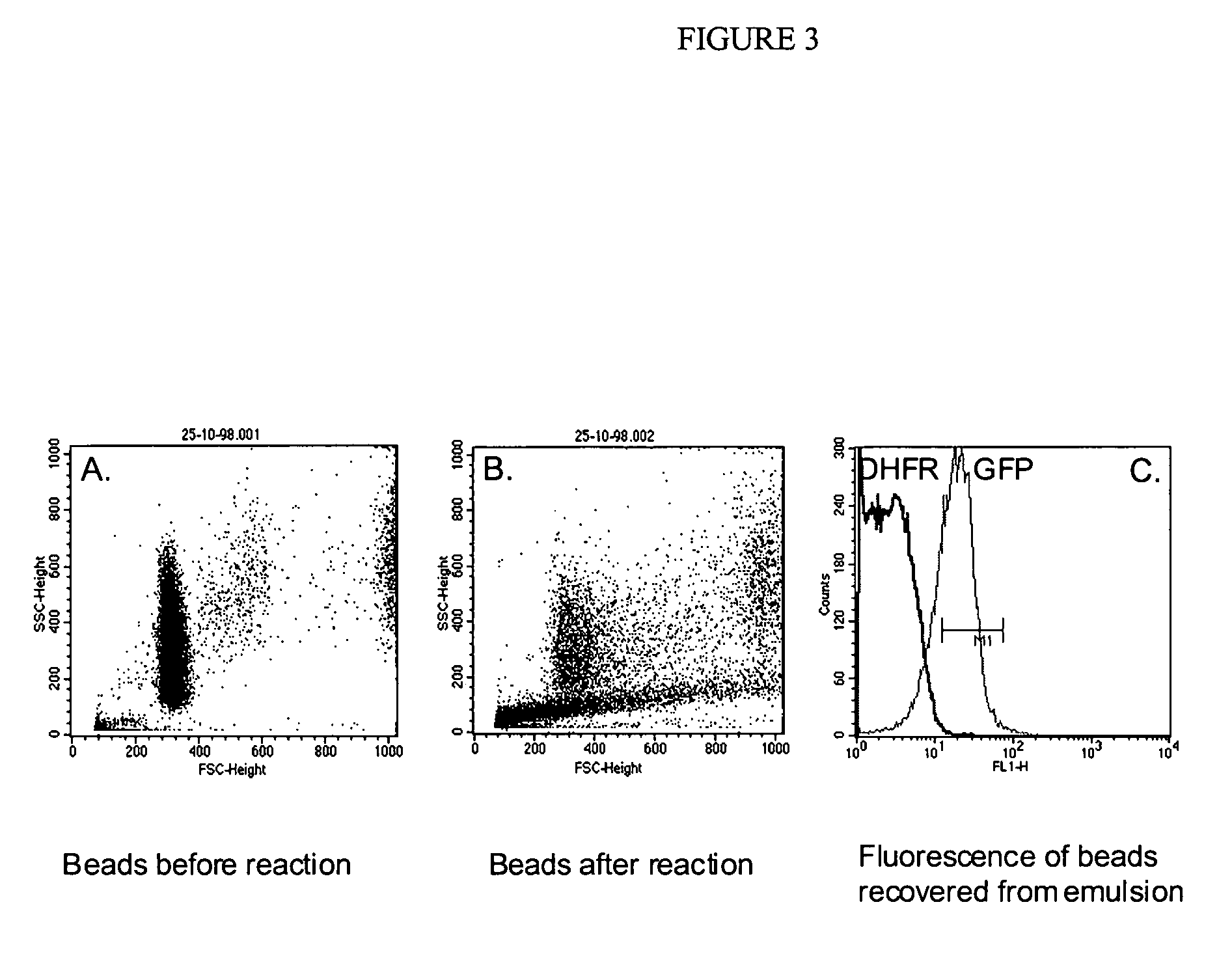
[0245] A fluorescent protein (GFP) can be translated in vitro from genes attached to single microbeads encapsulated in the aqueous compartments of a water-in-oil emulsion, the translated gene-product bound back the microbeads and the increased fluorescence of the microbeads detected by flow cytometry.

[0246] 150 μl streptavidin-coated polystyrene beads (diameter 1 μM; Bangs Laboratories, 2×10⁷ beads / μl) were suspended in 5 mM Tris 7.4 / 1M NaCl / 0.1% Tween20 and split into three aliquots of 50 μl. 0.5 μl of 0.2 μM DNA (T7-folA or T7-GFP) was added to each aliquot of beads, incubated at 43° C. for 15 min, washed three times in 25 mM NaH2PO4, 125 mM NaCl, 0.1% Tween20, pH 7.0 (PBS / 0.1% Tween20), resuspended in 40 μl TBST and 10 μl 80 μM biotinylated protein A (Sigma)was added (to give final concentration of 15 μM). After incubation for 30 minutes at room temperature, the beads were washed three times in PBS / 0.1% Tween20 and resuspended in 20 μl 1:10 dilution rabbit anti-GFP polyclonal an...


PUM

PropertyMeasurementUnit
diameteraaaaaaaaaa
diameteraaaaaaaaaa
diameteraaaaaaaaaa

Abstract

The invention describes a method for isolating one or more genetic elements encoding a gene product having a desired activity, comprising the steps of: (a) compartmentalising genetic elements into microcapsules; (b) expressing the genetic elements to produce their respective gene products within the microcapsules; (c) sorting the genetic elements which produce the gene product having the desired activity using a change in the optical properties of the genetic elements. The invention enables the in vitro evolution of nucleic acids and proteins by repeated mutagenesis and iterative applications of the method of the invention.

Description

[0001] This application is a Divisional of Ser. No. 09 / 896,915, filed Jun. 29, 2001, which was a Continuation-in-Part of International Application No. PCT / GB00 / 00030, filed Jan. 6, 2000, designating the United States, and claims the priority of United Kingdom Application No.: GB 9900298.2, filed Jan. 7, 1999.[0002] The present invention relates to methods for use in in vitro evolution of molecular libraries. In particular, the present invention relates to methods of selecting nucleic acids encoding gene products in which the nucleic acid and the activity of the encoded gene product are linked by compartmentation. [0003] Evolution requires the generation of genetic diversity (diversity in nucleic acid) followed by the selection of those nucleic acids which result in beneficial characteristics. Because the nucleic acid and the activity of the encoded gene product of an organism are physically linked (the nucleic acids being confined within the cells which they encode) multiple rounds ...

Claims


Application Information

Patent Timeline
Patent Type & Authority Applications(United States)
IPC IPC(8): G01N21/78C07K2/00C12N9/00C12N15/09C12N15/10C12P1/00C12P21/00C12P21/02C12Q1/00C12Q1/26C12Q1/68C12Q1/6811G01N21/64G01N33/15G01N33/50
CPCC12N15/1062C12N15/113C12Q1/6811C12N15/1075
Inventor GRIFFITHS, ANDREWTAWFIK, DANSEPP, ARMIN
Owner MEDICAL RESEARCH COUNCIL
Why Eureka
Try Eureka