

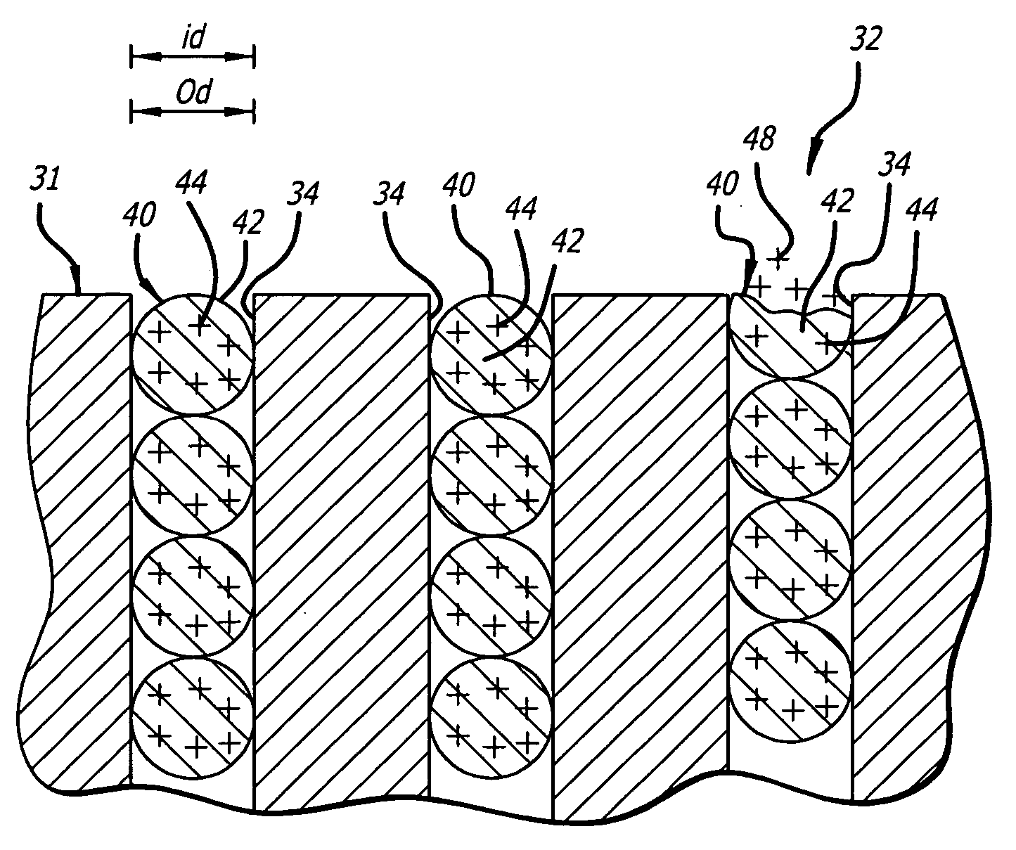
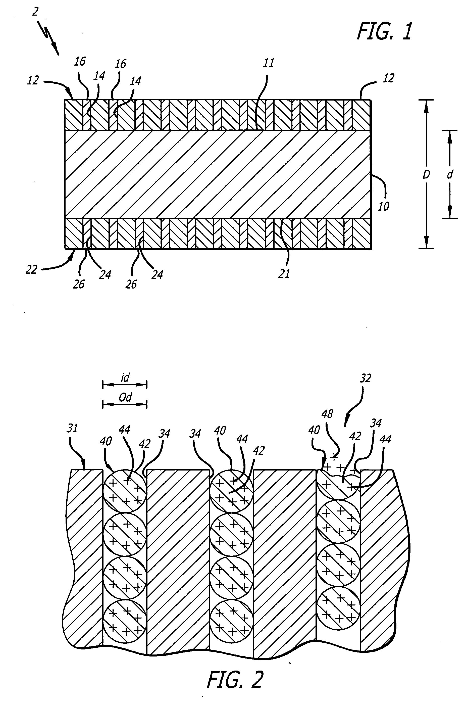
Medical device with porous surface containing bioerodable bioactive composites and related methods
a bioactive composite and medical device technology, applied in the field of implantable medical device surface, can solve the problems of body manifesting rejection of the implant, and significant re-narrowing of the lumen, so as to improve the device-tissue interface and lessen the reaction to the device
- Summary
- Abstract
- Description
- Claims
- Application Information
AI Technical Summary
Benefits of technology
Problems solved by technology
Method used
Image

Examples
example 1
[0136] Bioactive composite coatings were formed on the surface of stainless steel stents. Each stent had two tie layers of nickel struck on its surface prior to immersion in a nickel-phosphorous (Ni—P) electroless deposition bath—further including a de-oxidizing etching stent in-between such multiple nickel strikes. Sirolimus (Rapamycin™), paclitaxel (Taxol™), and des-aspartate angiotensin I (DAA-1) were dissolved / suspended in the various Ni—P baths and co-deposited on the tie layer.
[0137] More specifically, each stent was first prepared by immersion in a 37% hydrochloric (HCl) acid bath at room temperature for seven minutes. The stent was then rinsed with de-ionized and distilled water. After rinsing, the stent was immersed in an electrolytic bath containing nickel ions, which bath was concocted by dissolving nickel chloride (NiCl) in HCl and water. The nickel strike was conducted at room temperature. A negative electric charge was then applied to the stent causing the nickel ions...
example 2
[0141] Bioactive composite coatings were formed on the surface of nickel-titanium (Nitinol) self-expanding stents. Each stent had a tie layer of nickel struck on its surface prior to immersion in a Ni—P electroless deposition bath. Rapamycin, DM-1 and sialokinin (“HP-1”) were dissolved / suspended in the various Ni—P baths and co-deposited on the tie layer.
[0142] More specifically, each stent was first prepared by immersion in a bath of about 2% hydrofluoric (HF) and about 21% nitric (HNO3) acid bath at room temperature for about 2 minutes. The stents were then rinsed with deionized and distilled water, and immersed in an about 37% HCl acid bath at room temperature for about 7 minutes. Each stent was then rinsed with deionized and distilled water. After rinsing, each stent was immersed in an electrolytic bath containing nickel ions, which bath included NiCl dissolved in HCl and water. The nickel strike was conducted at room temperature. A negative electric charge was then applied to ...
PUM
| Property | Measurement | Unit |
|---|---|---|
| outer diameter | aaaaa | aaaaa |
| outer diameter | aaaaa | aaaaa |
| outer diameter | aaaaa | aaaaa |
Abstract
Description
Claims
Application Information