
Artificial vessel scaffold and artifical organs therefrom

Inactive Publication Date: 2005-09-22
BIOARTTIS
View PDF20 Cites 48 Cited by
  • Summary
  • Abstract
  • Description
  • Claims
  • Application Information

AI Technical Summary

Benefits of technology

[0010] One object of the present invention is to provide a synthetic scaffold that is durable yet flexible and will withstand wear and tear of use but be biocompatable.
[0023] The present invention involves a novel scaffold, which is reproducibly made and easily tailored to specific needs, and is fabricated, for example, using porous membrane tubing. The porosity is made and controlled by selective solubilities of the components of the tubing. In one embodiment of the invention, co-extruded or cast tubing can be made from two or more polymers, wherein one of the polymers and / or other material in the tubing is dissolved away upon exposure to solvent. The design allows initial growth of cells in a sterile laboratory setting, with subsequent implantation of the device with viable, functional cells into a host recipient. To date, implantation has been accomplished with similar devices in multiple animals of each cell type (see citations below). This will allow the use of the inventive devices and organs as functional replacement of blood vessels, and other listed organs, for medically indicated purposes in mammals, and in humans.
[0026] Nylon-11 is particularly useful as a matrix material and in bioartifical organ fabrication, because of its non-swelling properties compared to other nylons. It is autoclavable, hydrolytically resistant, water-insoluble, and durable. Combinations of nylon-11 and thermoplastic urethanes (TPU's) and / or TPU as the water-insoluble polymer are also useful to enhance the final flexibility of the porous tubing. The discovery of this type of porous tubing, which can be used as a matrix scaffold onto which and into which human cells can be grown, has led to a whole new arena of bioreactive devices.
[0031] After the porous polymer scaffold has been prepared, it is then intercalated with specific desired cells. This is achieved by using a bioreactor to selectively grow desired cells on the matrix to form an artificial organ. For example, an artificial artery may be produced by growing smooth muscle cells on the exterior and endothelial cells on the interior of the porous tube. The process builds up wall thickness using cells and eliminates direct blood contact with the matrix under high shear conditions. The presence of a smooth layer of natural cells on the inside of the artificial vessel is important because over the course of several months, high shear contact with noncellular material leads to adverse long term effects.
[0032] Cells enter the pores of the matrix and interconnect to form a uniform and continuous layer outside of the polymer tube. The cells are locked into the matrix because of the pore structure permits intimate cell-cell contact through the inner and outer walls of the tubing. The polar amide moieties of the porous polymer, which are at relatively low densities, serve as contact points for cellular adhesion. The uniform cell coverage isolates the polymer tube from the host environment, further enhancing the device's non-thrombogenicity. The small pore size and high porosity of the matrix permits improved cell-cell contact, which leads to enhanced in vivo response to environmental stimuli.
[0034] The invention can thus produce a variety of synthetic devices which can be implanted in a patient to facilitate sustained growth of species specific (i.e., human or animal) cells to provide a desired, species specific, medically applicable treatment, such as an artificial organ.

Problems solved by technology

No single approach has been completely successful in being able to deliver a product that achieves the performance requirements and the need for rapid fabrication.
Veins and bioartificial veins have been employed to replace arteries, but they lack the strength and durability needed for general use in the high shear environment of the arteries.
The disadvantage to this approach is that bovine collagen is not the most suitable material, since it is expensive and it must be harvested from an animal, and may precipitate immune response inflammation.
However, attempts to create synthetic scaffolds, for example from porous polymers, often prove to be too rigid, leading to hyperplasia and increased thrombosis frequencies over time.
However, tubing made from this approach lacks control of micro-porosity (uniform size and spacing) needed for optimal cell entrainment, and also lacks the flexibility and toughness achievable with high porosities.
Accordingly, the synthetic scaffolds of the prior art fail to achieve the optimal morphology and flexural / tensile properties of natural vessels.
High shear contact with noncellular material leads to adverse long term (greater than months) effects.
In addition to the shortcomings presently felt in the synthetic vessel art, there are similar deficiencies in presently-available synthetic organs.
While the demand for replacement organs, particularly in humans, has far outpaced available transplant organs, synthetic devices have met with limited success and use.
Compatibility remains an issue, as well as the concern regarding contamination and infection, especially for xenotransplantation.
In the transplant organ field, this delay may be fatal.

Method used


Image

  • Artificial vessel scaffold and artifical organs therefrom
  • Artificial vessel scaffold and artifical organs therefrom
  • Artificial vessel scaffold and artifical organs therefrom

Examples


example i

Fabrication of an Artificial Vessel Scaffold

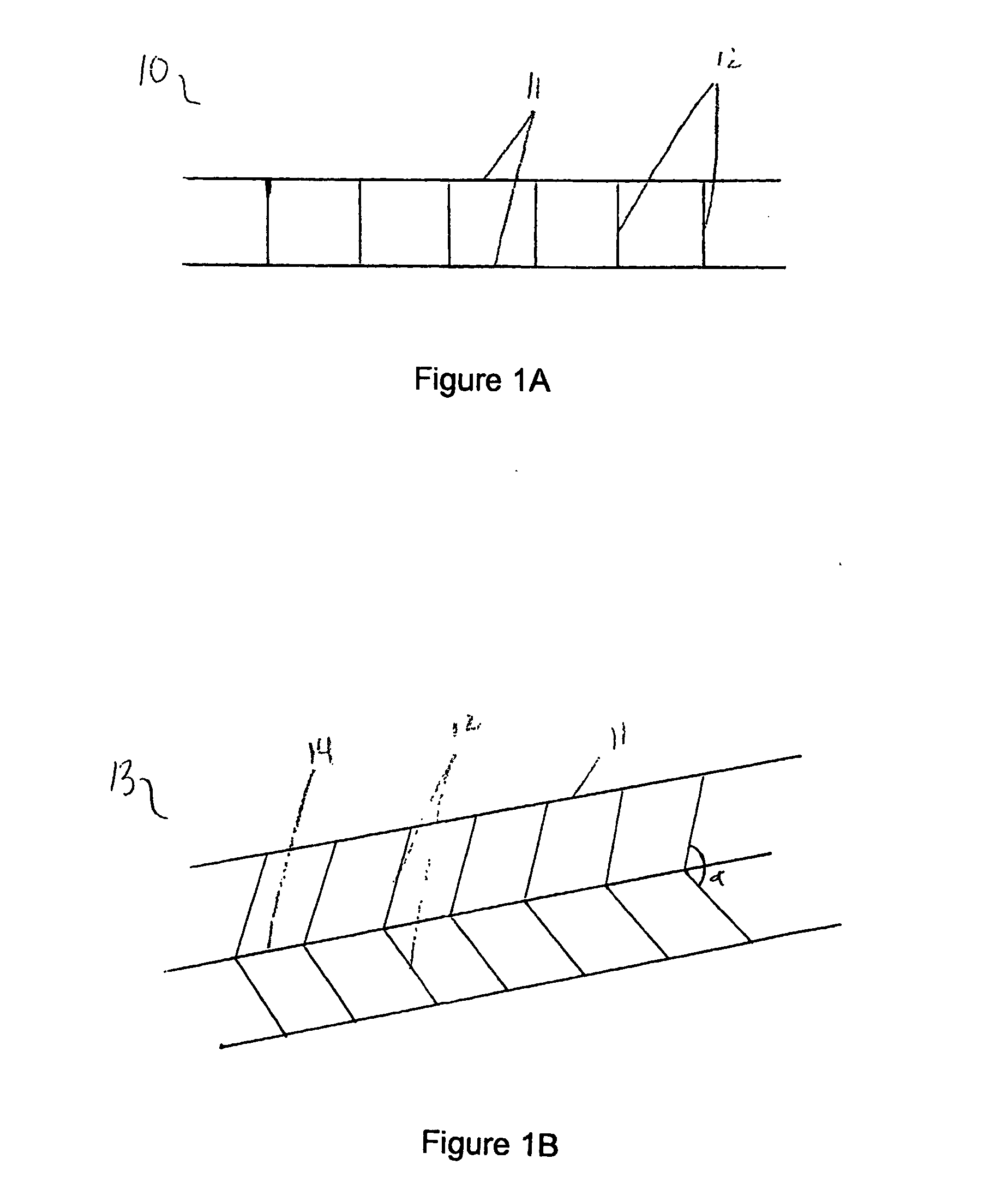
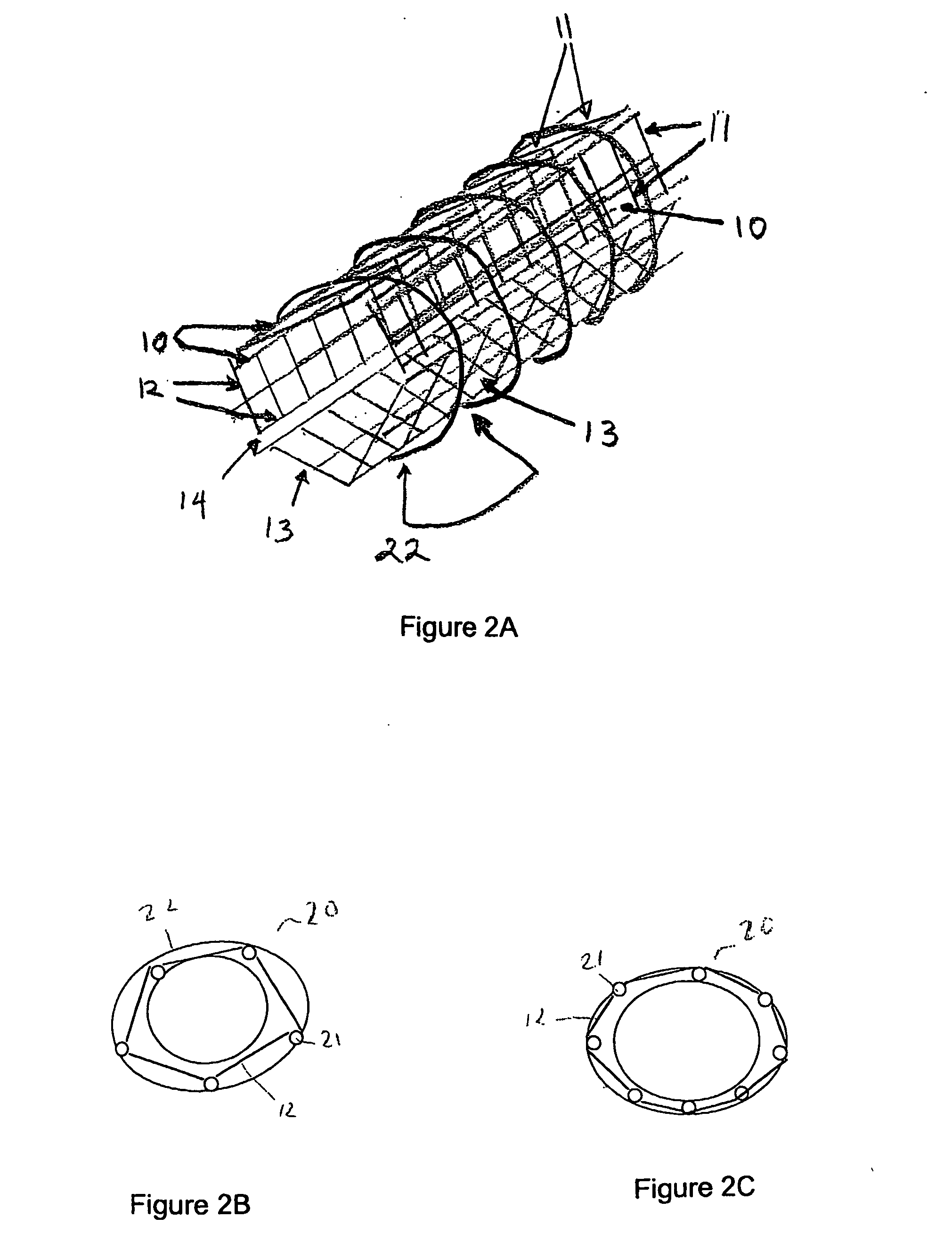
[0080] As depicted in FIG. 1A, a panel 10 is formed by connecting two or more, preferably two or three, parallel strands 11 of nonabsorbable material, preferably non-immunogenic, with minimal elastic potential, for example, #10 nylon fibers, with shorter, perpendicular strands 12 of the same diameter material. Perpendicular strands 12 are spaced approximately 0.5 to 1.0 μM apart and are oriented at approximately 90° to parallel strands 11. All strands are fixedly connected to one another by means appropriate for the selected material. Such means include, but are not limited to, heat and adhesive. For example, high temperatures induce the binding capability and anneal certain fibers such as SILASTIC™, or microscopic amounts of sealant substances such as silicone, polyurethane, or polyethylene can permanently join such fibers as would be used in the present invention.

[0081] In the embodiment illustrated in FIG. 1A, the panel is shown as ha...

example ii

Fabrication of a Bioartificial Vascular Device

[0088] Prior to implanting a prosthetic vessel as described above, vascular endothelial cells and vascular smooth muscle cells must be grown upon a permanent structure. This process begins with fabrication of an artificial vessel scaffold, optionally scaffold 20 according to Example I. A layer of vascular smooth muscle cells is grown along the outer surface of scaffold 20. A layer of endothelial cells is grown along the inner surface of scaffold 20. The addition of cellular layers can be accomplished by placing scaffold 20 in a vascular cell growth chamber 30, as shown in FIG. 3, so that scaffold 20 forms an outer chamber 31 and an inner chamber 32 of vascular cell growth chamber 30. Outer chamber 31 is filled with a cell culture solution containing vascular smooth muscle cells and incubated to allow the cells to attach to the outer surface of scaffold 20, approximately two days.

[0089] Following the deposit of an outer layer of cells, ...

example iii

Fabrication of an Internal Bioartificial Hepatic Organ

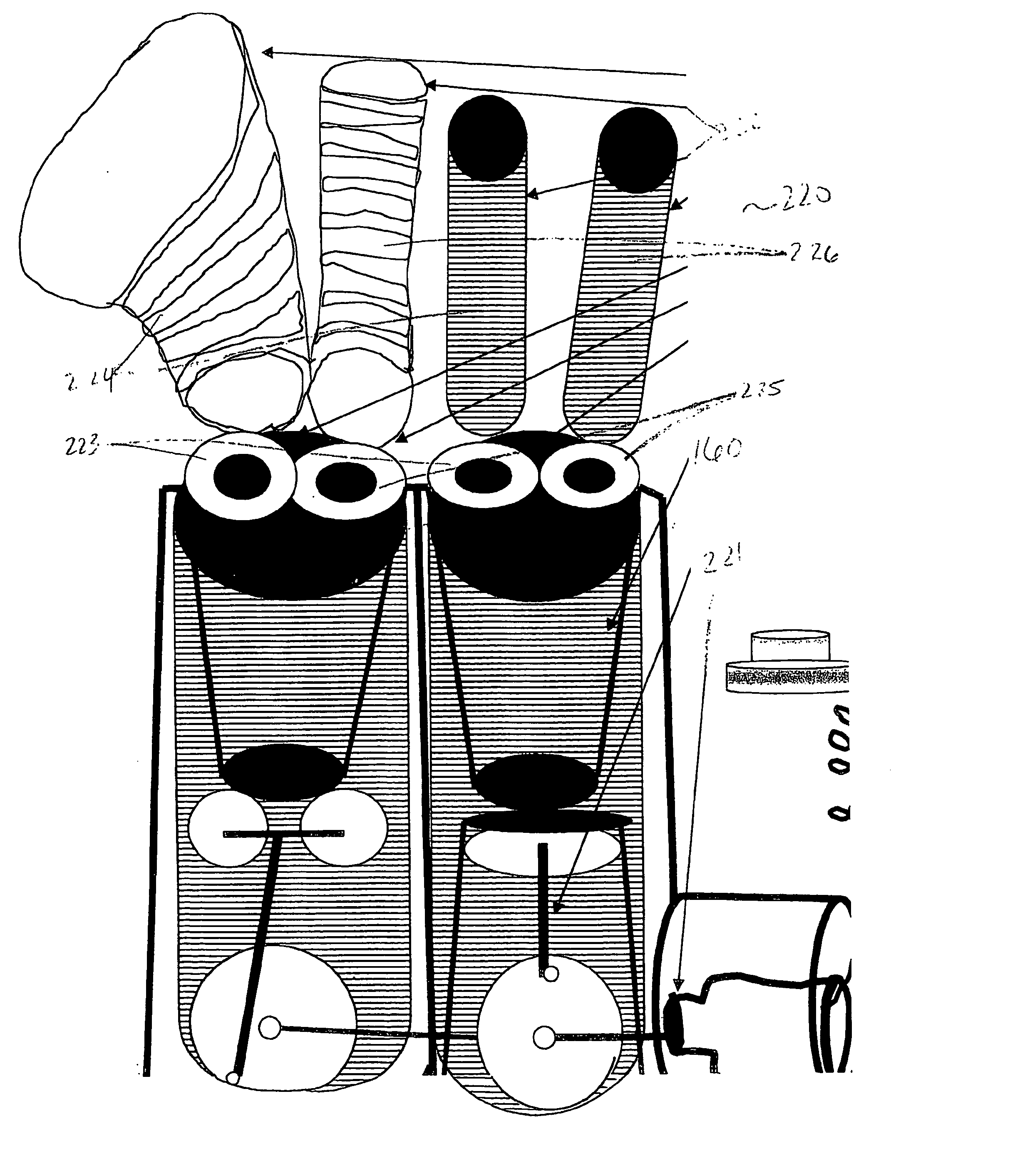
[0099]FIG. 6A shows a schematic overview of an artificial internal hepatic organ unit 60. A common entry portion 61 of artificial internal hepatic organ unit 60 is sutured to an abdominal artery (not shown). Common entry portion 61 has an inner diameter of approximately 2 cm and a length of approximately 1 to 2 cm. Extending from the end of common entry portion 61 opposite the artery are a plurality of 2-20 individual entry portions 62. Each individual entry portion 62 has a diameter of about 80-100 μm. Each individual entry portion 62 leads to the first end of a plurality of 4-8 inner vessels 63, each having an inner diameter of about 30-50 μm and a length of 8-12 cm. The plurality of inner vessels 63 originating from one individual entry portion 62 join at their second end to an individual exit portion 64. The individual exit portion 64 has a diameter of about 80-100 μm. Each individual exit portion 64 is joined together at a ...


PUM


Abstract

An artificial vessel scaffold is provided, of biocompatible materials and capable of being coated with selected cell types. A plurality of artificial organs are provided, formed of a biocompatible scaffold material and coated with selected cell types.

Description

CROSS-REFERENCE TO RELATED APPLICATION [0001] This applications claims benefit of priority of U.S. Provisional Application No. 60 / 357,118, filed Feb. 19, 2002.FIELD OF THE INVENTION [0002] The present invention relates generally to the field of biomaterials, implantable medical devices and cell biology. In particular, the invention relates to an artificial vessel scaffold, methods of making the same, and artificial organs made therefrom. The invention includes the manufacture of artificial organs and vessels, resulting in various devices for sustained growth of species specific (human, animal) cells, for species specific, medically suitable purposes. For example, biomaterial devices according to the invention include, but are not limited to transplantable organs, such as blood vessels, liver, bone, tendon / ligaments, and / or skin; transplantable endocrine glands, such as thyroid, parathyroid, pancreas, adrenal, pituitary, testis, and / or ovaries. The invention also encompasses transpla...

Claims


Application Information

Patent Timeline
IPC IPC(8): A61BA61F2/02A61F2/06A61F2/24A61F2/86A61M1/12C12N5/08
CPCA61F2/02A61F2/022A61F2/06A61F2/2415A61F2/844A61F2/86A61F2230/005C12M23/06C12M25/02A61F2210/0076A61F2220/0008A61F2220/005A61F2230/0017C12M21/08C12M25/14
Inventor SITZMANN, JAMES V.SITZMANN, EUGENE V.
Owner BIOARTTIS
Learn More