Nitric-oxide detection using Raman spectroscopy

a raman spectroscopy and nitric oxide technology, applied in the field of emergency medicine, can solve the problems of relative singularity of measurement, further exacerbate, and altered organ perfusion state, and achieve the effect of accurate characterizing the condition of patients, high degree of accuracy, and accurate measuremen

Inactive Publication Date: 2006-04-06
VIRGINIA COMMONWEALTH UNIV
View PDF9 Cites 86 Cited by
  • Summary
  • Abstract
  • Description
  • Claims
  • Application Information

AI Technical Summary

Benefits of technology

[0019] The invention realizes methods, profiles, medical measurement devices, and other products for accurate measurement of change or lack thereof from non-shock, non-ischemic, non-inflammation, non-tissue injury, non-immune dysfunction conditions which are referred to herein as “baseline conditions”. In attention to advantageous accuracy in such measurement, the invention provides practical, real-time approaches for accurately characterizing a patient's condition with respect to baseline conditions. With Raman and / or fluorescence spectroscopy according to the invention, change from baseline conditions is measured, characterized, monitored, identified and / or followed with a high degree of accuracy with measurement times on the order of seconds. Such high-accuracy measurement is achieved with Raman spectroscopy (such as resonance Raman spectroscopy) interrogation of tissue, optionally with simultaneous interrogation by fluorescence spectroscopy of compounds such as NADH. The tissue interrogation advantageously may be non-invasive to minimally-invasive to totally invasive. With methods and products according to the present invention, advantageously preclinical (ultra-early) states of shock, tissue ischemia, tissue injury, and tissue inflammation can be detected, severity can be determined, and the effectiveness of various treatments aimed at resolving the shock state can be determined, and other beneficial effects for patient care can be achieved.

Problems solved by technology

This mismatch commonly results from altered states of organ perfusion such as hemorrhage.
Shock additionally involves complex inflammatory and immune mediated events which result from, and may further exacerbate, this initial metabolic mismatch.
While these techniques have respective advantages, each is plagued by the relative singularity of its measure, lack of tissue specificity, inability to quantitate, or inability to easily apply or adapt for field use.
Information about biochemistry in shock states and disease states has not yet fully found its way and been used in practical applications.
Rather, currently emergency medicine is left to rely on physical examination not much advanced by conventional, relatively limited spectroscopic measurement technology.
Yet, the diagnosis of shock and its severity can be difficult, and cannot be accomplished with certainty, from simple vital signs.
A physical exam, including vital signs, is inadequate in detecting states of uncompensated shock.
In addition, resuscitation of victims of uncompensated shock back to “normal” vital signs is inadequate as a resuscitation endpoint.
Unrecognized continued accumulation of additional oxygen debt is still possible and may contribute to later development of multisystem organ failure and death.
Adding, to a physical exam, global measures of oxygen transport still does not ensure detection of early shock states or provide adequate information to act as sole end-points of resuscitation once shock is recognized and therapy instituted.
For various reasons, all have been problematic.
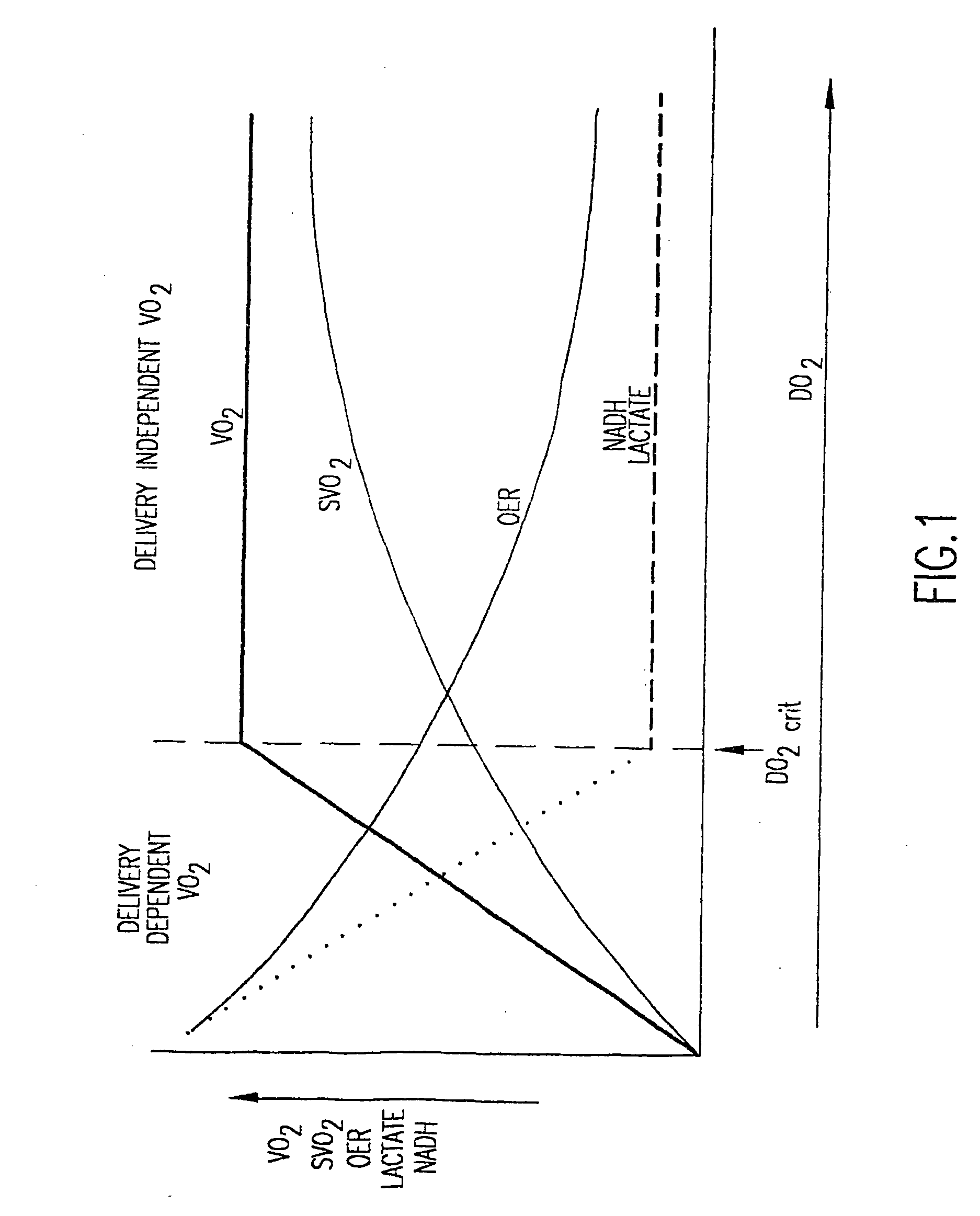
However, there is a point at which OER cannot keep pace with reductions in delivery.
Conventional monitoring and measuring used in emergency medicine do not adequately take into account such biochemistry of shock states and the like.
Knowing the biochemistry of shock states and the like but not being able to measure and monitor pertinent information thereto has been a frustrating, unresolved problem m emergency medicine.
However, problems with IR technology arise because water strongly absorbs IR radiation.
However, disadvantageously, NIR signals are so broad as to not be well-suited to quantification of overlapping species.
Conventional NADH-fluorescence techniques are more specific and quantitative than classical NIR absorption spectra but can only measure a single marker.
The major limitations of these devices are that they are limited to monitoring those specific gases and cannot provide additional information that, if provided, could be useful in diagnosis and stratification of patients.
Methods such as tonometry can be cumbersome due to its invasive nature.
These methods are also prone to deviations through changes either in minute ventilation or inspired oxygen concentration.
Transcutaneous gas monitoring, gastric tonometry, and even sublingual tonometry are one-dimensional and are prone to non-flow related changes caused by hypo or hyper ventilation.
Also, with the exception of sublingual tonometry, application of these methods in the field is problematic.
Again, broad overlap of signals in addition to needing to know the pathlength of light presents challenges in quantification and differentiation of signals.
For example it is difficult to distinguish hemoglobin and myoglobin making NIR use in hemorrhage problematic since myoglobin has a p50 of only 5 mmHg.
Monitoring the redox state of cytochrome oxidase is also difficult unless baseline absorptions are known.
Although some manufacturers of NIR absorption spectroscopy equipment claim to differentiate between the two species of oxygen hemoglobin and myoglobin, no work to this effect exists in the medical literature.
Another problem for NIR is that in terms of use on hollow organ systems such as the stomach, data from NIR absorption spectroscopy would likely include signals from non-stomach organs and thus not reflect data from the mucosal surface of the stomach.
However, such conventional methods do not necessarily provide optimum resolution.
However, satisfactory measurement of such compounds in vivo without invasive probing has not yet been provided.
No such technology is without a substantial disadvantage.
Civilian prehospital emergency medical services systems, emergency physicians, trauma surgeons, intensive care physicians, cardiologists, anethesiologists, and military medical personnel continue to be plagued by the insensitivity of the physical exam, lack of readily available physiologic and metabolic markers to judge the presence and severity of shock states, and lack of real-time relevant measurement approaches.
In addition, it has been difficult to use singular measures to guide treatment or predict outcome.
These problems are greatly magnified as the scale of the wounded population increases (such as on the battlefield and the various pre-definitive echelons of care provided to wounded soldiers or in a natural disaster).
To the inventors' knowledge, currently no conventional techniques are available for real-time monitoring of a broad range of potentially valuable emergency medicine markers of shock, tissue ischemia, tissue injury, tissue inflammation, or tissue immune dysfunction.

Method used


Image

  • Nitric-oxide detection using Raman spectroscopy
  • Nitric-oxide detection using Raman spectroscopy
  • Nitric-oxide detection using Raman spectroscopy

Examples


Embodiment Construction

[0057] The present invention provides methods and products in which resonance Raman spectroscopy interrogates biological material (such as tissue or a bodily fluid) at near-UV excitation. The Raman spectroscopy may proceed with or without simultaneous fluorescence spectroscopy (such as NADH fluorescence spectroscopy). The interrogation advantageously may be in a non-invasive to minimally-invasive manner, but is not required to be so and if desired may be invasive. Data from interrogating tissue according to the invention may be used to detect preclinical (ultra-early) states of shock and other tissue injury and disease states, determine severity, and determine the effectiveness of various treatments aimed at resolving the shock or tissue disease / injury state of a patient.

[0058] In a preferred embodiment of the invention, a tissue analysis method comprises interrogating a biological tissue with Raman spectroscopy and fluorescence spectroscopy to obtain spectroscopy results. The Rama...


PUM

PropertyMeasurementUnit
selectable wavelengthsaaaaaaaaaa
wavelengthaaaaaaaaaa
wavelengthsaaaaaaaaaa

Abstract

In an emergency medicine patient, accurate measurement of change or lack thereof from non-shock, non-ischemimc, non-inflammation, non-tissue injury, non-immune dysfunction conditions is important and is provided, as practical, real-time approaches for accurately characterizing a patient's condition, using Raman spectroscopy with a high degree of accuracy, Resonance Raman spectroscopy is used to monitor tissue nitric oxide activity either in vivo or in vitro, especially as a function of its interaction with hemoglobin or other metalloproteins. Measurement times are on the order of seconds. High-accuracy measurement is achieved with Raman spectroscopy interrogation of tissue. Measurements may be non-invasive to minimally invasive. The invention may be used to monitor the effect of instituting therapies using nitric oxide or disease processes that produce nitric oxide.

Description

CROSS-REFERENCE TO RELATED APPLICATIONS [0001] This is a continuation-in-part of U.S. patent application Ser. No. 10 / 332,613 (allowed) filed Jul. 29, 2003, which claims benefit of PCT application Ser. No. PCT / US01 / 22187 filed Jul. 13, 2001, which claims benefit of U.S. provisional application Ser. No. 60 / 218,055 filed Jul. 13, 2000.[0002] This invention may have received finding under Office of Naval Research Grant No. N00014-02-10344.FIELD OF THE INVENTION [0003] The invention generally relates to emergency medicine, and especially relates to medical conditions and states in which nitric oxide is involved. BACKGROUND OF THE INVENTION [0004] Shock is a complex entity, which traditionally has been defined as a state in which the metabolic demands of tissues are not matched by sufficient delivery of metabolic substrates, with the major substrate being oxygen. This mismatch commonly results from altered states of organ perfusion such as hemorrhage. Shock additionally involves complex i...

Claims


Application Information

Patent Timeline
Patent Type & Authority Applications(United States)
IPC IPC(8): A61B5/00A61B6/00G01N21/65
CPCA61B5/0071A61B5/0075A61B5/0084A61B5/0086A61B5/14546A61B5/1455A61B5/412A61B5/445G01N21/658
Inventor WARD, KEVIN R.BARBEE, R. WAYNEFILHO, IVO PONTES TORRESPITTMAN, ROLAND N.TERNER, JAMES
Owner VIRGINIA COMMONWEALTH UNIV
Why Eureka
Try Eureka