Bone enhancing composite
a composite and bone technology, applied in the field of bone enhancing composites, can solve the problems that none has proven entirely satisfactory in meeting the criteria required for successful tissue engineering, and achieve the effect of reducing the incidence of osteolytic debris
- Summary
- Abstract
- Description
- Claims
- Application Information
AI Technical Summary
Benefits of technology
Problems solved by technology
Method used
Image
Examples
example 1
Preparation of Bone Raft Powder
[0154] A method for preparation of a bone substitute is disclosed in U.S. Pat. No. 6,231,607. The bone substitute prepared by this method has an X-ray diffraction pattern similar to natural bone. The present inventors now disclose that at least one supplementary agent such as a biocompatible polymer and / or an anti-resorptive agent as may be added during the formation of the synthetic apatite crystals. The powder can be used per se or formulated into a fluid composition having advantageous properties for use as a bone graft material.
[0155] Materials and Methods
[0156] Graduated bottles, 2-liter capacity and 500 ml capacity
[0157] Glass ice bath
[0158] Microwave Oven (700W)
[0159] Vacuum Filter
[0160] Millipore Filter 0.45 μm-9 cm diameter Z29078-5 (Sigma)
[0161] Microwave oven
[0162] Trizma buffer PRE-SET Crystals Type 7.4-FT (Sigma)
[0163] Sodium dihydrogen phosphate monohydrate NaH2PO4 . H2O (MERCK)
[0164] Sodium Bicarbonate NaHCO3 (ICN)
[0165] L-As...
example 2
X-Ray Diffraction Analysis
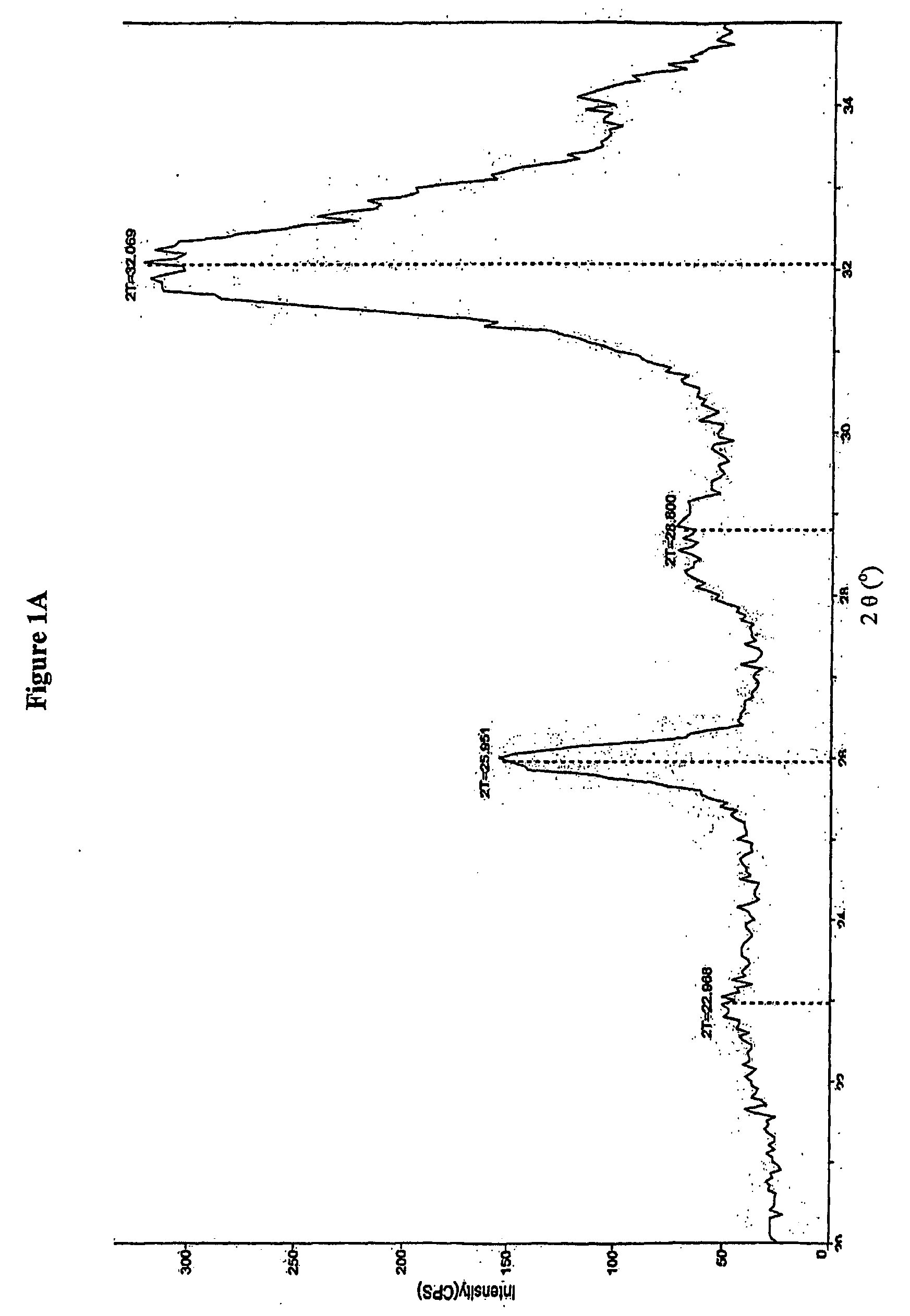
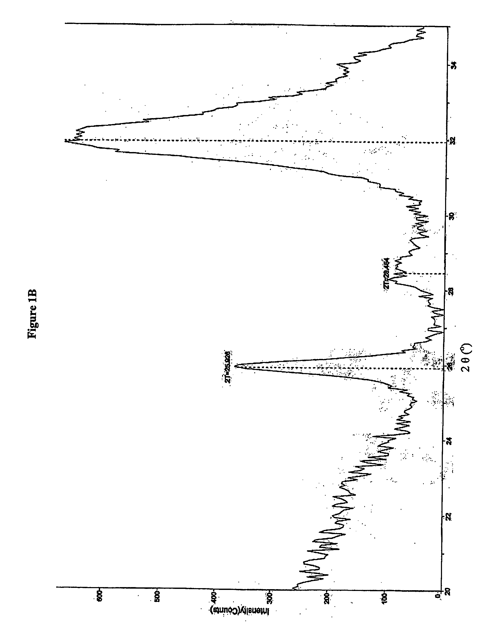
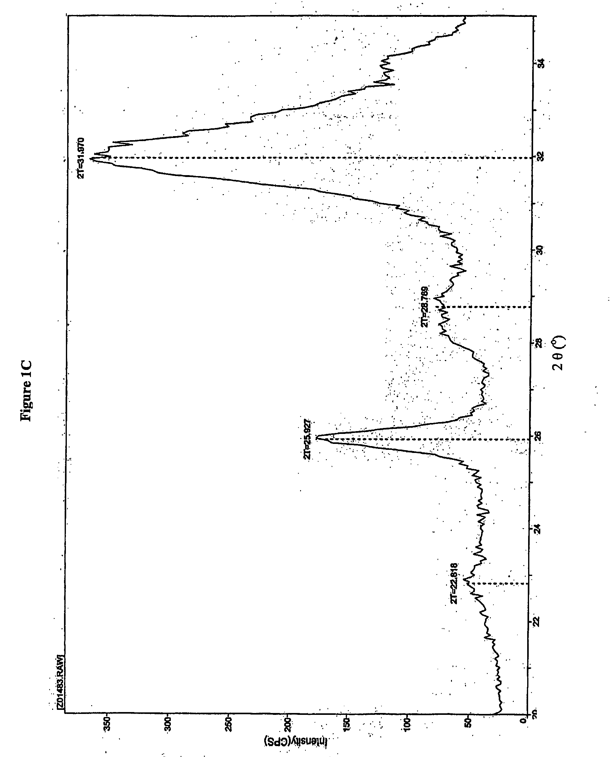
[0181] Bone substitute, material prepared according to U.S. Pat. No. 6,231,607 and composite prepared according to Example 1 and sterilized by heat sterilization or γ-irradiation was exposed to X-ray. An X-ray diffraction (XRD) pattern was obtained from a packed powder sample of the material pulverized in a mortar and pestle. X-rays were performed using an X-ray powder diffractometer, Rigaku, Japan. The scan rate was set to 0.5 degree / minute over the 2 theta (2θ) angular range from 20°-35°. The step size was set to 0.05°. FIG. 1A shows the X-ray diffraction pattern of the bone substitute powder prepared according to U.S. Pat. No. 6,231,607. A peak at 2θ about 26° and a large undifferentiated peak at 2θ about 31° to about 33° are noticeable. The X-ray diffraction pattern of a composite comprising heparin, wherein the heparin was added to 0.5 parts ug / ml is presented in FIG. 1B. The X-ray diffraction pattern of a composite comprising dextran sulfate added to...
example 3
Scanning Electron Microscope Analysis
[0183] Bone substitute composites prepared with various amounts of different polysaccharides was subject to Scanning Electron Microscope Analysis (SEM). FIGS. 2B and 2C show the aggregate formation of the synthetic apatite-heparin composite (0.5 ug / ml and 100 ug / ml, respectively) as compared to the synthetic apatite (PCA) alone shown in FIG. 2A. The composite prepared from the liquid mixture comprising 0.5% W / W Heparin MW 6,000 exhibited a distinct structure as seen by SEM. All figures are 39570× magnification.
PUM
| Property | Measurement | Unit |
|---|---|---|
| Angle | aaaaa | aaaaa |
| Angle | aaaaa | aaaaa |
| Angle | aaaaa | aaaaa |
Abstract
Description
Claims
Application Information
 - R&D
- Intellectual Property
- Life Sciences
- Materials
- Tech Scout
- Unparalleled Data Quality
- Higher Quality Content
- 60% Fewer Hallucinations
Browse by: Latest US Patents, China's latest patents, Technical Efficacy Thesaurus, Application Domain, Technology Topic, Popular Technical Reports.
© 2025 PatSnap. All rights reserved.Legal|Privacy policy|Modern Slavery Act Transparency Statement|Sitemap|About US| Contact US: help@patsnap.com


