Protein cages for the delivery of medical imaging and therapeutic agents

a technology for applied in the field of protein cages for the delivery of medical imaging and therapeutic agents, can solve the problems of soluble material inside the cage, and achieve the effects of facilitating the aggregation and crystallization of ions, increasing the number of introduced materials present, and being highly mobil

Inactive Publication Date: 2006-09-14
MONTANA STATE UNIVERSITY
View PDF6 Cites 25 Cited by
  • Summary
  • Abstract
  • Description
  • Claims
  • Application Information

AI Technical Summary

Benefits of technology

[0025] One advantage of the present invention is that a combination of medical imaging agents can be loaded into the cage. For example imaging agents for magnetic resonance imaging and x-ray imaging can be combined in one cage thereby allowing the resulting agent to be used with a multiple imaging methods. Another substantial advantage over the prior art is that protein cages are capable of encapsulating a larger number of molecules than other vehicles, i.e. liposomes, commonly used for the delivery of therapeutic agents. For example, up to 29,600 molecules of H2WO4210− have been packaged as a nano size crystalline solid within the cowpea chlorotic mottle virus (CCMV) protein cage. The size and shape of the crystallized nano material is determined by the size and shape of the cavity created by the CCMV protein cage. One other advantage, is that the protein cage can be used to increase the number of introduced materials present in the interior of the cage via crystallization. The crystallization of introduced materials can controlled because the protein cage provides a charged protein interface (on the interior) which can facilitate the aggregation and crystallization of ions.
[0026] Accordingly, the present invention provides compositions comprising a plurality of delivery agents. By “delivery agent” herein is meant a proteinaceous shell that self-assembles to form a protein cage (e.g. a structure with an interior cavity which is either naturally accessible to a solvent or can be made to be so by altering solvent concentration, pH, equilibria ratios, etc.), and contains imaging and therapeutic agents as discussed below. The protein cage may be obtained from a non-viral or viral source.
[0027] Preferred non-viral protein cages include ferritins and apoferritins, both eukaryotic and prokaryotic species, in particular mammalian and bacteria, with 12 and 24 subunit ferritins being especially preferred. In addition, 24 subunit heat shock proteins forming an internal core space are included. In particular, the heat shock protein of Methanococcus jannaschii assembles into a 24 subunit cage with 432 symmetry (see Kim, K. K. et al., 1998, Nature 394:595-599; Kim, K. K. et al., 1998, J. Struct. Biol. 121:76-80; and Kim, K. K. et al., 1998, PNAS 95:9129-9133).
[0028] Preferred viral protein cages can be obtained from any animal or plant virus from which empty viral particles can be produced. For example, empty viral particle can be obtained from viruses belonging to the bromovirus group of the Bromoviridae (Ahlquist, P., 1992, Curr. Opin. Gen. and Dev. 2:71-76; Dasgupta, R., and P. Kaesberg, 1982, Nucleic Acid Res. 5:987-998; and Lane, L. C., 1981, The Bromoviruses. In E. Kurstak (ed.), “Handbook of plant virus infection and comparative diagnosis”, Elsevier / North-Holland, Amsterdam) and from the family Caliciviridae. Viruses suitable for use in the invention include cowpea chlorotic mottle virus (CCMV) and the Norwalk virus.
[0029] In a preferred embodiment, empty viral particles are obtained from CCMV. A 3.2 Å resolution structure of CCMV is available that can be used to predict the role of individual amino acids in controlling virion assembly, stability, and disassembly (Speir, J. A., et al., 1995, Structure 3:63-78). The virion is made up of 180 copies of the coat protein subunit arranged with a T=3 quasi-symmetry and organized in 20 hexamer and 12 pentameric capsomers. A striking feature of the coat protein subunit is the presence of N— and C-terminal ‘arms’ that extend away from the central, eight-stranded, antiparallel b-barrel core. Each coat protein consists of a canonical β-barrel fold (formed by amino acids 52-176) from which long N-terminal (residues 1-51; 1-27 are not ordered in the crystal structure) and C-terminal arms (residues 176-190) extend in opposite directions. These N— and C-terminal arms provide an intricate network of ‘ropes’ which ‘tie’ subunits together. The first 25 amino acids are found lining the interior surface of the virion (Rao, A. L. and G. L. Grantham, 1996, Virology 226:294-305; and, Zhao, X., et al., 1995, Virology, 207:486-494). These 25 amino acids are thought to be highly mobile and to be required for viral RNA packaging. Nine of the first 25 amino acids are basic (Arg, Lys) and are thought to neutralize the negatively charged RNA. The first 25 amino acids are not required for empty virion assembly (devoid of viral RNA) and thus can be modified to change the electrostatic nature of the virion's interior surface, etc. The orientation of the coat protein β-barrel fold is nearly parallel to the five-fold and quasi six-fold axes. This orientation results in five exterior surface-exposed loops, βB-βC, βD-βE, βF-βG, βC-αCD1, βH-βI. Surrounding each of the 60 quasi three-fold axes located on the interface between hexamer and pentamer capsomers are Ca2+ binding sites. There are 180 Ca2+ binding sites per virion. Each Ca2+ binding site consists of five residues (Glu81, Gln85, Glu148 from one subunit; Gln 149 and Asp 153 from an adjacent subunit) in an ideal position to coordinate Ca2+ binding.
[0030] The protein cage may be unmodified or modified. By “unmodified” or “native” herein is meant a protein cage that has not been genetically altered or modified by other physical, chemical or biochemical means. By “modified” or “altered” herein is meant a protein cage that has been genetically altered or modified by a physical, chemical or biochemical means.

Problems solved by technology

Shifting the cage back to the closed form results in the entrapment of the soluble material inside of the cage.

Method used


Image

  • Protein cages for the delivery of medical imaging and therapeutic agents
  • Protein cages for the delivery of medical imaging and therapeutic agents
  • Protein cages for the delivery of medical imaging and therapeutic agents

Examples


example 1

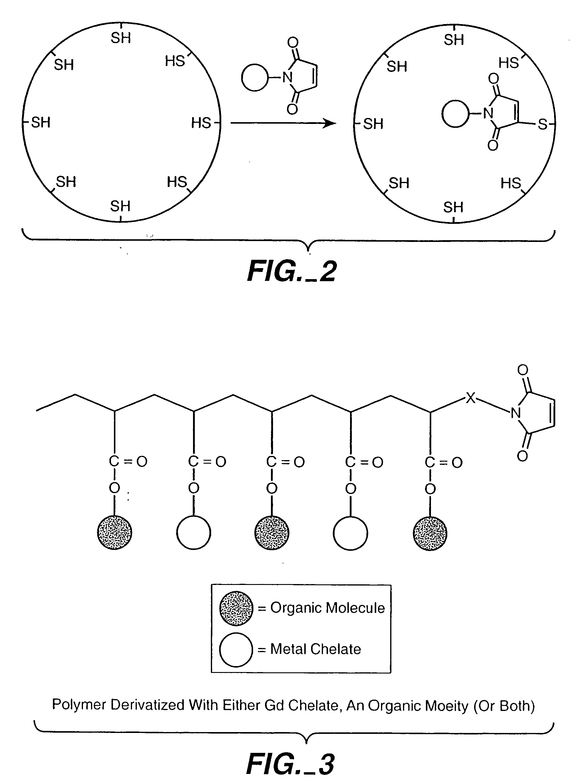
Modifications to Protein Cages for Enhanced Gd3+ Binding

[0136] We have taken advantage of our structural knowledge of the Ca2+ binding in wild type virions in an attempt to enhance binding of gadolinium (Gd3+) for eventual use as a possible MRI contrast agent. The Ca2+ binding sites in wild type virions results from the precise orientation of acidic residues contributed from adjacent coat protein subunits at the quasi three-fold axis (Speir, J. A., et al., 1995, Structure 3:63-78; and Zhao, X., 1998, Ph.D. Purdue University). There are 180 Ca2+ binding sites per virion. Ca2+ binding at these sites is thought to satisfy the charge repulsion created at pH 6.5 by the cluster of acidic residues, and to assist with creating shell curvature during virion assembly. Ca2+ is normally required for in vitro assembly of CCMV at >pH 6.5. We have demonstrated that Gd3+ can act as a substitute for Ca2+ in the pH-dependent assembly assay. We are attempting to enhance assembly-dependent Gd3+ bindi...

example 2

Electrostatic Modifications to Protein Cages

Entrapment and Growth of Anionic Metal Species

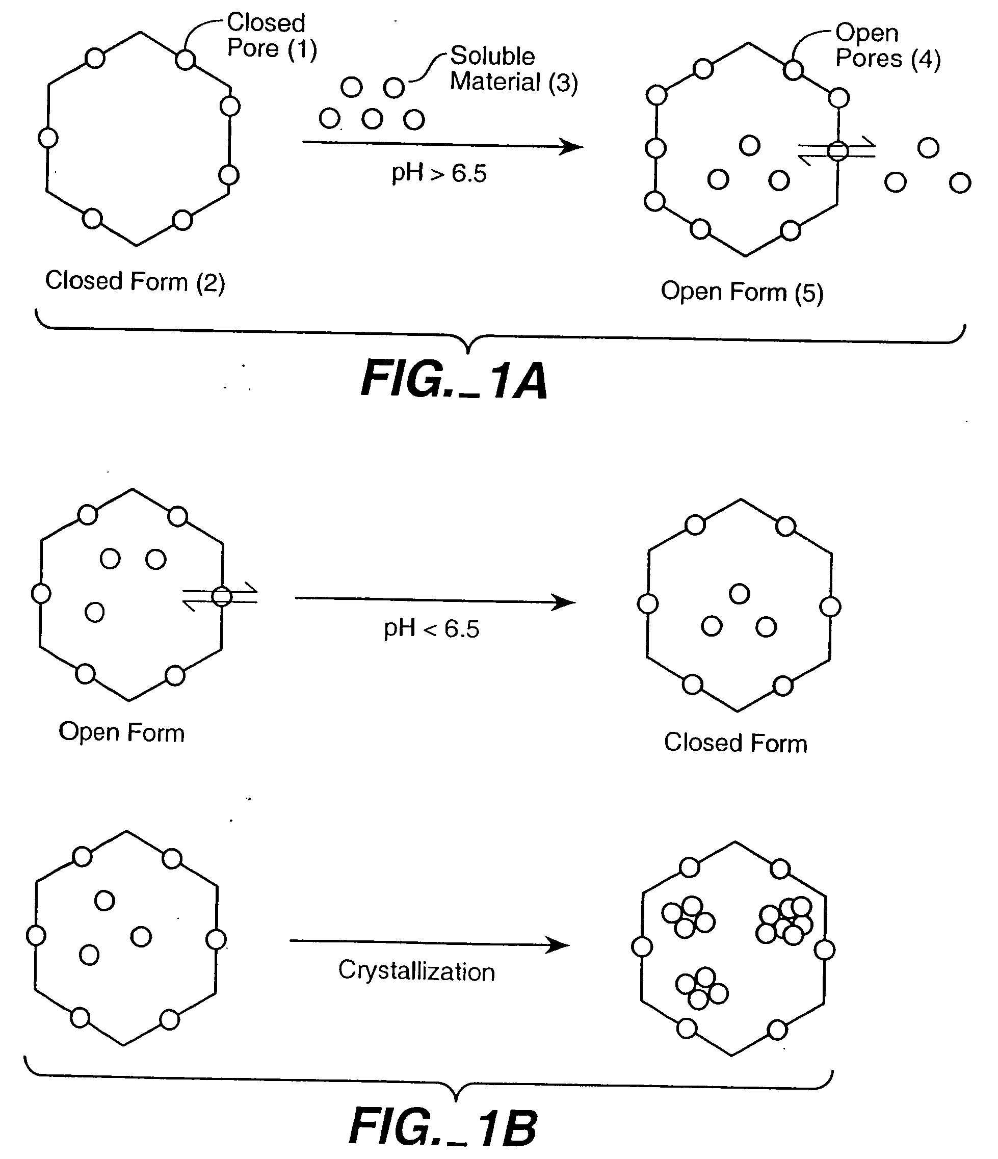
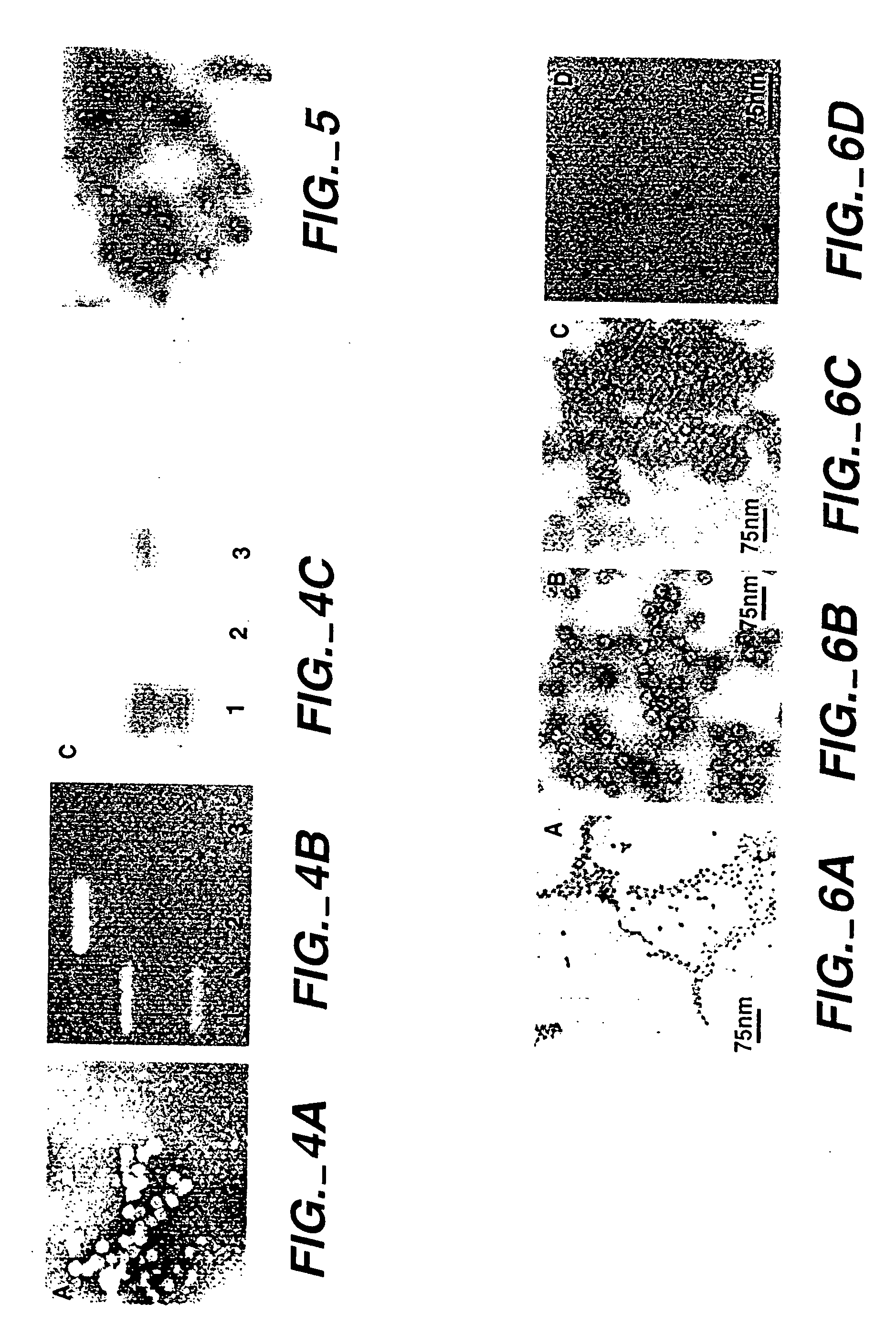
[0145] We have crystallized a range of polyoxometalate species in CCMV and the Norwalk Virus. This was accomplished by providing an interface for molecular aggregation, based on complementary electrostatic interactions between the protein and the anion metal species, which creates a locally high concentration at the protein interface. Briefly outlined, the empty virions were incubated with the precursor ions (WO42−, VO3−, MoO42−) at approximately neutral pH. Under these conditions the virus exists in its open (swollen) form and allows all ions access to the cavity. The pH of the virus solution was then lowered to approximately pH 5.0. This induced two important complementary effects i) The inorganic species underwent a pH dependent oligomerization to form large polyoxometalate species such as H2WO4210− (Douglas, T., and M. J. Young., 1998, Nature 393:152-155) which were readily crystallized...

example 3

Bioengineering of New Chemical Switches for the Regulated Entrapment / Release of Materials

[0162] We have demonstrated that pH dependent expansion at the quasi three-fold axes is the result of deprotination of the acidic residues comprising the Ca2+ binding sites. The loss of protons at the elevated pH results in a close cluster of negative charges that must be accommodated either by the binding of Ca2+ or by the physical expansion (i.e. swelling) induced by electrostatic repulsion. We have taken advantage of CCMV's reversible swelling properties as a control mechanism to introduce and to release materials from the central cavity of the protein cage (see e.g. Examples 1 and 2). This reversible switching property of CCMV provides an exciting opportunity for development of elegant control mechanisms for entrapment and release of therapeutic agents.

pH Activated Chemical Switches

[0163] Gating in the wild-type virion results from electrostatic repulsion of carboxylate groups in the ab...


PUM

PropertyMeasurementUnit
sizeaaaaaaaaaa
sizeaaaaaaaaaa
temperatureaaaaaaaaaa

Abstract

The present invention is directed to novel compositions and methods utilizing delivery agents comprising protein cages, medical imaging agents and therapeutic agents.

Description

CROSS REFERENCE TO RELATED APPLICATIONS [0001] This application is a divisional application of U.S. application Ser. No. 10 / 441,962, filed May 19, 2003, which claims the benefit of the filing date of Ser. No. 60 / 380,942, filed on May 17, 2002 under 35 U.S.C. §119(e), which is expressly incorporated by reference in its entirety.GOVERNMENTAL SUPPORT OF APPLICATION [0002] This invention was made with governmental support under grant number GM61 340, awarded by the National Institutes of Health. The United States government has certain rights in the invention.FIELD OF THE INVENTION [0003] The present invention is directed to novel compositions and methods utilizing delivery agents comprising protein cages, medical imaging agents and therapeutic agents. BACKGROUND OF THE INVENTION [0004] There is considerable interest in the chemical design and construction of self-assembling systems that can be used as delivery vehicles for encapsulated “guest” molecules. For example, viral capsid prote...

Claims


Application Information

Patent Timeline
Patent Type & Authority Applications(United States)
IPC IPC(8): A61K49/00A61K31/70A61K49/18
CPCA61K31/70A61K49/0002A61K49/189B82Y5/00A61P35/00
Inventor YOUNG, MARK J.DOUGLAS, TREVORIDZERDA, YVES U.
Owner MONTANA STATE UNIVERSITY
Why Eureka
Try Eureka