
Three-dimensional structure of DNA recombination/repair protein and use thereof

a recombination/repair protein, three-dimensional structure technology, applied in the direction of molecular structures, instruments, drug compositions, etc., can solve the problem of insufficient understanding of the precise molecular mechanism, particularly the catalytic mechanism of the enzyme including the dna-binding mode during the homologous pairing step, and the difficulty of directly assaying the homologous recombination activity of the rad52 in eukaryotic cells, etc. problem

Inactive Publication Date: 2007-02-08
RIKEN
View PDF1 Cites 0 Cited by
  • Summary
  • Abstract
  • Description
  • Claims
  • Application Information

AI Technical Summary

Benefits of technology

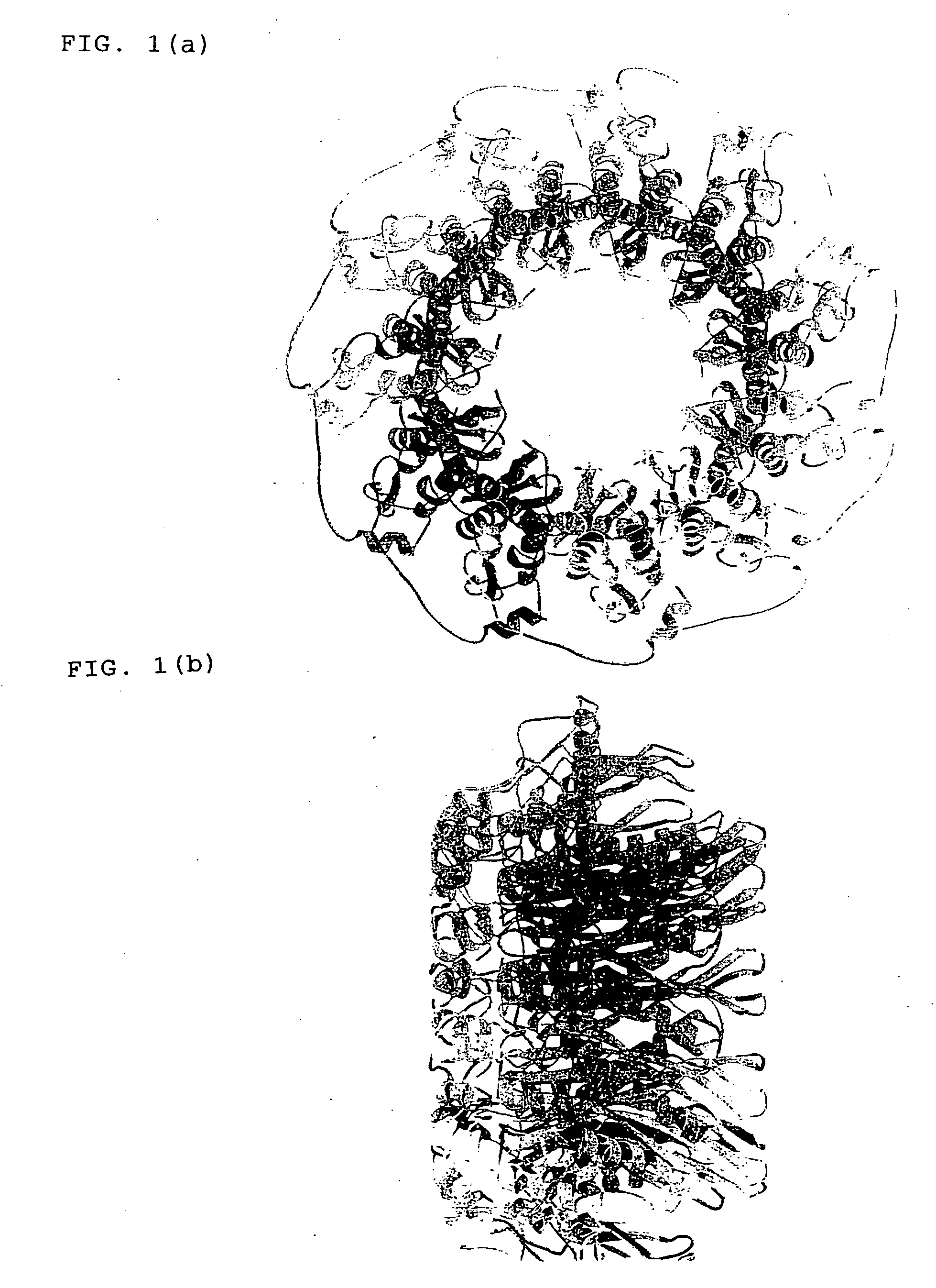
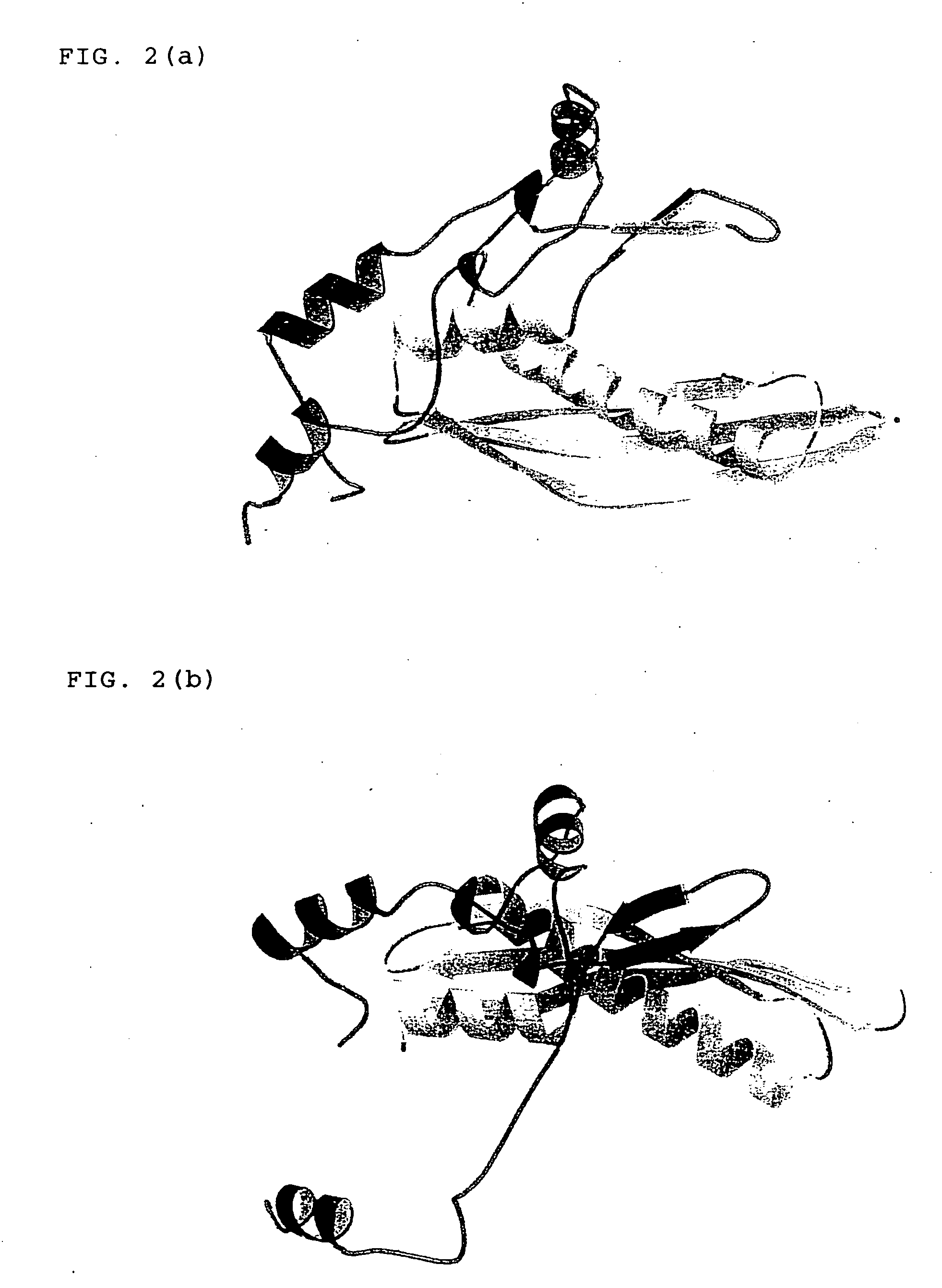
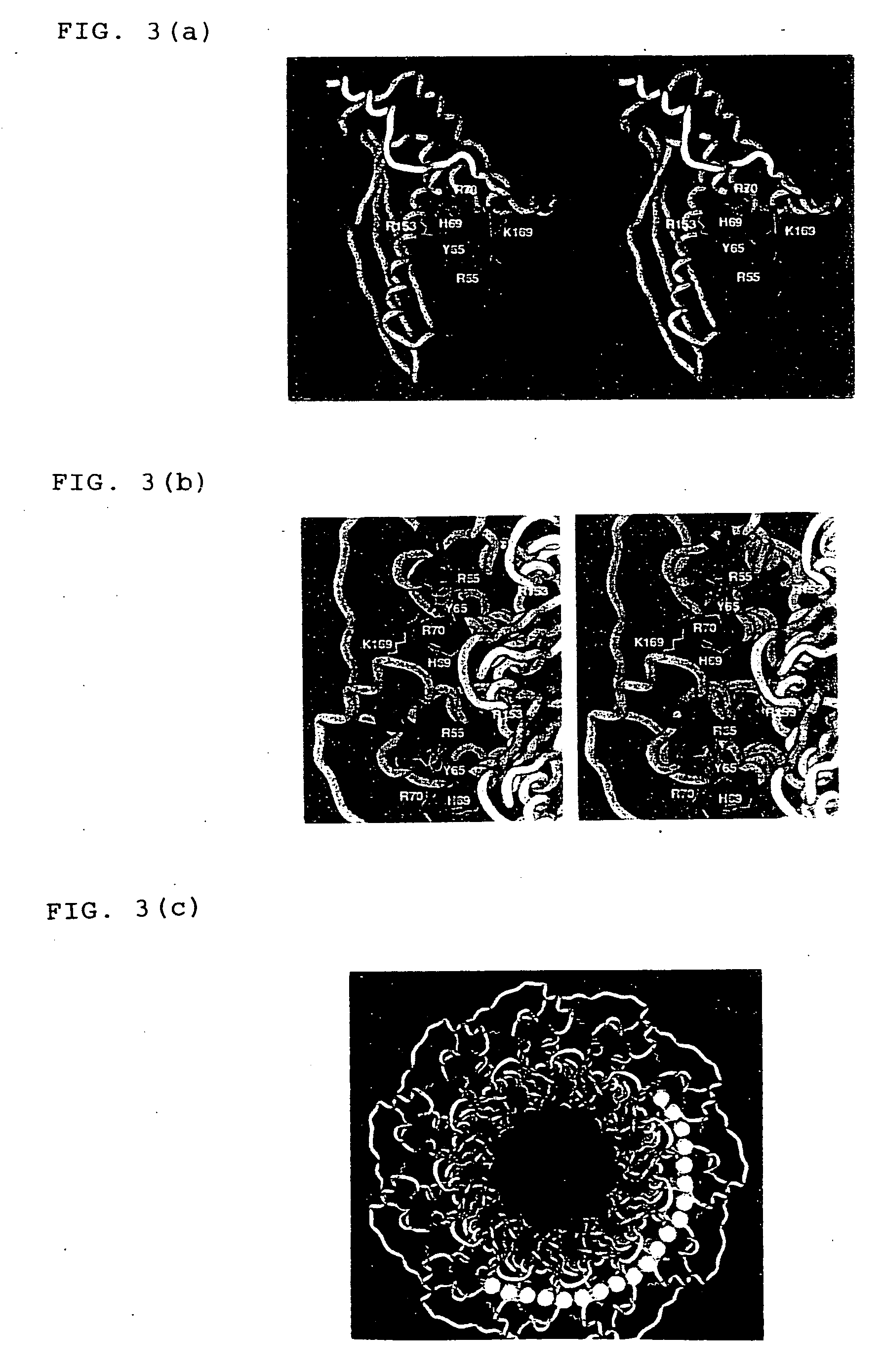
[0048] (16) The method for identifying a mutant DNA recombination / repair protein of (15), wherein the amino acid residues selected at the step (b) are one or two or more amino acid residues selected from arginine at position 55, tyrosine at position 65, histidine at position 69, arginine at position 70, arginine at position 153 and lysine at position 169 in the amino acid sequence of SEQ ID NO:1.

Problems solved by technology

However its precise molecular mechanism, particularly the catalytic mechanism of the enzyme including the DNA-binding mode during the homologous pairing step has not yet sufficiently been understood.
Thus, it is difficult to directly assay the homologous recombination activity of the Rad52 in eukaryotic cells.
Because the assay procedure for the homologous recombination activity is complicated, screening of numerous compounds using the complex is extremely inefficient.
Although the role of the DNA recombinational repair mechanism and its applications are so significant as described above, it is very difficult to assay the Rad52-specific DNA recombination activity or to elucidate the physiological role thereof, because various DNA recombination / repair proteins exist in cells as described above.
Additionally, the relationship between the structure and functions of Rad52 in homologous DNA recombinational repair has not yet been elucidated.

Method used


Image

  • Three-dimensional structure of DNA recombination/repair protein and use thereof
  • Three-dimensional structure of DNA recombination/repair protein and use thereof
  • Three-dimensional structure of DNA recombination/repair protein and use thereof

Examples


example 1

Limited Proteolysis of HsRad52

[0110] 5 μl of HsRad52 protein (1 to 3 mg / ml) prepared by a known method (Kagawa et al., J. Biol. Chem., 2001, 276, 35201-8) was mixed with 5 μl of proteinase K (30 μl / ml) and the mixture was incubated at 25° C. for 4 to 6 hours. The proteinase K solution was diluted in 10 mM Tris-HCl solution, pH 7.5. The reaction mixture was fractionated by 12% SDS polyacrylamide gel electrophoresis (SDS-PAGE), and the resulting protein bands were transferred to a PVDF membrane using a semi-wet blotting apparatus (Bio-Rad). Proteolytic bands were visually detected by staining with a Coomassie Brilliant Blue staining solution, followed by destaining of the membrane with methanol. The protein was eluted from a portion of the PVDF membrane containing the protein bands, to determine the N-terminal amino acid sequence. To measure the molecular weight by mass spectrometry, the reaction mixture containing proteolyzed HsRad52 was treated with 1 μl of 2N HCl to stop further ...

example 2

Analysis of HsRad521-237 by Deletion

[0111] Plural deletion mutants of HsRad521-237 as prepared by deleting sequentially every 5 amino acid residues from the C terminus thereof, namely HsRad521-232, HsRad521-227, HsRad521-222, HsRad521-217, HsRad521-212, HsRad521-207, HsRad521-202, HsRad521-197, HsRad521-192 and HsRad521-187 were subjected to a combination of both of crystallization and biochemical experiments. HsRad521-192 and deletion mutants larger than the HsRad521-192 had absolutely the same DNA binding activity as that of HsRad521-237. Among all of the deletion mutants, the highest amount of crystal was obtained from HsRad521-212 in a system using a screening kit (Crystal Screens 1 and 2 manufactured by Hampton Research Co., Ltd.). Thus, it was determined from these results that HsRad521-212 was the most suitable for structural analysis.

example 3

Purification of HsRad521-212

[0112] HsRad521-212 was purified by three steps of affinity adsorption on Ni-NTA agarose, hexahistidine-tag cleavage and Heparin-Sepharose column chromatography. A DNA encoding HsRad521-212 was cloned in pET15-b (Novagen), an expression vector transcribed by T7 polymerase. The HsRad521-212 protein was expressed at a large scale, using E. coli strain JM109 (DE3) together with tRNAArg3 recognizing the CGG codon and RNAArg4 recognizing the AGA / AGG codon. In case that the expression plasmid did not contain the tRNA genes, the expression level of the HsRad521-212 protein was extremely low. Generally, the HsRad521-212 protein was purified from a culture of one liter. The recombinant E. coli was cultured at 30° C., to which IPTG was added to a final concentration of 1 mM in the logarithmic growth phase (OD600=0.6), to induce the expression of the protein. Subsequently, the bacteria were cultured for 4 to 6 hours to collect the bacterial cells, which were disru...


PUM

PropertyMeasurementUnit
concentrationaaaaaaaaaa
pHaaaaaaaaaa
temperatureaaaaaaaaaa

Abstract

A crystal of DNA recombination / repair protein, Rad52 protein is prepared and the three-dimensional structure thereof is determined by X-ray crystallography with the use of the same. The obtained three-dimensional structure of Rad52 protein can be used for screening a variety of mutant enzymes or compounds which binds to the enzyme complex. The mutant Rad52 protein complex, or the compound that binds to the complex so as to modulate the DNA recombinational repairing activity, which are screened by the method of the present invention, can be used for diagnosis and treatment of diseases induced by DNA damages, and detection of the DNA recombinational repairing activity in vivo. Moreover, these substances are usable in drug discovery targeting to Rad52, and gene transfer to a specific site by homologous recombination, etc.

Description

[0001] The present specification includes a Sequence Listing of 2 sequences on three (3) pages. A copy of Table 1 is also included on a CD-ROM filed with the present application. LENGTHY TABLES FILED ON CDThe patent application contains a lengthy table section. A copy of the table is available in electronic form from the USPTO web site () An electronic copy of the table will also be available from the USPTO upon request and payment of the fee set forth in 37 CFR 1.19(b)(3).BACKGROUND OF THE INVENTION [0002] 1. Field of the Invention [0003] The present invention relates to the crystallization of DNA recombination / repair protein, the determination of the three-dimensional structure of DNA recombination / repair protein by X-ray crystallography using the crystal, and the use thereof. More specifically, the invention relates to the designing and identification of a mutant enzyme based on the three-dimensional structure, and a rational method for screening a compound binding to the enzyme....

Claims


Application Information

Patent Timeline
Patent Type & Authority Applications(United States)
IPC IPC(8): C12Q1/68C07H21/04C12N9/22C12Q1/25G01N33/68G16B15/30
CPCC07K2299/00C12Q1/25G06F19/16G01N2500/00G01N33/6803G16B15/00A61P35/00A61P43/00G16B15/30
Inventor KURUMIZAKA, HITOSHIKAGAWA, WATARUYOKOYAMA, SHIGEYUKI
Owner RIKEN
Learn More