Decellularized Tissue and Method of Preparing the Same

Inactive Publication Date: 2007-10-18
CARDIO +1
View PDF3 Cites 59 Cited by
  • Summary
  • Abstract
  • Description
  • Claims
  • Application Information

AI Technical Summary

Benefits of technology

[0176] The present invention is fundamentally different from conventional decellularization methods in that a radical reaction (in particular, exposure to a radical) is used in addition to a chemical treatment. In particular, it should be noted that use of a non-micellar molecule allows unexpected improvement in decellularization efficiency in decellularization technology in a manner similar to the machinery of cellular component extraction.
[0177] The decellularized tissue thus prepared are minimized with respect to the damage to extracellular matrices. The decellularized tissue of the present invention may be used as an artificial vessel which can be used in a permanent manner. The decellularized tissue of the present invention may be used as a transplant with or without use of cells, as such, the utility of the decellularized tissue of the present invention ranges to a wide extent. In particular, the efficiency of decellularization is improved to an extent which has not been conventionally achieved (i.e., cell residual rate is less than 5%, preferably less than 1%), and thus decellularized tissue substantially free of cells are provided. Therefore, the decellularization

Problems solved by technology

Rejection to artery grafts pathologically leads either to enlargement (up to rupture) or obstruction of the grafts.
However, complete elimination of all antigens is considerably difficult to perform and verify.
In the case of small-diameter blood vessels, it is difficult to apply replacement therapy.
Although venous and arterial grafts currently yield the best results, disadvantages include the need for complicated operations and no suitable blood vessels available in patients with certain diseases.
Howev

Method used


Image

  • Decellularized Tissue and Method of Preparing the Same
  • Decellularized Tissue and Method of Preparing the Same
  • Decellularized Tissue and Method of Preparing the Same

Examples


example 1

[0431] (Materials and Methods)

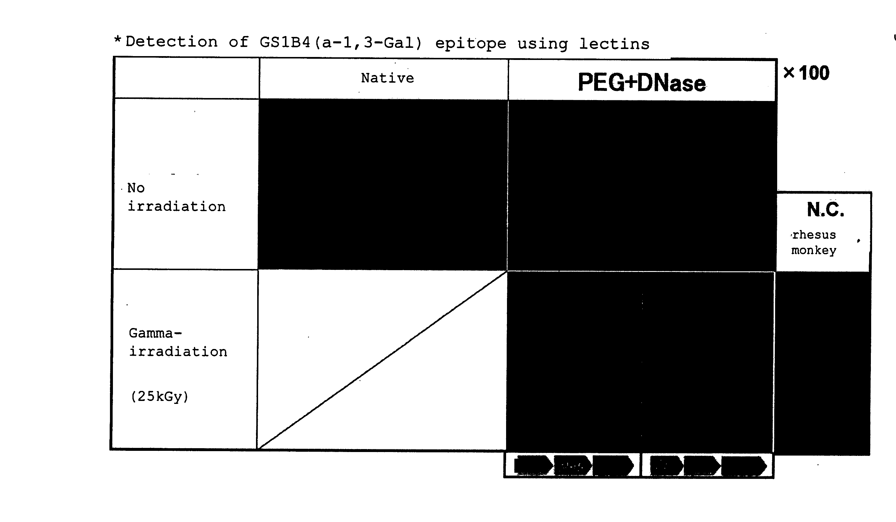
[0432] (Decellularization by PEG)

[0433] Porcine carotid arteries were prepared from Hybrid (Labo Products Co. Ltd., Osaka, Japan), and rat aortas were prepared from SD rats (male, 5 weeks old, Nippon Animal Co., Ltd., Tokyo, Japan) under sterile conditions. Animal experiments were conducted in accordance with the guidelines for ethics established by Osaka University.

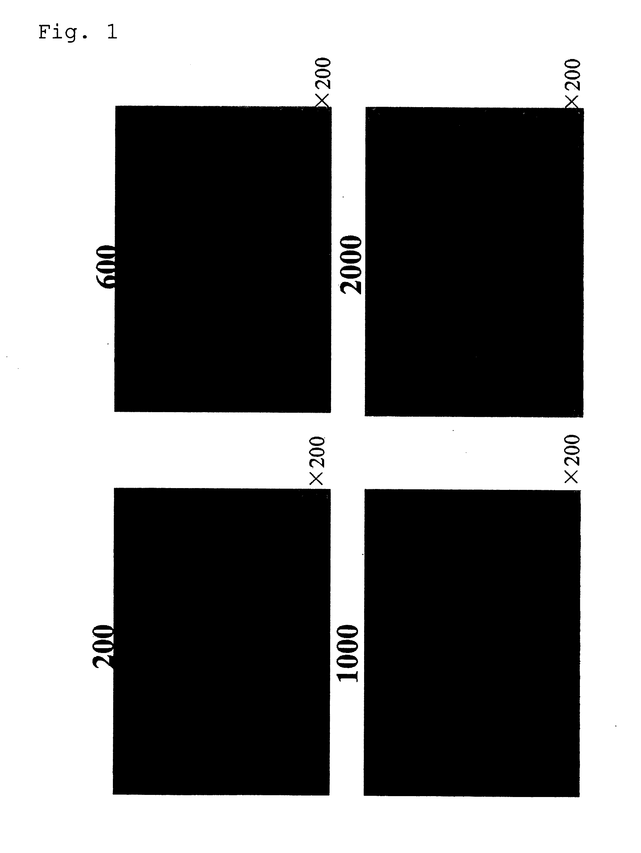
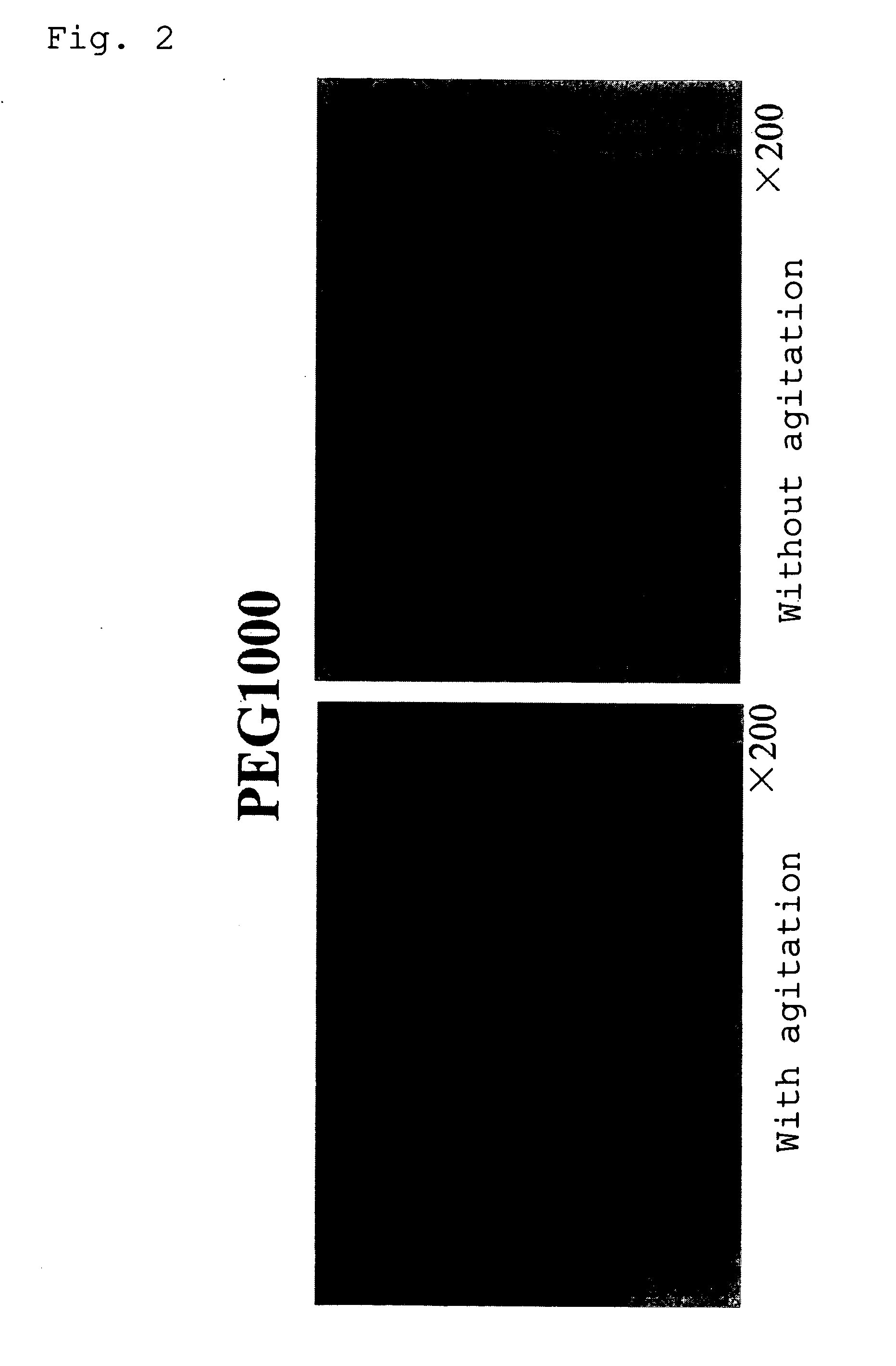
[0434] Freshly collected porcine carotid arteries and rat aortas were placed in PBS (referred to as PBS (−) in this example; Gibco BRL, Life Technologies Inc. Rockville, Md., USA) containing antibiotics (Gibco BRL, Life Technologies Inc. Rockville, Md., USA) to wash out blood components. The blood vessels were then placed in a decellularizing aqueous solution containing polyethylene glycol (1 g / ml, Nacalai Tesque Inc., Kyoto, Japan) (average molecular weight: 1000), and allowed to stand for 0.5 h. Because of high viscosity of the solution, the blood vessels were gently pressed several tim...

example 2

Comparison of Reactions within Biological Tissue)

[0527] (Method)

[0528] (Immunological Response)

[0529] Aorta wall portions (1×1 cm) of a gamma-ray irradiated and decellularization treated valve according to Example 1 and an SDS-treated decellularization tissue (the artificial valve prepared by SDS decellularization III) according to Example 1 were implanted under the skins of the dorsal portions of SD rats. After one week and two months, the animals were sacrificed. The degree of inflammatory cellular infiltration was scored for evaluation. In this example, porcine native valves was used as controls for comparison.

[0530] (Calcification)

[0531] The specimens were collected one week and two months after subcutaneous implantation, followed by von Kossa staining for evaluation of calcification. Also, Ca concentration within the tissue was measured with an atomic absorption spectrometry. The Ca concentration was measured and quantified as follows. The tissue was placed in concentrated...

example 3

Confirmation of Cell Replacement

[0541] Comparison of Reactions in Biological Tissue Between Each Valve

[0542] Decellularization treated porcine forearm arteries according to Example 1 are implanted into dog femoral aortas. The animals are sacrificed after 10 days. The degree of inflammatory cellular infiltration is compared and studied.

[0543] (Results)

[0544] It is found that there is substantially no rejection reaction in the decellularized tissue of the present invention, and thus it is understood that global structure is not impaired. Further, by observing the implanted tissue, it is also possible to confirm that the decellularized tissue is replaced with self cells.


PUM


Abstract

Decellularization of tissue by means of an amphipathic solvent a well-established practice. However, situations exist where the provision of enhanced decellularization is preferred. There is a demand for treating methods for coping with such situations. Thus, it is intended to provide a method for enhancing decellularization. The method comprises not only the immersing of a tissue in a solution containing an amphiphilic molecule in non-micellar form (for example, 1,2-epoxide polymer) but also performing a radical reaction (for example, treatment selected from the group consisting of exposure to gamma-ray irradiation, ultraviolet irradiation, a free radical supply source, ultrasonication, electron beam irradiation, and X-ray irradiation).

Description

TECHNICAL FIELD [0001] The present invention relates to a method and system for decellularizing tissue, tissue prepared by the decellularization method, and a pharmaceutical and therapeutic method utilizing a tissue graft or the like. BACKGROUND ART [0002] Implantation of organs (e.g., heart, blood vessel, etc.) derived from exogenous tissue is mainly hindered by immunological rejections. Changes occurring in allografts and xenografts were first described at least 90 years ago (Carrel A., 1907, J. Exp. Med. 9:226-8; Carrel A., 1912., J. Exp. Med. 9:389-92; Guthrie C. C., 1908, J. Am. Med. Assoc; Calne R. Y., 1970, Transplant Proc. 2:550; and Auchincloss 1988, Transplantation 46:1). Rejection to artery grafts pathologically leads either to enlargement (up to rupture) or obstruction of the grafts. The former is caused by decomposition of extracellular matrices, while the latter is caused by the proliferation of cells in a blood vessel (Uretsky B. F., Mulari S., Reddy S., et al., 1987,...

Claims


Application Information

Patent Timeline
IPC IPC(8): A61F2/02A61K35/12A61L27/00A61L27/36
CPCA61L27/3683A61L27/3604
Inventor MATSUDA, HIKARUSAWA, YOSHIKITAKETANI, SATOSHIMIYAGAWA, SHIGERUIWAI, SHIGEMITAUOTA, TAKEYOSHIMIYAKE, JUNHARA, MASAYUKIFURUTA, MASAKAZUUCHIMURA, EIICHIRO
Owner CARDIO
Why Eureka
Try Eureka