
Multi-layer braided structures for occluding vascular defects

a vascular defect and multi-layer technology, applied in textiles and papermaking, medical science, surgery, etc., can solve the problems of limiting the effectiveness of the procedure, affecting the efficiency of the device, and affecting the patient's blood circulation, etc., to achieve quick hemostasis, reduce the risk of vascular disease, and reduce the effect of vascular diseas

Active Publication Date: 2007-11-15
ST JUDE MEDICAL CARDILOGY DIV INC
View PDF99 Cites 536 Cited by
  • Summary
  • Abstract
  • Description
  • Claims
  • Application Information

AI Technical Summary

Benefits of technology

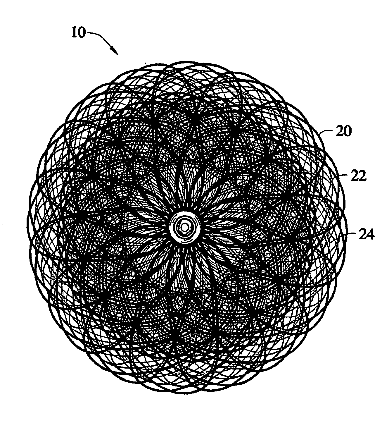
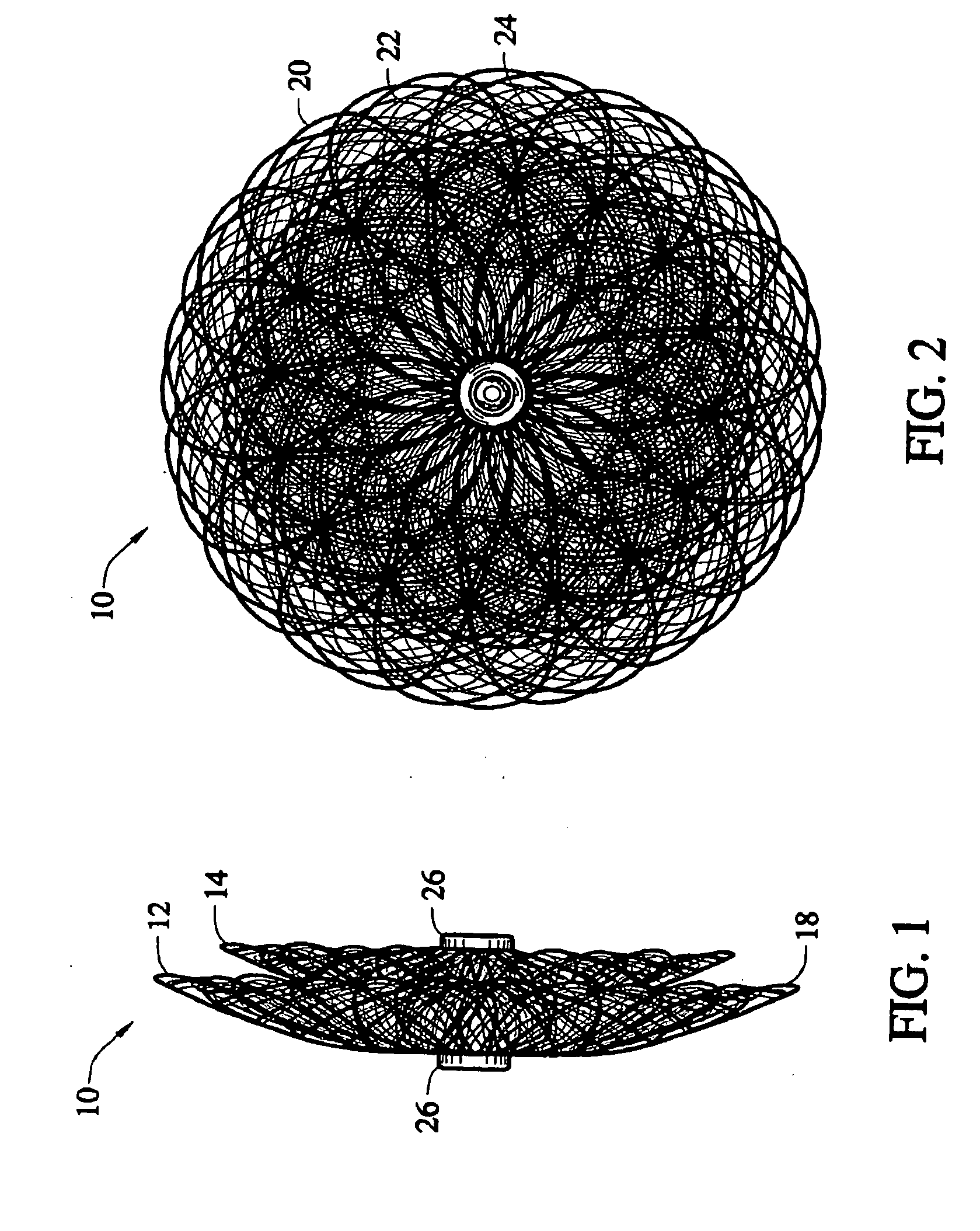
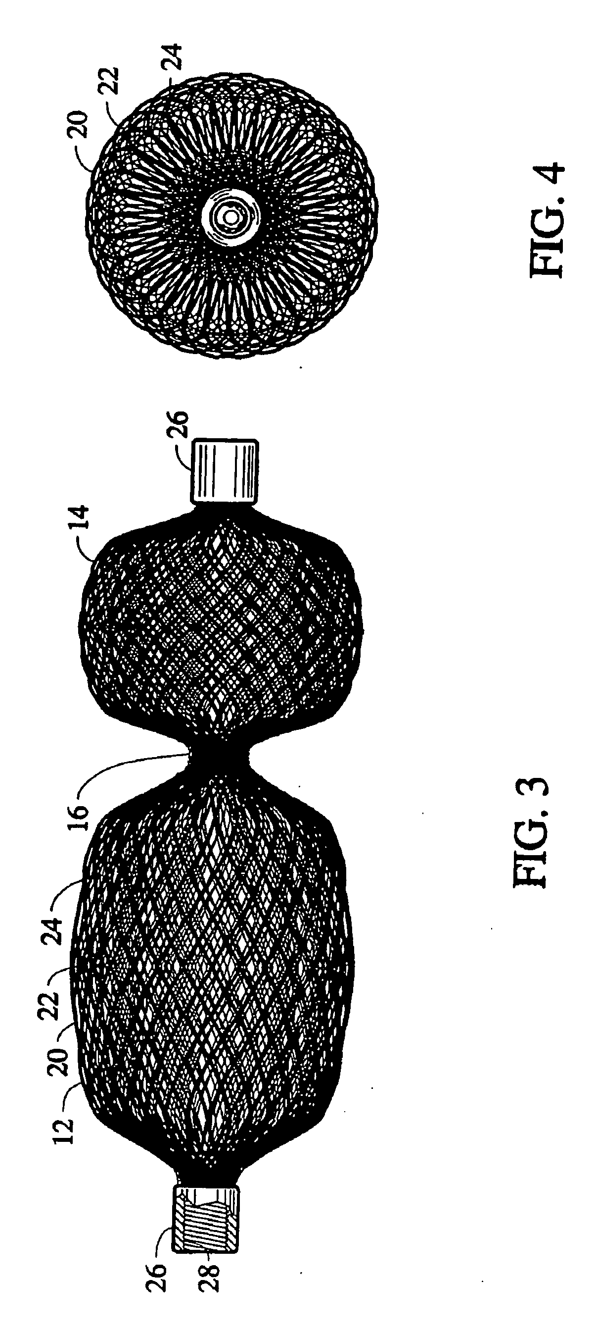
[0030] In other embodiments the various layers have different pre-set shapes in concentric co-axial arrangement. In another embodiment the inner layers are side by side instead of coaxial with the outer layer. In still another embodiment an outer layer, that defines a pre-shaped but conformable volume, surrounds a concentric very much longer braid, pre set into a bead & chain type shape. In this embodiment the internal beaded chain braid is inserted into the outer braid volume to fill the volume and cause the volume to take the shape of the cavity it is placed in. The filled volume results in quick hemostasis due to high metal density while maintaining a small diameter delivery profile.

Problems solved by technology

This procedure is often limited in its utility, in part, due to the inability to precisely position the embolization agents.
Overfilling the balloon is an equally undesirable occurrence, which may lead to the rupture of the balloon and release of resins into the patient's bloodstream.
During deployment of these devices, recapture into the delivery catheter is difficult if not impossible, thereby limiting the effectiveness of these devices.
Significantly, the size of these devices is inherently limited by the structure and form of the device.
In a membranous type septal defect, it is difficult, if not impossible to be able to effectively position the '388, '217, '089, or '420 device without at least partially closing off the aorta.
Also, these disclosed devices tend to be rather expensive and time-consuming to manufacture.
It the case of a membranous ventricular septal defect, if the central diameter of he occluder is exerting too much pressure on the septum, heart block may occur, and if the retention skirt is too large, it may interfere with the opening and closing of the aortic valve.
The stiffness required to retain the current devices in place against blood pressure makes them more difficult to deliver.
These patients are current sent to surgery because the use of coils to occlude the PDA, are not suitable due to the size of the PDA anatomy.
Each time the atria contracts (approximately 100,000 times per day), internal wires within the prior art devices, such as described in the Das '217 patent, are flexed, creating structural fatigue fractures in approximately 30 percent of all cases.
The sharp corners of these devices resulted in a high percentage of cardiac perforations and they were, therefore, withdrawn from the market.
Furthermore, the previous devices require a 14-16 French introducing catheter, making it impossible to treat children affected with congenital defects with these devices.
However, a practical limit exists on just how many such strands can be braided.
For example, if 72 bobbins are used on the braiding machine, the resulting pick size is such that a prolonged period of time must elapse before total thrombosis takes place and blood flow through the device is totally occluded.
However, the resulting braiding machine becomes impractical from a size and cost standpoint.
This occluding fiber material or fabric is generally hand sewn in place, which adds significantly to the manufacturing cost of the medical devices.
Perhaps more importantly, adding polyester fiber or fabric in the interior of the device interferes with the ability to reduce the effective diameter of the device upon stretching prior to loading the device into the lumen of a delivery catheter.
Another limitation of the bell-shaped occlusion device described in the Kotula et al '261 patent regards its use in occluding a Patent Ductus Arteriosus (PDA) This passage way between the pulmonary artery and the aorta is variable in diameter and length and the passageway is not always perpendicular to the connected vessels.
The bell-shaped design also does not accommodate passageway length and route variation ideally and it is possible for the device to partially extrude out of the PDA.
A further limitation is that the device must be delivered from the more difficult to reach pulmonary artery side of the PDA.
For a number of reasons, the suture may occasionally pull out from weak tissue or suture may break or suture may not have been sewn ideally.
In any event this loss of connective tissue to the valve cuff results in open holes (para-valvular leak, PVL) along the cuff causing valve leakage and poor valve performance from regurgitation of blood between the ventricle and the atrium and a lowering of blood pressure.
Today there is no ideal means of closing these valve leaks other than by surgery.
Attempts have been made by physicians to deploy devices as herein described by the Kotula el al '261 patent but this device has not been ideal for such variable sized and shaped leaks.
One of the most time consuming aspects of a percutaneous endoluminal approach to closing a PVL is locating the closure device in the hole along the valve cuff.

Method used


Image

  • Multi-layer braided structures for occluding vascular defects
  • Multi-layer braided structures for occluding vascular defects
  • Multi-layer braided structures for occluding vascular defects

Examples


Embodiment Construction

[0048] The present invention provides a percutaneous catheter directed occlusion device for use in occluding an abnormal opening in a patients' body, such as an Atrial Septal Defect (ASD), a ventricular septal defect (VSD), a Patent Ductus arteriosus (PDA), a Patent Foramen Ovale (PFO), and the like. It may also be used in fabricating a flow restrictor or an aneurysm bridge or other types of occluders for placement in the vascular system. In forming a medical device, via the method of the invention, a planar or tubular metal fabric is provided. The planar and tubular fabrics are formed of a plurality of wire strands having a predetermined relative orientation between the strands. The tubular fabric has metal strands which define two sets of essentially parallel generally helical strands, with the strands of one set having a “hand”, i.e. a direction of rotation, opposite that of the other set. This tubular fabric is known in the fabric industry as a tubular braid.

[0049] The pitch of...


PUM


Abstract

A collapsible medical device and associated methods of occluding an abnormal opening in, for example, a body organ, wherein the medical device is shaped from plural layers of a heat-treatable metal fabric. Each of the fabric layers is formed from a plurality of metal strands and the assembly is heat-treated within a mold in order to substantially set a desired shape of the device. By incorporating plural layers in the thus-formed medical device, the ability of the device to rapidly occlude an abnormal opening in a body organ is significantly improved.

Description

[0001] CROSS-REFERENCE TO RELATED APPLICATIONS [0002] This application is a continuation-in-part of application Ser. No. 11 / 473,971, filed Jun. 23, 2006, which is a continuation-in-part of application Ser. No. 10 / 804,993, filed Mar. 19, 2004.BACKGROUND OF THE INVENTION [0003] I. Field of the Invention [0004] The present invention generally relates to intravascular devices for treating certain medical conditions and, more particularly, relates to a low profile intravascular occlusion devices for treating congenital defects including Atrial and Ventricular Septal Defects (ASD and VSD respectively), Patent Ductus Arteriosus (PDA) and Patent Foramen Ovale (PFO) as well as conditions that result from previous medical procedures such as Para-Valvular Leaks (PVL) following surgical valve repair or replacement. The devices made in accordance with the invention are particularly well suited for delivery through a catheter or the like to a remote location in a patient's heart or in analogous v...

Claims


Application Information

Patent Timeline
IPC IPC(8): A61M29/00
CPCA61B2017/00606A61B17/0057A61B17/12031A61B2017/00592A61B2017/00632A61B17/12163A61B2017/00867A61B2017/00575A61B17/12109A61B2017/12127A61B17/12122A61B17/12172A61B17/12159A61F2/24A61F2250/0069D04C1/02D10B2401/046D10B2509/06A61M29/00A61B17/08A61L31/02A61F2/06
Inventor AMPLATZ, KURTGLIMSDALE, MATTSANTER, JANAWISE, DEREKOSLUND, JOHNANDERSON, KENTADAMS, DANIEL O.
Owner ST JUDE MEDICAL CARDILOGY DIV INC
Learn More