Cryopreservation of human embryonic stem cells in microwells

a technology of embryonic stem cells and cryopreservation, which is applied in the direction of biomass after-treatment, specific use of bioreactors/fermenters, biochemistry apparatus and processes, etc., can solve the problems of reducing the viability of cells, reducing the viability of pluripotent stem cells, and imposing resistance to drying, so as to achieve direct positive effect on cell viability

Inactive Publication Date: 2008-09-11
WISCONSIN ALUMNI RES FOUND
View PDF8 Cites 21 Cited by
  • Summary
  • Abstract
  • Description
  • Claims
  • Application Information

AI Technical Summary

Benefits of technology

[0022]In some embodiments, the first and second matrix portions that support growth of undifferentiated cells can be layers beneath and above the cells, and in certain embodiments, the second matrix layer can be thinner than the first matrix layer. The second matrix portion maintains the attachment of the cells to the first matrix portion during the freezing process, with direct positive impact on viability of cells after thawing. Preferably, the first and the second matrix portions are porous. For example, the matrices can contain an extracellular matrix material, such as Matrigel® (BD Biosciences; a basement membrane preparation extracted from Engelbreth-Holm-Swarm (EHS) mouse sarcoma) in conditioned medium (CM), or other matrices that support growth of undifferentiated cells, such as a feeder layer of irradiated mouse embryonic fibroblasts (MEFs), especially as the first matrix. Use of MEFs can permit continuous undifferentiated

Problems solved by technology

As temperatures increase, cellular reaction and oxidative stress rates increase, shortening the time cells remain viable.
Cryopreservation and lyophilization of eukaryotic cells, such as hESCs, poses challenges that are not present with prokaryotic cells, such as bacteria.
In contrast, eukaryotic cells possess intracellular membranes that increase the number of structures requiring preservation and may provide additional barriers to protectant transport.
Furthermore, Garcia de Castro & Tunnacliffe reported that trehalose concentrations of 80 mM increased the survival rate of a mouse fibrobias

Method used


Image

  • Cryopreservation of human embryonic stem cells in microwells
  • Cryopreservation of human embryonic stem cells in microwells
  • Cryopreservation of human embryonic stem cells in microwells

Examples


example 1

3-D Microwells for Culturing Embryonic Stem Cells

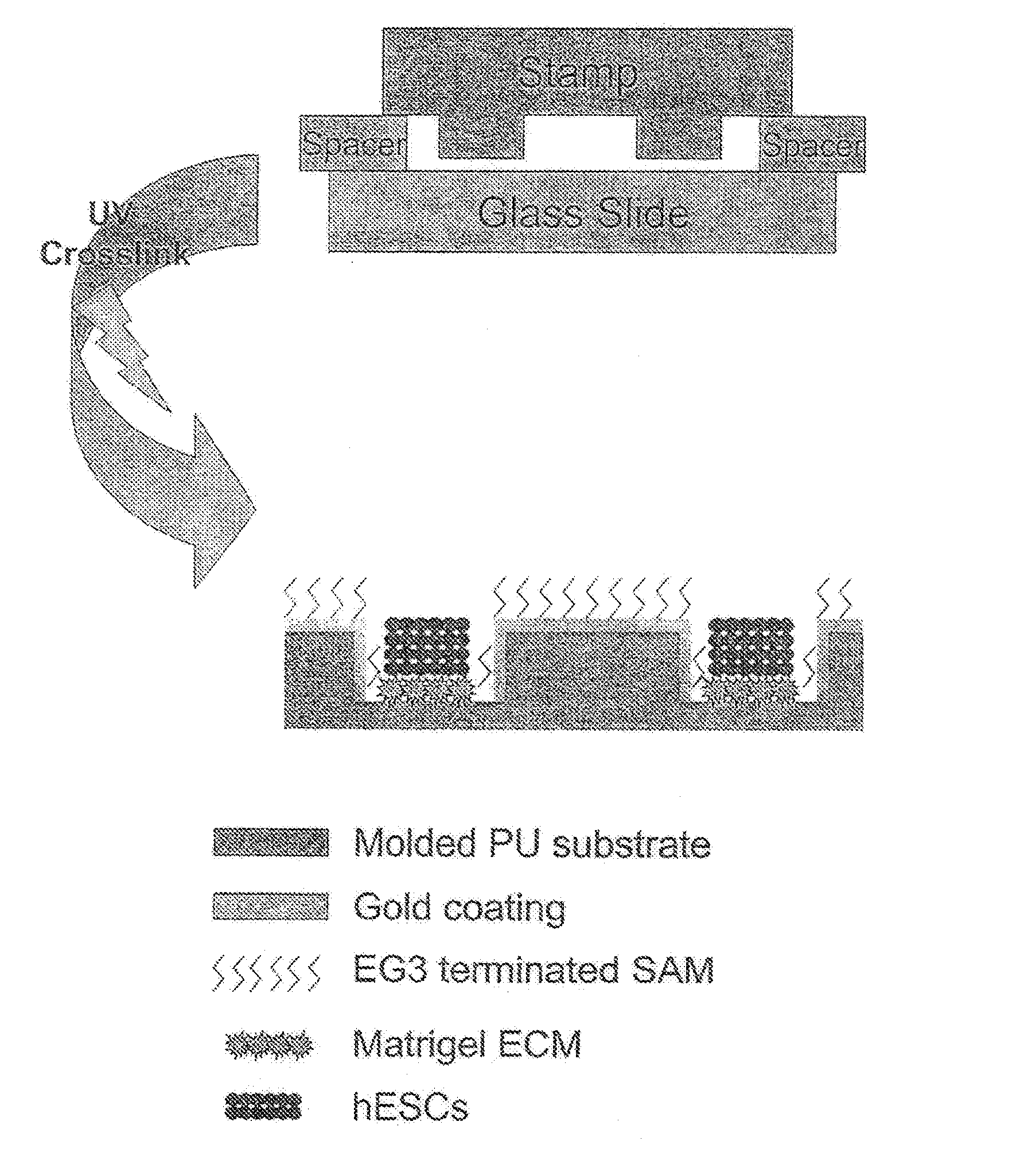
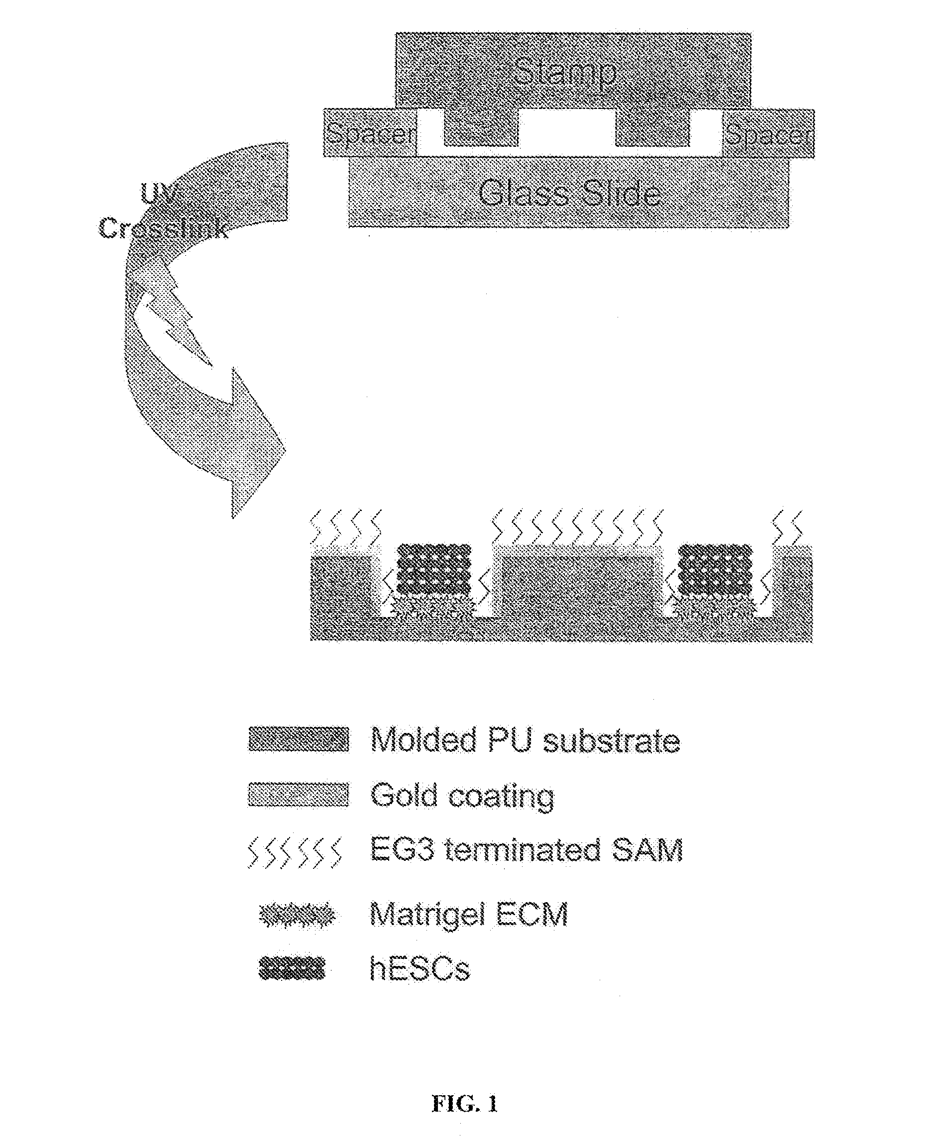
[0038]Reference is made to FIG. 1. Microscope slides having a homogeneous distribution of wells of identical size and shape were constructed in three steps using a polydimethylsiloxane (PDMS) stamp to shape a surface of a UV-crosslinkable polyurethane polymer matrix.

[0039]First, silicon masters each having desired microwell patterns formed into a surface thereof were prepared using photolithography and plasma etching techniques similar to those used by Chen et al. Chen C, et al., “Using self-assembled monolayers to pattern ECM proteins and cells on substrates,” Methods Mol. Biol. 139:209-219 (2000), incorporated herein by reference as if set forth in its entirety. The surfaces were passivated by fluorination with (tridecafluoro-1,1,2,2,-tetrahydrooctyl)-1-trichlorosilane vapor. Second, a mixture of PDMS elastomer prepolymer with curing agent (10:1) (Sylgard 184 Silicon Elastomer; Dow Corning; Midland, Mich.) was poured over the silico...

example 2

Cryopreservation of Embryonic Stem Cells

[0045]To prepare a Matrigel® plate, a tube of Matrigel® stock (2 mg) was taken directly from storage at −20° C. A Matrigel® pellet was immediately re-suspended in 6 ml ice-cold DMEM / F12. All chunks in the mixture were eliminated by vigorous pipetting. A 1 ml aliquot of the mixture was added to each well of a 6-well plate. The plate was maintained at room temperature for 1 hour or overnight at 4° C. before use.

[0046]To prepare conditioned medium, a flask was coated with 0.1% gelatin solution, 10 ml to a T75 flask. After the flask was coated, it was incubated overnight in a 37° C., humidified incubator with 5% CO2 for 24 hours prior to plating irradiated MEF cells. 15 ml of irradiated MEF cells a concentration of 2.12×10⁵ cells / ml MEF medium (90% DMEM, 10% FBS and 1% MEM non-essential amino acids solution) were added to a T75 flask and incubated overnight. The MEF medium was aspirated away and 20 ml HES medium without bFGF (80% DMEM / F12 medium, ...

example 3

Cryopreservation of Encapsulated hESCs in 3-D Microwells

[0052]3-D microwells were created as described in Example 1 and treated in accord with the method of Example 2. Microwells were treated with Matrigel®, which selectively absorbs to the bottom of the wells. hESCs were seeded at 1-5 ×10⁻⁵ cells / micro well and allowed to grow until they filled the microwells. Culture conditions were as described above. Although CM / F+ was changed daily, the cells were not passaged. Prior to freezing, the hESCs were treated as described above in Example 2 to form matrix-colony-matrix constructs (i.e., the microwells were covered with a top layer of Matrigel® and treated with a carbohydrate-based cryopreservation medium, followed by a freezing medium). The hESCs were frozen and stored at −80° C. or in liquid nitrogen.

[0053]hESCs were thawed and then cultured in the microwells or harvested by dispase treatment and either transferred to new microwells or to MEF monolayers or Matrigel®-coated plates. Vi...


PUM


Abstract

The present invention relates to methods and structures for preparing stem cells for use in cryopreservation methods. Stem cell colonies are provided between first and second matrix portions and are exposed to a carbohydrate-containing cryoprotecting medium and a freezing medium. The methods of the invention yield cryopreserved cells that maintain cell viability and exhibit limited cell differentiation after freezing and thawing, facilitating storage, shipping and handling of embryonic stem cell stocks and lines for research and therapeutics.

Description

CROSS-REFERENCE TO RELATED APPLICATIONS[0001]This application is a continuation-in-part of U.S. patent application Ser. No. 10 / 993,468, filed Nov. 19, 2004, which claims the benefit of U.S. Provisional Patent Application No. 60 / 523,343, filed Nov. 19, 2003. This application is also a continuation-in-part of U.S. patent application Ser. No. 11 / 765,831, filed Jun. 20, 2007, which claims the benefit of U.S. Provisional Patent Application No. 60 / 814,975, filed Jun. 20, 2006. Each application is incorporated herein by reference as if set forth in its entirety.STATEMENT REGARDING FEDERALLY SPONSORED RESEARCH OR DEVELOPMENT[0002]This invention was made with United States government support awarded by the following agencies: NAVY / ONR N66001-02-C-8051; NSF-MRSEC 0520527; and NIH HHSN309200582085C. The United States government has certain rights in this invention.BACKGROUND[0003]The invention relates generally to methods of preparing embryonic stem cells for cryopreservation, to structures fo...

Claims


Application Information

Patent Timeline
IPC IPC(8): C12N5/06C12M1/00
CPCA01N1/02A01N1/0231A01N1/0221
Inventor PALECEK, SEAN P.DEPABLO, JUAN J.MOHR, JEFFREY C.JI, LIN
Owner WISCONSIN ALUMNI RES FOUND
Why Eureka
Try Eureka