
Method of Detecting Kidney Dysfunction

Inactive Publication Date: 2009-04-09
UNIVERSITY OF MANITOBA
View PDF0 Cites 19 Cited by
  • Summary
  • Abstract
  • Description
  • Claims
  • Application Information

AI Technical Summary

Benefits of technology

[0020]The methods of the invention are advantageously non-invasive. The β2-microglobulin protein fragments of the invention are associated with kidney dysfunction and can be detected in urine samples. This allows for frequent measurement, which may further improve clinical outcome by better individualization of therapeutic interventions.
[0035]The present inventors have also shown that protease activity is elevated in the urine of patients with kidney dysfunction.

Problems solved by technology

Furthermore, nephrotoxicity can be secondary to environmental toxic agents such as lead, cadmium, mercury and perchlorethilene as well as pharmaceutical drug toxicity.
This was attributed to “a higher proportion of acute rejection episodes which have not resolved with full functional recovery in recent years” (2), but it may also be due to undetected—and therefore untreated—rejection episodes (i.e. subclinical rejection) which harm the allograft over time.
Currently about 50% of renal allografts are lost due to patient death with a functioning graft.
The problem with such strategies, however, is that there has been to date no way other than a renal allograft biopsy of ascertaining whether the graft is free of rejection, and several attempts at reducing immunosuppression have been followed by acute rejection episodes.
However, this is complicated by the complex and often redundant biology of allograft rejection.
However, studies by the Winnipeg Transplant Group have demonstrated that the serum creatinine is an insensitive method for the early detection of renal allograft pathology.
With the advent of new immunosuppressive agents it is becoming apparent that a limitation to the ‘gold standard’ (i.e. renal biopsy) is the extent of heterogeneity of inflammation within the allograft resulting in sampling error (49).
However, clearly this is restricted by patient risk for complications that limits the frequency with which they can be performed, not to mention the associated cost.
An alternative is to further increase the baseline immunosuppression for all patients, but this carries the known risks of infection in the short-term, and of drug toxicity and malignancy in the long-term.
However, their utility to monitor for acute rejection post-transplant has been rather limited (53).
While highly specific for donor antigens, the main disadvantages of these assays are: [I] the need for a repository of donor cells (limits frequency of testing possible), [II] the need for cell expansion (time consuming and labour intensive), [III] reproducibility is poor, [IV] complex interpretation, [V] low sensitivity, [VI] in the case of tetramers requires availability of a diverse panel with a number of potential donor-recipient disparities, and [VII] in the case of trans-vivo DTH the need for a large number of animals (50).
However, it is unclear whether these assays will reliably detect the more subtle (subclinical) forms of acute and / or chronic rejection.
This study highlighted the difficulty in using antigen non-specific biomarkers; it is difficult to ensure specificity since activation of immune markers in blood can reflect inflammation generated through multiple pathways (i.e. rejection versus infection) and occurring at multiple sites within the patient.
However, urine can be very heterogeneous concerning the amount of cells, the concentration of proteins and the pH.
Yet again, the major problem in these studies was the insufficient sensitivity and specificity, which limits the clinical usefulness of such assays.
This fact largely limited the usefulness of measuring intact urinary β2-microglobulin.
However, the need for administration of alkali prior to urine analysis and the restriction of using only urine samples with pH≧6 made the measurement of intact urinary β2-microglobulin unattractive for routine clinical use.

Method used


Image

  • Method of Detecting Kidney Dysfunction
  • Method of Detecting Kidney Dysfunction
  • Method of Detecting Kidney Dysfunction

Examples


example 1

Methods and Materials

Patient Characteristics

Transplanted Patients

[0146]All patient data (e.g. allograft function measured by serum creatinine, biopsies) and urine data were stored and managed in a central access database. From July 1997 to March 2003, 2400 serial mid-stream urine samples from 212 renal transplant patients were collected. These 212 patients underwent a total of 693 protocol or clinically indicated core needle allograft biopsies. All patient charts were reviewed and additional information extracted as needed. Biopsies were analysed by experienced renal pathologists, and scored according to the Banff 1997 classification (Table 3) (23). The acute Banff score determines acute interstitial (ai 0-3), tubular (at 0-3), vascular (av 0-3) and glomerular (ag 0-3) changes, whereas the chronic Banff score assesses chronic interstitial (ci 0-3), tubular (ct 0-3), vascular (cv 0-3) and glomerular (cg 0-3) changes. The individual scores are added to a total acute (a 0-12) and total...

example 2

Urine Protein Profiling

[0161]Healthy people secrete less than 150 mg of protein in urine each day. Depending on the kidney or urinary tract system disease, proteinuria can reach more than 10 g per day. Basically, there are four different pathophysiological pathways that influence the protein content and composition of urine.

[0162][I] Filtration from serum: The major part of urine proteins is derived from serum by filtration through the glomerular barrier. The glomerular barrier consists of the fenestrated endothelial cells, the glomerular basement membrane and the slit-diaphragm of the podocytes. The latter is considered to be predominantly responsible for the characteristics of the barrier. Proteins are thought to be retained from filtration into the urine based on their molecular weight, size, shape and net charge (75). Normally, proteins below 20 kDa are completely filtrated into urine, whereas larger proteins are generally retained in the serum. Albumin (66 kDa), for instance, w...

example 3

Impact of Extrinsic Factors on Reproducibility and Peak Detection of Urine Protein Profiles

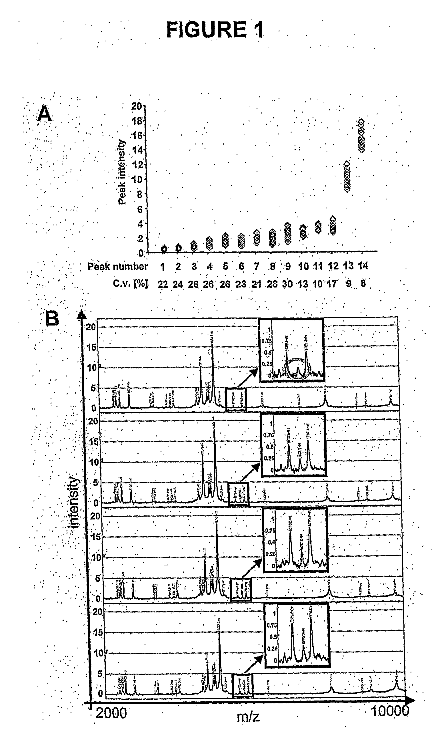
[0174]The most important extrinsic factors that influence reproducibility and peak detection are the matrix composition and the instrument settings. Matrix allows for efficient ionization and vaporization of proteins (82). The most popular matrices for the SELDI-TOF-MS system are SPA and CHCA. Saturated SPA is preferable for looking at masses above 10-20 kDa, while 10-20% CHCA provides the best resolution for proteins / peptides up to about 5 kDa. For urine protein profiling from 2-25 kDa, more peaks and a higher degree of resolution were observed with 35% CHCA. Instrument settings such as detector sensitivity, detector voltage, and laser intensity have to be determined individually. The higher the detector sensitivity and voltage or the laser intensity, the better the detection of high mass proteins. This is accompanied by an increase in background noise, which limits detection of low intensity...


PUM


Abstract

Methods and kits for monitoring kidney function, and detecting kidney dysfunction and transplant related disease and rejection are disclosed. The method involves analyzing a sample, such as a urine sample, containing protein from an animal for fragments of β2-microglobulin, wherein the presence of specific β2-microglobulin fragments is indicative of kidney dysfunction and transplant rejection. In another embodiement, urine samples from an animal are tested for protease activity, such as cathepsin D or napsin A, wherein increased protease activity compared to a control sample is indicative of kidney dysfunction.

Description

FIELD OF THE INVENTION[0001]The present invention relates to methods and kits for monitoring kidney function and detecting kidney dysfunction.BACKGROUND OF THE INVENTION[0002]Renal insufficiency is associated with many pathological conditions. Decreased kidney function can be indicative of renal transplant rejection, as well as other organ rejection. Acute tubular necrosis, transient hypertension and preeclampsia during pregnancy, and chronic glomerular diseases can also result in increased proteinuria and enzymuria indicative of decreased kidney function. Furthermore, nephrotoxicity can be secondary to environmental toxic agents such as lead, cadmium, mercury and perchlorethilene as well as pharmaceutical drug toxicity. Therefore, accurate assessment of kidney function has application and significant prognostic value in the clinic.Kidney Transplant and Transplant Related Disease[0003]Although short and long-term kidney allograft survival has improved substantially from 1988-1996 (1...

Claims


Application Information

Patent Timeline
IPC IPC(8): C12Q1/37C12Q1/02
CPCC12Q1/37G01N33/6848G01N2800/347G01N2800/245G01N33/6893
Inventor NICKERSON, PETERWILKINS, JOHNRUSH, DAVIDSCHAUB, STEFAN
Owner UNIVERSITY OF MANITOBA
Learn More