

Compounds for fluorescence imaging
- Summary
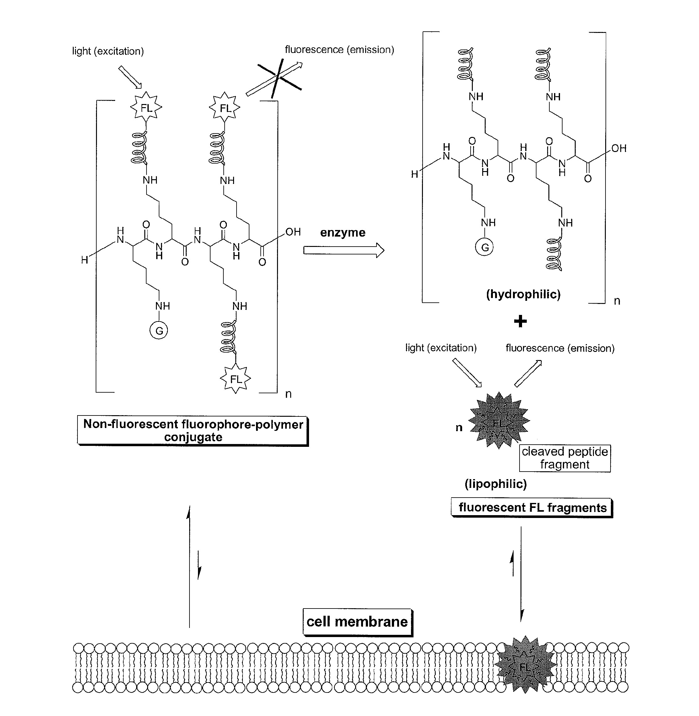
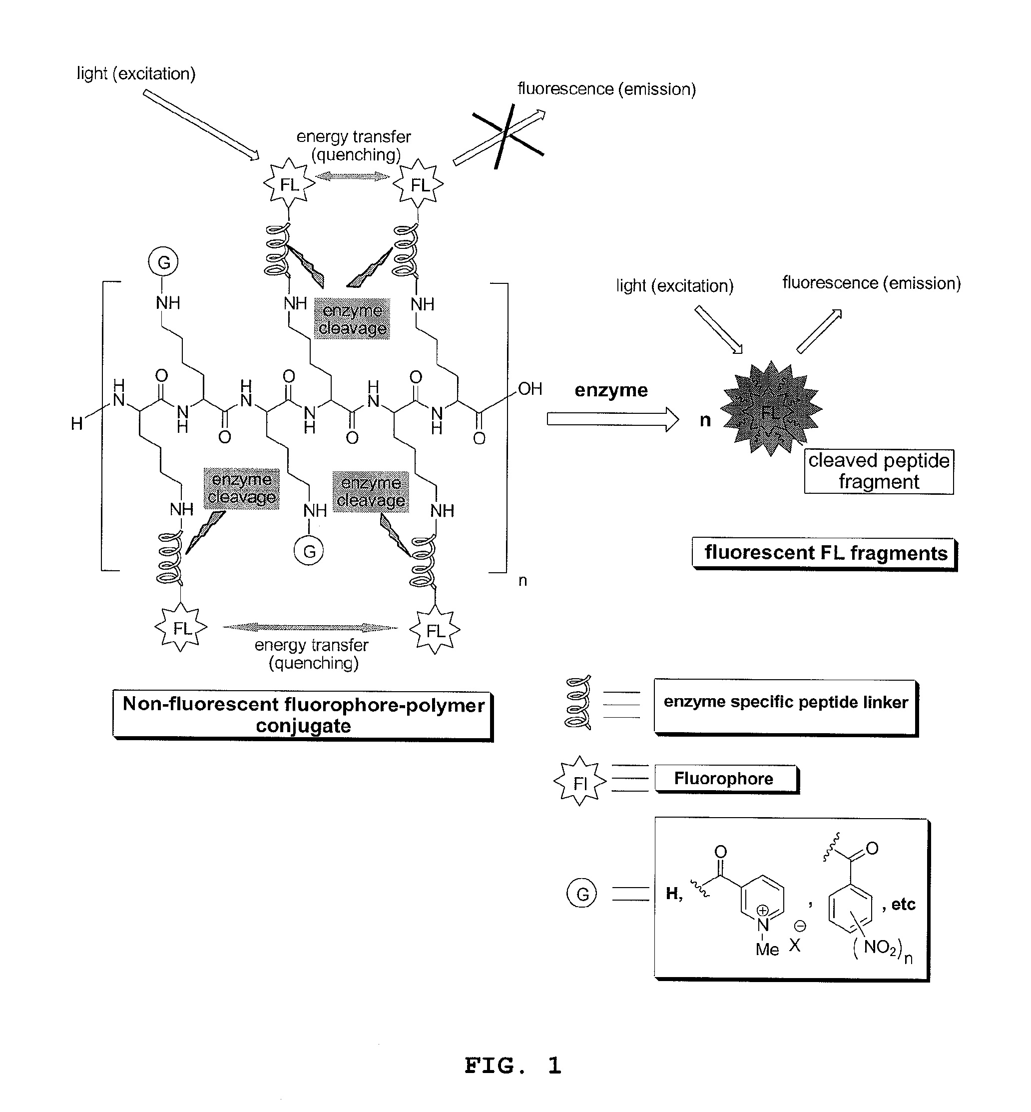
- Abstract
- Description
- Claims
- Application Information
AI Technical Summary
Benefits of technology
Problems solved by technology
Method used
Image

Examples
example 1
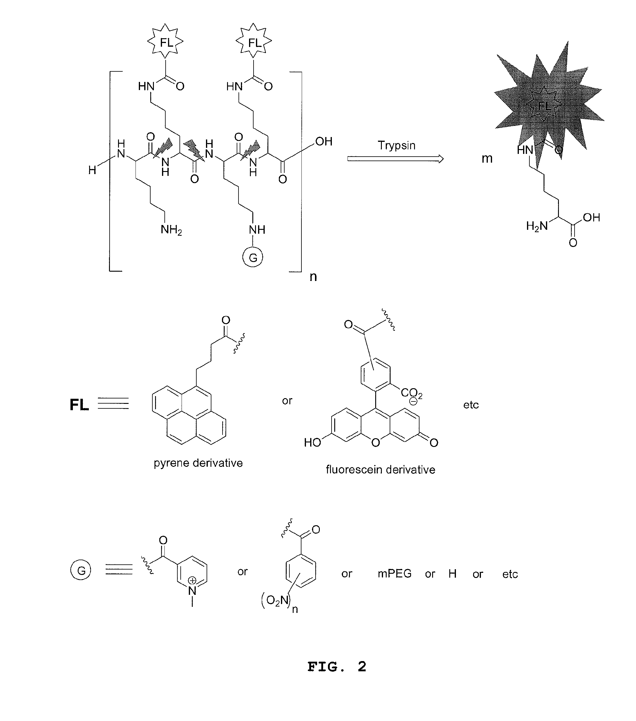
[0170]Preparation of a fluorophore-polymer conjugate comprised of a poly-L-lysine backbone with 15% loading of 5(6)-carboxylfluorescein via N-epsilon amide bonds: in a small vial fitted with a strong magnetic stirrer was dissolved PLL.HBr (8.0 mg, 3.23×10⁻⁴ mmol) in dry DMSO (0.9 mL) then was added DIPEA (3 equiv. per NH2 side chain, 14.9 mg). This solution was stirred for 10 min. before adding drop wise and under vigorous stirring 5(6)carboxylfluorescein NHS ester (0.15 equiv. per NH2 side chain) in DMSO (0.3 mL). The resulting solution was stirred in the dark for 2 h, then, the product, which selectively precipitates out of DMSO, was filtered and washed repeatedly with acetone to yield the desired product as an orange solid.
example 2
[0171]Preparation of a fluorophore-polymer conjugate comprised of a poly-L-lysine backbone with 15% loading of 5(6)-carboxylfluorescein and 85% loading of moieties derived from 1-methylnicotinic acid via N-epsilon amide bonds: in a small vial fitted with a strong magnetic stirrer was dissolved PLL.HBr (8.0 mg, 3.23×10⁻⁴ mmol) in dry DMSO (0.9 mL) then was added DIPEA (3 equiv. per NH2 side chain, 14.9 mg). This solution was stirred for 10 min. before adding drop wise and under vigorous stirring succinimidyl (1-methyl-3-pyridinio)formate iodide (0.30 equiv per epsilon NH2 group in PLL) in DMSO (0.2 mL) and the solution stirred for 30 minutes, then was added under vigorous stirring 5(6)carboxylfluorescein NHS ester (0.15 equiv. per NH2 side chain) in DMSO (0.3 mL). The resulting solution was stirred in the dark for 2 h, then an additional amount of N-succinimidyl (1-methyl-3-pyridinio)formate iodide (0.55 equiv per epsilon NH2 group in PLL) in DMSO (0.4 mL) was added under vigorous st...
example 3
[0172]Preparation of a fluorophore-polymer conjugate comprised of a poly-L-lysine backbone with 15% loading of 5(6)-carboxylfluorescein and 85% loading of 5 KDa mPEG chains via N-epsilon amide bonds: in a small vial fitted with a strong magnetic stirrer was dissolved PLL.HBr (8.0 mg, 3.23×10⁻⁴ mmol) in dry DMSO (0.9 mL) then was added DIPEA (3 equiv. per NH2 side chain, 14.9 mg). This solution was stirred for 10 min. before adding drop wise and under vigorous stirring mPEG-NHS (Nektar Therapeutics) (0.10 equiv per epsilon NH2 group in PLL) in DMSO (0.1 mL) and the solution stirred for 3 h. Then, 5(6)carboxylfluorescein NHS ester (0.15 equiv. per NH2 side chain) in DMSO (0.3 mL) were added under stirring. The resulting solution was stirred in the dark for 3 h, then mPEG-NHS (Nektar Therapeutics) (0.75 equiv per epsilon NH2 group in PLL) in DMSO (1.0 mL) was added under vigorous stirring. The reaction solution was stirred for an additional 8 hours then was diluted in water to make 4.0...
PUM
| Property | Measurement | Unit |
|---|---|---|
| Fraction | aaaaa | aaaaa |
| Fraction | aaaaa | aaaaa |
| Fraction | aaaaa | aaaaa |
Abstract
Description
Claims
Application Information