
Pufa covered implants
a technology of metal implants and spherical layers, applied in the field of metal implants, can solve the problems of variation in the bond strength between the coating and the metallic substaret, affecting the success of the implant, and non-uniform layer thickness, so as to facilitate the control of hard and/or mineralized tissue adhesion, improve the adhesion of hard and/or mineralized tissue, and improve the effect of bone addition to the implant surfa
- Summary
- Abstract
- Description
- Claims
- Application Information
AI Technical Summary
Benefits of technology
Problems solved by technology
Method used
Image

Examples
example 1
Matrix:
[0132]
TypeConcentrationof coinof EPATreatmentNo of coinsPolished 0 mMirradiated with UV lightin vitro 6 · 5 = 3010 mMno irradiationchar. 6 · 3 = 1850 mMGrit- 0 mMirradiated with UV lightin vitro 6 · 5 = 30blasted10 mMno irradiationchar. 6 · 3 = 1850 mMTotal number of coins96
Cleaning
[0133]1. Coins rinsed with pure water
2. Coins washed with ethanol
3. Ultrasonication of coins in water for 5 min
4. Again coins rinsed with pure water
5. Coins allowed to dry on sterile bench
6. Autoclaving of coins
[0134]1. Work in sterile bench
2. EPA solution is filtered (0.2 μm pore size) to sterilise it
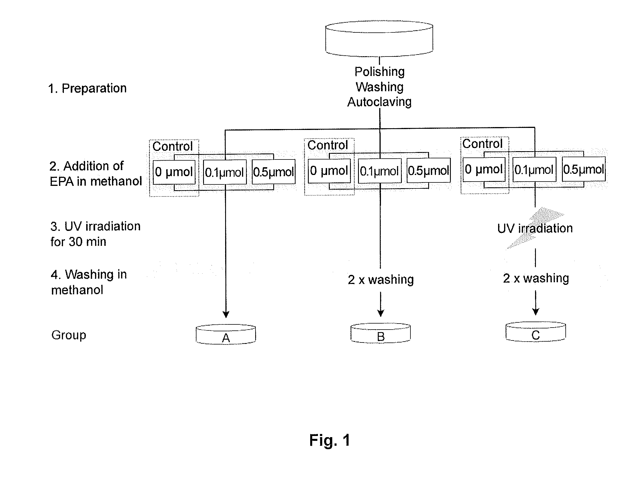
3. 10 μl of respective solution of EPA in methanol is given on the surface of the coins
4. Coin is allowed to dry on sterile bench
5. Part of the coins are irradiated with UV light after the solvent evaporated
→Fluo.link, λ=312 nm
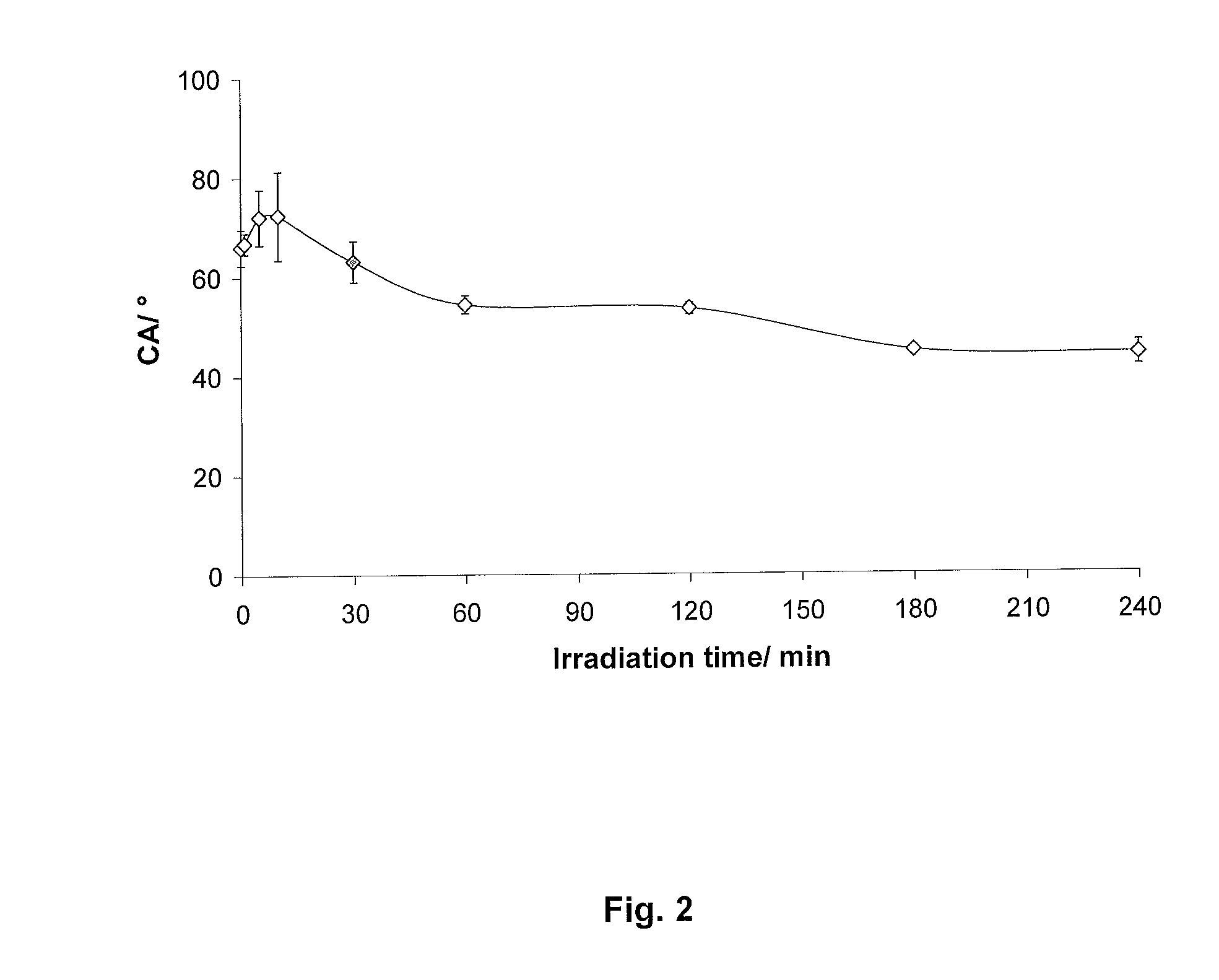
→Time of irradiation: 30 min (first test showed that the contact angle is increasing up to
10 min irradiation and decreases subsequently)...
example 2
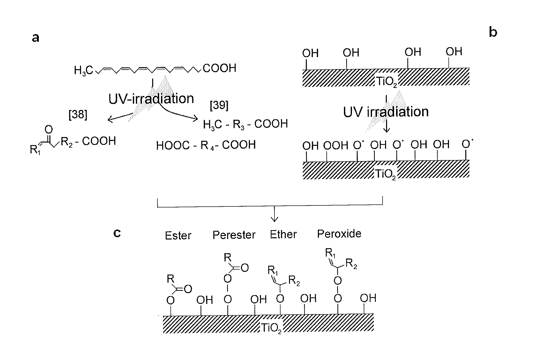
[0136]The aim of this study was to investigate the effect of EPA coating on Ti surface on osteoblast cell response in vitro. Methods to either physically adsorb or chemically bind the layer with EPA were compared, the latter one being produced by UV irradiation of TiO2 surfaces and EPA. Those surface coatings were detected and characterized by physical and chemical analytical methods. In vitro experiments were carried out to test cytotoxicity and ability of EPA-modified Ti surfaces to promote cell attachment and osteogenic differentiation of MC3T3-E1 cells.
Materials and Methods
Chemicals
[0137]EPA was purchased from Sigma (St. Louis, Mo., USA), radioactive labelled [14C]-EPA was obtained from American Radiolabeled Chemicals, Inc. (St. Louis, Mo., USA).
Titanium Implants and Treatments
[0138]Cp Ti implants with a diameter of 6.25 mm and a height of 2 mm were machined from cp Ti rods and subsequently grinded and polished (Phoenix 4000, Buehler GmbH, Düsseldorf, Germa...
example 3
[0176]Experiments performed in vivo with titanium implants containing a high dosage of physical adsorbed EPA and a low dosage of chemically-bound EPA.
Materials and Methods
Titanium Coins
[0177]Commercially pure (cp) machined titanium implants with a diameter of 6.25 mm and a height of 1.95 mm were cleaned and sterilized before use. Briefly, implants were washed together in a glass beaker with deionised water for 30 s, then with 70% ethanol for 30 s, and then with ultrasonic bath at 40° C. for 5 min in deionised water. The implants were subsequently placed in 40% NaOH solution in a water bath of 40° C. for 10 min, sonicated in deionised water for 5 min, and then washed with deionised water until the pH reached 6. Afterwards the implants were sonicated in deionised water at 50° C. for 5 min, placed in 50% HNO3 solution at 50° C. for 10 min, and sonicated in deionised water for another 5 min. The implants were washed with deionised water until reached pH=6 and were stored in 70% ethanol....
PUM
| Property | Measurement | Unit |
|---|---|---|
| Time | aaaaa | aaaaa |
| Percent by mass | aaaaa | aaaaa |
| Concentration | aaaaa | aaaaa |
Abstract
Description
Claims
Application Information