Immunogenic escherichia coli heat stable enterotoxin
Active Publication Date: 2011-12-08
BOARD OF TRUSTEES OPERATING MICHIGAN STATE UNIV
26 Cites 3 Cited by
AI-Extracted Technical Summary
Problems solved by technology
The development of effective strategies to reduce the incidence and severity of ETEC-caused diarrhea has b...
View moreMethod used
[0067]A carrier protein is an antigenic polypeptide entity that induces the formation of antibodies directed against an antigen conjugated to it, by the immune system of an organism into which the carrier-antigen conjugate is introduced. Although many short epitopes are protective, they are poorly immunogenic. By conjugating an immunogenic carrier protein to a molecule that is poorly immunogenic, it is possible to confer higher immunogenicity. Such conjugate molecules stimulate the generation of an immune response and thus have been effectively used in vaccines that protect against pathogens for which protective immunity could not otherwise be generated.
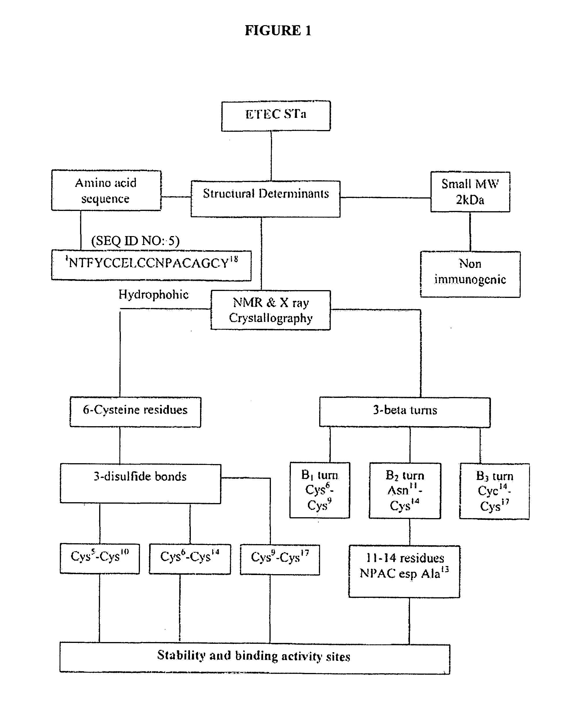
[0080]However, this approach has been a challenge, partly because of the haptenic nature of STa (molecular weight of less than 2 kDa), which fails to elicit an antibody response as provided for in Boedeker (2005) Current Opinions in Gastroenterology 21, 15-19, incorporated herein by reference. Additionally, the correlation between STa toxicity and antigenicity as disclosed in Takeda et al. (1993) Infection and Immunity 61, 289-294, hereby incorporated by reference, hampers the ability to produce a safe STa/CFAs vaccine. However, it was hypothesized that the poor immunogencity associated with the STa molecule could be improved by conjugation of the STa to a suitable macromolecule (carrier protein) as provided for in Pauillac et al. (1998) Journal of Immunological Methods 220, 105-114, incorporated herein by reference. While not limiting the scope of the present invention, it is believed that antibody-based therapy (passive immunization) targeting the STa antigen could be used to reduce the impact of ETEC-STa induced diarrhea and avoid the safety issue associated with active immunization with CFAltoxin based-vaccine. Attempts to conjugate the STa to a carrier protein have been disclosed in Clements (1990) Infection and Immunity 58, 1159-1166, incorporated herein by reference. However, previous disclosures were unclear regarding the efficiency and characteristics of these conjugates.
[0082]In some embodiments, the invention relates to immunotherapy using antibodies raised against the conjugated STa toxin. In some embodiments, the invention relates to the using the STa-neutralizing antibodies as a prophylactic to prevent diarrhea. In some embodiments, the invention relates to the using the STa-neutralizing antibodies (or antibody fragments) to relieve symptoms of traveler's diarrhea and speed recovery. In some embodiments, the invention relates to the using the STa-neutralizing antibodies in a pill, powder, or injectible form.
[0089]In some embodiments the invention relates to compositions of the enterotoxigenic Escherichia coli heat stable enterotoxin (STa) through its unique coupling to a modified protein carrier (such as a modified BSA or “MBSA”) as a treatment of post-anaesthesia urine retention in humans, which would lower the need for post-operative catheterization.
[0092]In a preferred embodiment, the active compound and optionally another therapeutic or prophylactic agent are formulated in accordance with routine procedures as pharmaceutical compositions adapted for intravenous administration to human beings. Typically, the active compounds for intravenous administration are solutions in sterile isotonic aqueous buffer. Where necessary, the compositions can also include a solubilizing agent. Compositions for intravenous administration can optionally include a local anesthetic such as lignocaine to ease pain at the site of the injection. Generally, the ingredients are supplied either separately or mixed together in unit dosage form, for example, as a dry lyophilized powder or water free concentrate in a hermetically sealed container such as an ampoule or sachette indicating the quantity of active agent. Where the active compound is to be administered by infusion, it can be dispensed, for example, with an infusion bottle containing sterile pharmaceutical grade water or saline. Where the active compound is administered by injection, an ampoule of sterile water for injection or saline can be provided so that the ingredients can be mixed prior to administration.
[0108]The amount of the active compound that is effective in the treatment or prevention of macular degeneration or angiogenesis can be determined by standard research techniques. For example, the dosage of the active compound which will be effective in the treatment or prevention of age-related macular degeneration can be determined by administering the active compound to an animal in a model such as, e.g., the animal models known to those skilled in the art. In addition, in vitro assays can optionally be employed to help identify optimal dosage ranges.
[0111]In a preferred embodiment, BSA was modified by introducing succinic moieties. Extensive modification of the free amino groups was achieved with the addition of succinic anhydride as disclosed in Habeeb (1967) Journal of Immunology 99, 1264-1276. The subsequent step in the design of STa-BSA conjugate was the cross-linking of STa to the modified BSA. In one embodiment, this reaction was initiated by incubation of the modified BSA with p-nitrophenol and DCC (i.e. a carbodiimide) for three hours to provide reactive ester groups that could easily attach the STa from its amino terminal, forming amide linkages. The use of DMF was shown to enhance the solubility of reactants including peptides and carrier proteins as disclosed in Lateef (2007) Journal of Biomolecular Techniques 18, 173-176, incorporated herein by reference. We believe that the use of DMF as a solvent reagent may have facilitated the solubility of the hydrophobic STa molecules, solving a problem encountered with the other solvents and coupling media. The STa-conjugate was tested for its protein content and biological activity. Based on the protein estimation, there was a conjugation efficiency of 52-64%. Moreover, this conjugate showed a higher biological activity than any activity reported in the previous STa-conjugates
[0123]Preparation of cell free filtrate. After 24 hours of incubation, the growth medium was immediately filtered by tangential flow filtration through a 0.2 micron cassette in Millipore Pellicon System (Millipore Crop, Bedford, Mass.). Cell free filtrate was kept on ice throughout the time of filtration to minimize bacterial growth and enzymatic activity. Samples from the cell-free filtrate were collected for determination of total protein and STa content using suckling mouse assay.
[0126]Reversed-Phase Batch Adsorption Chromatography (MCI-gel). An intermediate purification step was applied to the acetone STa-rich fraction to achieve a further level of STa purity. The lyophilized crude STa was solubilized in 100 ml of 0.1% of 20% HPLC-grade methanol. To this solution, 100 grams of Reverse-Phase Methacrylate Adsorbent Polymer Resin (340° A, 30 μm; Mitsubishi Chemical Corporation, Catalog #CHP2MGY-01 L) was slowly added under gentle mixing and the slurry was kept at 4° C. for 2 hours under gentle shaking. The slurry was poured into a 10-mm (inner diameter)×25-cm-long glass column. The column was washed with 300 ml 0.1% TFA/H2O (v/v). Stepwise elution of the proteins was performed with 100 ml of 0.1% TFA of 20, 40, 60, 80 and 100% MeOH (v/v). Fractions were collected separately from each elution step. The methanol and TFA were evaporated and the residues were tested for protein and STa biological activity.
[0135]Reversed Phase-Batch Adsorption Chromatography (MCI-gel). Specific activity of the STa at this step of purification increased from 88.7×10³ to 112×103 MU/mg protein. This step allowed for a larger sample load on preparative RP-HPLC. Up to 15 mg of the crude STa cleaned by this procedure could be used as a single load in RP-HPLC without overloading the column or losing the resolution. This has led to a considerable reduction in the number of HPLC runs needed to purify STa.
[0155]Protocol 3: Hyper-Succinylated BSA-Based Protocol. Fuentes et al. (2005) Journal of Immunological Methods 307, 144-149, reported that an increase in the numbers of succinyl groups [COO—] on the carrier protein can enhance the cross linking of the peptides from their amino terminal. The procedure for hyper-succinylation of BSA was previously described herein.
[0171]STa Conjugate Analysis by MALDI-TOF/MS. Lyophilized conjugate samples based on DMF and HS-BSA protocols were subjected to MALDI-TOF/MS to accurately determine the molecular weight (Mr Value). Both samples showed median Mr Values over 80 kDa (FIG. 14 and FIG. 15). FIG. 16 shows the molecular weight differences for the modified BSA before and after STa peptide conjugation (ΔM.W.=8342.5 Da). This difference was attributed to the contribution of ...
View moreBenefits of technology
[0071]In one embodiment, the present invention contemplates modified carrier protein, such as modified BSA (MBSA). In one embodiment, the modified BSA is chemically modified, e.g. succinylated which results in “suBSA” succinylated BSA. In suBSA, Bovine serum albumin is modified with succinic anhydride so that lysine residues are acylated. The free amino groups are modified. The degree of modification can vary based upon how many amino groups are acylated. Succinic anhydride (SA) reacts rapidly with the E-amino groups of lysines and the a-amino groups of the N-termini of proteins at pH 7-9, forming an amide bond by replacing the amino group with a carboxyl. Thus, introducing a succinic anhydride moiety on BSA will afford a protein derivative with more carboxyl groups and hence increase the possibility to link STa from its amino terminal and preserve its antigenic determinants accosiated with the carboxyl termina...
View moreAbstract
The present invention relates to methods and compositions for the treatment and prevention of diarrhea and diarrheal related diseases and disorders in both animals and humans. In some embodiments, the invention relates to the treatment of said diarrhea and diarrheal related diseases and disorders with a vaccine. In still further embodiments, the invention relates to the treatment of constipation using the disclosed methods and compositions.
Application Domain
Technology Topic
Image
Examples
- Experimental program(3)
Example
Example I
Materials and Methods
Animals
[0114]Swiss Webster Mice: A group of 20 Swiss-Webster (fifteen females and five males) was used to establish a colony as a source of suckling mice for STa bioassay. Exhausted females and males were continuously replaced with younger animals to ensure production efficiency of infant mice litters by the colony.
Reagents
[0115]All reagents were obtained from commercial sources and were of analytical grade. Mobile phases used for purification of STa include HPLC-grade methanol, trifluoroacetic acid, as well as the other chemical ingredients listed under this section. Verifying the ETEC K99+ Strain: The PCR protocol disclosed in Olsvick et al. (1993) Diagnostic Molecular Biology, American Society for Microbiology (Washington, D.C.) and Salvadori et al. (2003) Journal of Microbiology 34, 230-235, both of which are hereby incorporated by reference, was used to detect the STa gene and verify the strain as STa-producing E. coli.
[0116]Bacterial Strains: An ETEC strain was isolated from a clinically diarrheic neonatal calf and was provided by A. M. Saeed (Molecular Epidemiology Laboratory, National Food Safety Toxicology Center (NFST), Michigan State University (MSU), East Lansing, Mich.). A control strain (K-12 E. coli) was kindly obtained from the Bacterial Evolution Laboratory, NFST, MSU, East Lansing-Mich.
[0117]DNA Extraction (Template) by boiling lysis: ETEC and K-12 strains were grown on Tripticase Soy Agar slants overnight at 37° C. A uniform bacterial colony from both strains was taken and suspended in 1 ml sterile Milli Qwater and boiled for 10 minutes, then left in ice for 5 minutes, followed by centrifugation at 13,000 rpm for 4 minutes. The supernatant was taken and kept at −20° C. until use as provided for in Holmes et al. (1981) Analytical Biochemistry 114, 193-197, hereby incorporated by reference.
[0118]Primer selection and preparation: Two different sizes of STa primer, 244 bp and 127 bp (Table III), were obtained from Integrated DNA Technology Inc. (Coralville, Iowa). The STa primers for 244 base pair product (SEQ ID NO: 1) 5′-TCC GTG AAA CAA CAT GAC GG-3′ and (SEQ ID NO: 2) 5′-ATA ACA TCC AGC ACA GGC AG-3′. The STI primers for 127 base pair product (SEQ ID NO: 3) 5′-TTA ATA GCA CCC GGT ACA AGC AGG-3′ and (SEQ ID NO: 4) 5′-CTT GAC TCT TCA AAA GAG AAA ATT-3′. Both primers were prepared according to the manufacturer's instructions. PCR program: PCR running conditions for detection of the STa gene is presented in Table IV using PCT-100 Programmable Thermal Controller (MJ Research, Inc). PCR reaction: The PCR reaction was performed as described in Table V using a Fisher exACTGene Complete PCR kit.
[0119]Agarose Gel Electrophoresis Analysis of PCR Products: The analysis of the PCR products was performed in 2% agarose gel electrophoresis using the Horizontal Gel Electrophoresis System, Life Technology (Cat #11068-012). Briefly, two percent of agarose was prepared (1.5 gm/75 ml 1×TAE electrophoretic sequence grade) and ethidium bromide was added at a concentration of 3 μl/50 ml. The reagent was poured into the electrophoretic chamber and filled with 1×TAE. Five volumes of PCR product were mixed with 1 volume of gel loading buffer and loaded into the wells along with a 1.5 kb ladder. The agarose gel was left to run at the appropriate voltage (100-160 volts) for 30-45 minutes and examined via UV irradiation.
Purification and Characterization of E. Coli STa
[0120]STa was purified as disclosed in Staples et al. (1980) Journal of Biology Chemistry 155, 4716-4721, Saeed et al. (1983) Infection and Immunity 40, 701-710 and Saeed et al. (1985) Analytical Biochemistry 151, 431-437, all of which are hereby incorporated by reference.
[0121]Seed culture and frozen stock of ETEC preparation. Casamino acid-yeast extract-salts (CAYE) seed culture was used for optimal growth of the ETEC strain as disclosed in Giannella (1976) Transactions of the American Clinical and Climatological Association 114, 67-85. The ETEC strain was grown on 500 ml of CAYE, incubated at 39° C. for 24 hours on a rotary shaker at 120 rpm, mixed with glycerol at a final concentration of 15%, then aliquoted into 10 ml samples and frozen at −80° C. (frozen stock).
[0122]Batch Medium (Asparagine Salt Medium) and Growth Conditions. Medium was prepared as described in Staples et al. (1980) Journal of Biology Chemistry 155, 4716-4721. The disclosed media was found to offer several advantages, including high level of STa· production along with minimal contaminating proteins that facilitated the STa-purification process. Each batch consisted of 30 liters of culture-innoculated ASM grown in a 36 L omni vessel under different pH conditions (7.4, 8 and 8.6) using a Bellco bioreactor (Bellco Glass Inc., Vineland N.J.). Preculture was prepared by inoculating 10 ml of frozen stock of ETEC into IL of CAYE broth and was incubated at 39° C. for 24 hours on a rotary shaker at 120 rpm. The preculture medium was then transferred into 30 liters of batch medium and kept at 39° C. under continuous agitation at 120 rpm, aeration and oxygenation were at a rate of 5 Umin and 600 ml/min, respectively, through a sintered metal dispersion ring. Foam, speed of agitation, temperature and O2 pressure was controlled using Bellco control modules. Samples were taken every two hours to determine the growth kinetics under various pH levels.
[0123]Preparation of cell free filtrate. After 24 hours of incubation, the growth medium was immediately filtered by tangential flow filtration through a 0.2 micron cassette in Millipore Pellicon System (Millipore Crop, Bedford, Mass.). Cell free filtrate was kept on ice throughout the time of filtration to minimize bacterial growth and enzymatic activity. Samples from the cell-free filtrate were collected for determination of total protein and STa content using suckling mouse assay.
[0124]Amberlite XAD-2 Batch Adsorption Chromatograph. Cell-free filtrate was desalted and the hydrophobic STa was concentrated using Amberlite XAD-2 batch adsorption chromatography. Amberlite XAD-2 resin was first washed extensively with purified water to remove any preservative and powdery contaminants. Then 500 grams were suspended into 15 L of cell free filtrate in a 20 L carboy and kept overnight at 4° C. under gentle stirring. Resin was poured from the carboy into a 40 cm long glass column and washed with 5 L of Milli Qwater. The contaminants loosely bound to the resin were eluted with 1 L of 1% acetic acid in 20% methanol/water (v/v). A stepwise elution system was applied to elute the STa ˜tarting with 1 L of 1% acetic acid in 80% methanol/water (v/v) followed by 1 L of 1% acetic acid in 99% methanol/water (v/v) and finally 1 L of 50% acetone/water (v/v). The last three fractions were pooled and concentrated by flash evaporation and freeze-drying. The resin was degassed for 5 minutes after each solvent was added to drain completely before further addition of solvent. Samples were collected for determination of the total protein and testing for STa biological activity in suckling mouse.
[0125]Acetone Fractionation. Lyophilized crude STa was dissolved in 20 ml of 25% of acetic acid. Acetone was added to bring the final volume to 100-150 ml. After standing 1 hour at 4° C., the sample was centrifuged at 10,000 g for 30 minutes at 4° C. The supernatant fraction was evaporated to remove the acetone and was then freeze-dried. Samples were collected for protein determination and STa biological activity.
[0126]Reversed-Phase Batch Adsorption Chromatography (MCI-gel). An intermediate purification step was applied to the acetone STa-rich fraction to achieve a further level of STa purity. The lyophilized crude STa was solubilized in 100 ml of 0.1% of 20% HPLC-grade methanol. To this solution, 100 grams of Reverse-Phase Methacrylate Adsorbent Polymer Resin (340° A, 30 μm; Mitsubishi Chemical Corporation, Catalog #CHP2MGY-01 L) was slowly added under gentle mixing and the slurry was kept at 4° C. for 2 hours under gentle shaking. The slurry was poured into a 10-mm (inner diameter)×25-cm-long glass column. The column was washed with 300 ml 0.1% TFA/H2O (v/v). Stepwise elution of the proteins was performed with 100 ml of 0.1% TFA of 20, 40, 60, 80 and 100% MeOH (v/v). Fractions were collected separately from each elution step. The methanol and TFA were evaporated and the residues were tested for protein and STa biological activity.
[0127]Preparative Reverse-Phase High Performance Liquid Chromatography (RP-HPLC). RP-HPLC was performed on Waters Associate Liquid Chromatography System equipped with multi-solvent delivery pumps, an automated gradient programmer 600S controller, Model 486 tunable absorbance detector using 7 μm, 300 Å, 25 cm×10 mm inner diameter Vydac C8 preparative columns (Sorbent Technologies, Inc., Atlanta, Ga.). Samples from RP-methacrylate adsorbent polymer resin were applied on an RP—C8 column and STa was eluted by gradient system with 0.1% TFA in water as solvent A and 0.1% TFA in 80% methanol as solvent B (0-30% for 5 min and 30-80% for 80 min). The UV absorbing peaks were detected at 214 nm. Peaks were collected separately and the methanol was evaporated then freeze-dried. The resulting freeze-dried substance was reconstituted into physiologically balanced saline and evaluated for protein content and STa biological activity.
[0128]STa Assessment for Biological Activity. Detecting and quantifying STa biological activity was done using a reference standard in vivo model test, suckling mouse assay, according to Dean et al. (1972) The Journal of Infectious Diseases 125, 407-411 and Giannella (1976) Infection and Immunity 14, 95-99, both of which are hereby incorporated by reference. Newborn Swiss Albino suckling mice (2-3 days old) were randomly divided into groups (three each). Samples from RP-HPLC were serially diluted 1/100; 1/10,000 & 100,000 and 10 μl of 0.2% Evans blue (w/v) was added per ml. Each suckling mouse was inoculated orally by 100-μl sample using a 1 ml syringe and a 20-μ-diameter polyethylene tube. Each sample dilution was tested in triplicate. After 2-hour incubation at room temperature, the mice were euthanized by carbon dioxide in a CO2 chamber and the intestine, not including the stomach, was removed from each newborn mouse and weighed. The ratios of intestinal weight to remaining body weight of the three mice were determined. Animals with no dye in the intestine or with dye within the peritoneal cavity at autopsy were discarded. One unit of ST activity (one mouse unit) is defined as the minimal amount of toxin that produces an intestinal weight/carcass ratio of greater than 0.083.
[0129]Criteria for homogeneity of purified STa. Homogeneity of the purified STa from preparative runs was validated by analytic aquapore RP-300A Perkin Elmer C8 column. Additionally, the exact molecular weight of STa was determined by matrix-assisted laser desorption ionization-time of flight mass spectroscopy (MALDI-TOF/MS). The purified STa was then submitted for amino acid sequencing.
Results and Discussion
[0130]Detection of STa gene. PCR amplification verified that the tested strain carried the gene encoding for STa after analyzing the product on gel electrophoresis. Two amplicon bands of 127 bp and 244 bp were detected under UV light for the tested strain, which were not apparent for the control strain (E. coli K-12) (FIG. 5).
[0131]Culture analysis. Growth kinetics experiments were conducted on 30 L batch cultures under various pH values (7.4, 8 and 8.5). Samples were taken every two hours and the growth pattern was determined by counting the total cell count (CFU/ml) using a robotic spiral plate and computer linked camera (Q counter). As FIG. 6 indicates, tested ETEC growth was maximal in medium in which the initial pH was adjusted to 7.4. This level of growth was associated with higher level of crude STa as verified by the suckling mouse assay.
[0132]Purification and Characterization of E. coli STa. Table VII shows the summary of the purification scheme of SrTa for the ETEC E. coli in 30 L batch culture.
[0133]Amberlite XAD-2 Batch Adsorption Chromatography. This step yields a high specific activity of the crude STa (8.70×10³ MU/mg protein) compared with the STa specific activity in the cell free filtrate (1.22×10³ MU/mg protein).
[0134]Acetone fractionation. Acetone fractionation resulted in further purification of the STa by removing additional amount of non-STa protein that was precipitated in acetone. Samples were taken for protein determination and STa biological activity. Specific activity of STa increased to 88.7×10³ MU/mg protein.
[0135]Reversed Phase-Batch Adsorption Chromatography (MCI-gel). Specific activity of the STa at this step of purification increased from 88.7×10³ to 112×103 MU/mg protein. This step allowed for a larger sample load on preparative RP-HPLC. Up to 15 mg of the crude STa cleaned by this procedure could be used as a single load in RP-HPLC without overloading the column or losing the resolution. This has led to a considerable reduction in the number of HPLC runs needed to purify STa.
[0136]Preparative Reverse-Phase HPLC Chromatography (RP-HPLC). Sixty percent methanol MCI-gel STa-rich fractions were loaded on a preparative C8 column for further purification. FIG. 7 and FIG. 8 describe the elution profiles of STa. Elution with an increasing methanol gradient resulted in number of absorbance peaks at 214 nm (FIG. 7), the last of which was found to contain enterotoxin activity. The enterotoxin peak began to elute at approximately 55-60% methanol after 35 minutes retention time. This peak was collected and after methanol evaporation, was freeze-dried. It was then reconstituted into physiological saline and tested for STa biological activity and protein concentration. Further improvement in the STa specific activity (885×104 MU/mg) was achieved in this step. The biological activity was demonstrated to be 0.113 ng per one mouse unit of STa minimal effective dose (MED) in 2-3 day-old inoculated Swiss Webster suckling mice.
[0137]Criteria for Homogeneity of Purified STa. [0138] Analytic C8 Column: Pooled peaks from several preparative RP-HPLC runs were tested on an analytic aquapore RP-300A Perkin Elmer C8 column to demonstrate a single symmetrical peak (FIG. 8). [0139] Mr-Value Determination Using Matrix Assisted Laser Desorption Ionization Time of Flight/Mass Spectroscopy: A lyophilized HPLC-purified sample was analyzed by MALDI-TOF/MS to determine the molecular weight and the result is shown in FIG. 9. The observed signal at 100% MS intensity with mlc=1972.1 indicates that the Mr of the purified product is 1972.1 Da, which is compatible with the Mr (1969-1972) value calculated from amino acid composition of the STa, confirming the purity and identity of the purified product as the STa molecule. This was in agreement with the findings of Takao et al. (1983) FEBS Lett. 152, 1-5, incorporated herein by reference. [0140] Amino Acid Sequence: Further confirmation of the homogeneity and identity of the purified product was performed by determination of amino acid sequence. A lyophilized HPLC-purified sample was submitted for amino acid sequence analysis and the results showed the 18 amino acid residues of the STa molecule were matching the reported sequence.
Conclusions
[0141]This protocol includes concentrating the cell free filtrate using Amberlite XAD-2 batch adsorption chromatography (BAC), acetone fractionation, methacrylate polymer resin BAC and finally through RP-HPLC. Chemical analysis of the purified preparations matched the reported structure for this type of enterotoxin. The biological activity was demonstrated to be less than 0.2 ng per one mouse unit of the STa in 2-3 day-old inoculated Swiss Webster suckling mice. In summary, purification of STa to homogeneity was accomplished and the purity of the produced STa was documented through amino acid sequencing, and mass spectroscopy.
Example
Example II
Methods and Materials
[0142]Reagents. All reagents were obtained from commercial sources and were of analytical grade. Bovine serum albumin (BSA), succinic anhydride (SA), dioxane, N,N-dicyclohexylcarbodiimide (DCC), 1-ethyl-3-(3-dimethylaminopropyl carbodiimide hydrochloride (EDAC), N-methyl-imidazole, dimethylormamide (DMF), 2-(N-morpholino) ethanesulfonic acid buffer (MES), triethylamine (ET3N), p-nitrophenol, sodium azide (NaN3) and phosphate buffer saline (PBS) tablets were obtained from Sigma Chemical Co. (St. Louis, Mo.). STa was purified as described in Example I.
[0143]Procedure for covalently cross-linking STa with modified BSA: Chemical modification of bovine serum albumin. Bovine serum albumin was chemically modified to introduce new carboxyl moieties using two different protocols: [0144] Succinylation [0145] Hyper-succinylation
Succinylation of Bovine Serum Albumin
[0146]Basis of reaction. Succinic anhydride (SA) reacts rapidly with the E-amino groups of lysines and the a-amino groups of the N-termini of proteins at pH 7-9, forming an amide bond by replacing the amino group with a carboxyl as disclosed in Riordan et al. (1964) Biochemistry 11, 1768-1774, Gounaris et al. (1976) Journal of Biological Chemistry 242, 2739-2745 and The Merck Index, 12th ed., Merck & Co., Inc. (1996), all of which are hereby incorporated by reference (FIG. 11). Thus, introducing a succinic anhydride moiety on BSA will afford a protein derivative with more carboxyl groups and hence increase the possibility to link STa from its amino terminal and preserve its antigenic determinants accosiated with the carboxyl terminal of the molecule.
[0147]Procedure: The methods followed were those described by Habeeb (1967) Biochemistry and Biophysics 121, 652 and Habeeb (1967) Journal of Immunology 99, 1264-1276, both of which are incorporated herein by reference. Briefly, one gram of BSA was dissolved in 200 ml of 0.2 M borate buffer, pH 9.3. A 20 ml solution of dioxane with 5.4 g succinic anhydride was added in small aliquots over a period 30 minutes, the reaction mixture was stirred magnetically while maintaining the pH at 9.3 through the addition of 3 M NaOH. Following the last addition of succinic anhydride, the acylation reaction was allowed to continue for 45 minutes. The solution was then dialyzed at 4° C. against several changes of 0.01 M triethylamine using dialysis tubing with a M.W. cutoff of 12-14 KD. The dialyzed preparation was first freeze-dried and then further dried in a dessecator over phosphorous pentoxide (P205). Samples were taken and reconstituted in PBS buffer (pH 6.8) for size exclusion chromatography and mass spectroscopy.
Hyper-Succinylated Bovine Serum Albumin (HS-BSA)
[0148]Basis of reaction. An extensive modification of the BSA by introducing a large) number of succinyl moieties [COO—] on the carrier protein could be achieved in the hyper-succinylation reaction, The hyper-succinylation reaction was carried out in two steps, The first step involves the production of hyperaminated BSA by conversion of all free carboxyl groups on the BSA (aspartic and glutamic acids) into amino groups. The second step was the production of hypersuccinylated BSA by addition of succinic anhydride to the hyperaminated BSA to convert all amino groups (newly introduced, free, and N-terminal) into carboxyl groups.
[0149]Procedure: Native BSA was treated with 1 mM of ethylenediamine at pH 4.75 in the presence of 10 mM EDAC. This hyperaminated protein molecule was then treated with 100 mmoles of succinic anhydride at pH 8.0 for two hours to produce a hyper-succinylated BSA molecule as disclosed in Fuentes et al. (2005) Journal of Immunological Methods 307, 144-149, incorporated herein by reference.
[0150]Coupling of E. coli STa to Modified BSA. Four different conjugation protocols were used to covalently cross-linking STa to modified BSA. They were evaluated on the basis of stability of covalent bond, retained STa biological activity and the conjugation efficiency.
[0151]Protocol 1: Using Dimethylormamide (DMF) as a solubilizer for the peptide and carrier protein. As described in Atassi (1981) Biochimica et Biophysica Acta 670, 300-330, incorporated herein by reference, DMF was used to solubilize several synthetic peptides before cross-linking them to carrier proteins. While not limiting the present invention to any particular theory, it is believed that this protocol enhances the coupling of the amino terminal ends of synthetic peptides to the carrier protein. In this study, the carrier protein (suBSA) was solubilized in DMF and then treated with p-nitrophenol to activate the carboxyl groups on the carrier.
[0152]Procedure: Applying this procedure, we have used the purified native STa peptide and have solubilized it in DMF prior to addition to the modified suBSA. The mixture was kept stirring overnight at room temperature. This design encourages the STa coupling through its amino terminals based on nucleophilic attack at the reactive ester groups of the modified suBSA forming amide linkages. In a typical reaction, 140 mg of suBSA was suspended in 10 ml of anhydrous DMF and stirred it magnetically in a tight-capped foil-wrapped bottle for 3-4 hours. A solution of p-nitrophenol (65 mg/0.5 ml) in DMF was added and magnetic stirring continued for 15 minutes. A solution of DCC (50 mg/0.5 ml) was added to suBSA at a molar ratio of 120:1 and the reaction was allowed to continue stirring at room temperature for three hours. 100 mg of STa in 1 ml DMF was added to the activated suBSA at a molar ratio of 24:1. Shortly after, 1 ml of triethylamine was added. The reaction mixture was stirred overnight, at room temperature, while protected from direct light. The next day, 30 ml of Milli Q water was added and the mixture was dialyzed extensively against distilled water at 4° C. using a dialysis membrane with a 12-14 kD M.W. cutoff and then freeze-dried. Samples were taken for measurement of the STa biological activity of the conjugate using suckling mice assay (SMA), protein determination, biochemical and molecular characterization.
[0153]Protocol 2: Imidazole-Based Protocol. The coupling procedure disclosed in Dean et al. (1990) Journal of Immunological Methods 129, 119-125, incorporated herein by reference, may be used to stabilize the carrier protein and minimize the formation of polymers due to the acylation process when the cross linker and the peptides intended for cross-linking are added.
[0154]Procedure: 0.5 M N-methyl-imidazole, pH 6.0, was used to dissolve the STa peptide and the carrier protein suBSA at a molar ratio (100:1). After the addition of EDAC (molar ratio: 50 mol EDAC/mol STa), the mixture was stirred for 30 minutes at room temperature followed by dialysis (M.W. cutoff: 12-14 kD) against distilled water at 4° C.
[0155]Protocol 3: Hyper-Succinylated BSA-Based Protocol. Fuentes et al. (2005) Journal of Immunological Methods 307, 144-149, reported that an increase in the numbers of succinyl groups [COO—] on the carrier protein can enhance the cross linking of the peptides from their amino terminal. The procedure for hyper-succinylation of BSA was previously described herein.
[0156]Procedure: In this coupling protocol, 3 mg hypersuccinylated-BSA was dissolved in 2.5 ml of 5 mM sodium phosphate buffer pH 7 and mixed with 2.5 ml of the STa peptide (0.5 mg/ml) in dioxane. EDAC was then added gradually to reach a concentration of 100 mM. After that, the conjugated composite was dialyzed using a tube with a M.W. cutoff 12-14 kD against distilled water at 4° C.
[0157]Protocol 4: Conventional Peptide-Carrier Coupling Protocol. In this coupling procedure, suBSA, EDAC and MES buffer were used as disclosed in the EDC Conjugation Protocol Technical Sheet, Uptima Interchim (2007), incorporated herein by reference.
[0158]Procedure: SuBSA carrier protein was dissolved in 0.1 M MES buffer, pH 5, to a final concentration of 10 mg/ml. Two milligrams of STa peptide were added to 2 mg of suBSA carrier protein. Then, EDAC (10 mg/ml in cold distilled water) was added at a ratio of 0.5 mg of EDAC per mg of total protein. The reaction was stirred for 2-3 hours at room temperature before dialysis at 4° C. against PBS using a tube of 12-14 kD molecular weight cutoff.
[0159]Dialysis. The products of BSA modification and STa-modified BSA conjugation reactions were subjected to extensive dialysis to remove the small molecular weight reactants <<14 kD). Dialysis tubing with nominal M.W. cut-off 12-14 kD was purchased from Fisher Scientific (Pittsburgh, Pa.). STa-SuBSA conjugate was subjected to extensive dialysis against Milli Q purified water using a dialysis membrane of 12-14 kD M.W. cutoff. Molecular species of 14 kD or higher were retained inside the dialysis tube and all other reactants below 14 kD including uncoupled (free) toxin were dialyzed out.
[0160]Gel Filtration Chromatography (GFC). PD-10 columns, Sephadex G-25M packed columns, of a nominal molecular mass exclusion limit of 5000 for protein were purchased from G.E. Healthcare (Buckinghamshire, UK). These columns are designed to separate proteins based on their molecular weight. The columns were equilibrated and developed by following the manufacturer's instructions. The dialyzed STa conjugate samples were passed through PD-10 Sephadex G-25 GFC column to purify the STa-suBSA conjugates from free STa, then assessed for biological activity and protein concentration. A freeze-dried STa-suBSA conjugate sample was reconstituted into 2.5 ml of PBS and passed through a PD-10 column to separate the unconjugated STa peptide from the portion that was successfully cross linked to the carrier protein. The STa-carrier conjugate was then eluted with 3.5 ml PBS and the effluent was collected and tested for biological activity in a suckling mouse assay.
[0161]Size-Exclusion High Performance Liquid Chromatography. SE-HPLC was performed to compare the molecular size of both native and modified BSA. Bio-Sil® SEC-125 HPLC 300×7.8 mm filtration column (Catalog Number 125-0060) was purchased from BioRad (Hercules, Calif.) and hooked to the Waters Associate Liquid Chromatography System equipped with multi-solvent delivery pumps, an automated gradient programmer 600S controller and a tunable absorbance detector (Model 486). The column was equilibrated with 0.05 M sodium phosphate (dibasic), 0.05 M sodium phosphate (monobasic), 0.15 M NaCl and 0.01 M NaN) at pH 6.8. Isocratic elution system was applied at flow rate of 1 ml/min. The peak absorbance was monitored at UV wavelength of 280 nm.
[0162]Amino Acid Compositional Analysis. To determine the conjugation ratio of STa to the modified BSA, a freeze-dried STa-suBSA conjugate sample was subjected to amino acid compositional analysis (Research Technology Support Facility (RTSF), Michigan State University).
[0163]Matrix-Assisted Laser Desorption Ionization/Time of Flight Mass Spectroscopy (MALDI-TOF/MS). 200 micrograms of freeze-dried STa-suBSA conjugate sample were subjected to MALDI-TOF/MS analysis at the MSU-RTSF laboratory to determine precisely the molecular weight of the conjugate and use this figure to calculate the number of STa molecules that covalently cross-linked to one BSA molecule (conjugation ratio).
[0164]Protein Assay. Protein assays were performed according to the Lowery method as described in Lowry et al. (1951) Journal of Biological Chemistry 193, 265-275, incorporated herein by reference, using a Perkin Elmer Lambda 3A UV/US spectrophotometer.
[0165]STa-suBSA Conjugates Activity Bioassay. The biological activity of the STa-suBSA conjugates was determined using the suckling mouse assay as described in Saeed et al. (1983) Infection and Immunity 40, 701, incorporated herein by reference.
Results
[0166]Table VIII shows the summary of conjugation experiments and their evaluation. More details on the DMF conjugation protocol are presented below.
[0167]Characteristics of the Modified BSA. The results from SE-HPLC showed that modified BSA with succinic anhydride was eluted faster (RT=2.99 min) than native BSA (RT=4.20 min), suggesting that the molecular weight of suBSA had undergone a significance change (FIG. 12). Change in the molecular weight of the succinylated BSA was also confirmed by MALDI-TOF/MS. It was found that suBSA has a M.W. of 72.40 kD in comparison to native BSA which has a M.W. of ˜67.00 kD (FIG. 13).
[0168]Characteristics of E. coli STa-suBSA Conjugates. Dialysis and Gel Filtration Chromatography. A summary of the results is presented in Table VIII. The conjugation protocol based on the DMF method showed a higher rate of conjugation efficiency and higher level of retained STa biological activity compared to other conjugates (Table VIII). Further characterization of the DMF-based conjugation protocol is described below.
[0169]Amino Acid Compositional Analysis. A dialyzed conjugate sample based on the DMF protocol was subjected to amino acid compositional analysis. Table IX shows the picomolar concentration and retention time of each amino acid residue.
[0170]Calculation of STa Peptide to SuBSA Ratio. The conjugation ratio is defined as the number of STa molecules covalently cross-linked to one molecule of succinylated BSA. The amino acid composition of the conjugate was empirically determined by measuring the picomoles of each amino acid detected in a known volume of sample. Well-recovered residues, arginine and methionine, were used to quantify the concentration of each residue (pmole) in the conjugate sample. The number of STa molecules in the conjugate sample was calculated using the Arg and Meth residues not present within the sequence of the STa peptide. Table X gives the approximate number of coupled STa molecules to one molecule of modified BSA. Based on the data of amino acid compositional analysis, it was found that approximately 4-5 STa molecules were coupled to each molecule of suBSA.
[0171]STa Conjugate Analysis by MALDI-TOF/MS. Lyophilized conjugate samples based on DMF and HS-BSA protocols were subjected to MALDI-TOF/MS to accurately determine the molecular weight (Mr Value). Both samples showed median Mr Values over 80 kDa (FIG. 14 and FIG. 15). FIG. 16 shows the molecular weight differences for the modified BSA before and after STa peptide conjugation (ΔM.W.=8342.5 Da). This difference was attributed to the contribution of the STa molecules. Based on this data, the median number of STa molecules successfully crossed linked to one molecule of suBSA was calculated from the following equation: ΔM.W./STa M.W.=8342.5/1959=4−5. Four to five STa molecules were successfully crossed linked to one molecule of modified BSA based on DMF and HS-BSA protocols.
[0172]STa Conjugate Activity Bioassay and Conjugation Efficiency. We concluded that the STa biological activity and the conjugation efficiency of the conjugate were highest in the DMF protocol. A summary of conjugation efficiency based on the tested protocols expressed largely by the conjugation ratios and the retained STa biological activities of the conjugates is presented in Table VIII.
Discussion
[0173]Numerous attempts have been made to render STa immunogenic, including chemical coupling and genetic fusions to appropriate carrier proteins as disclosed in Clements (1990) Infection and Immunity 58, 1159-1166, incorporated herein by reference. However, results of these studies showed limited success since the uncontrolled cross-linking process led to the loss of the biological activity of STa as a part of the conjugation process as disclosed in Pereira et al. (2001) Microbiology 147, 861-867. Additionally, these studies showed no sufficient details on the efficiency and the characteristics of the produced STa conjugates. The objective of this study was to design and characterize a well-defined, stable and active STa conjugate for further study of its immunogenicity in laboratory animal models. We have evaluated several conjugation protocols to achieve a stable biologically active STa conjugate through carefully planned cross-linking of the STa peptide to a modified carrier protein using BSA, carbodiimide derivatives and different solvents. Given the perceived molecular structure of the STa peptide and the desire to crosslink it through its amino terminus, we have selected carbodiimide coupling reagents. Other coupling reagents, glutaraldehyde and m-Maleimidobenzoyl-N-hydroxysuccinimide ester (MBS), may affect the 3-dimensional structure of the STa peptide. Glutaraldehyde binds non-specific amino groups and this leads to polymerization of peptide and/or carrier protein, which results in a poorly defined conjugate as disclosed in Molin et al. (1978) The Journal of Histochemistry and Cytochemistry 26, 412-414, incorporated herein by reference. Cysteine residues on the STa peptide play a crucial role in the biological activity and the stability of STa peptide. Thus, using MBS as a hetero-bifunctional reagent targeting thiol group on cysteine residue as disclosed in Carlson et al. (1978) Biochemical Journal 173, 723 and Yoshitaki et al. (1979) European Journal of Biochemistry 101, 395, both of which are incorporated by reference, may disrupt the disulfide bonds and affect the biological moieties on the STa peptide. BSA is widely used as a carrier protein in conjugation reactions because it is highly antigenic and can be easily modified to introduce a new moiety for specific coupling procedures as disclosed in Habeeb (1967) Biochemistry and Biophysics 121, 652 and Habeeb (1967) Journal of Immunology 99, 1264-1276. Therefore, the use of carbodiimide and BSA, in our conjugation reaction was justified based on a thorough understanding of the molecular structure of the STa peptide. In this study, BSA was modified by introducing succinic moieties, and its modification was confirmed using size exclusion chromatography and MALDI/TOF/MS. The data showed a 5000 Da difference in the molecular size between the modified and native BSA molecules, indicating an 8% increases in the M.W. of the modified BSA. This suggests that an extensive modification of the free amino groups was achieved with the addition of succinic anhydride as disclosed in Habeeb (1967) Journal of Immunology 99, 1264-1276. The subsequent step in the design of STa-BSA conjugate was the cross-linking of STa to the modified BSA. This reaction was initiated by incubation of the modified BSA with p-nitrophenol and DCC for three hours to provide reactive ester groups that could easily attach the STa from its amino terminal, forming amide linkages. The use of DMF was shown to enhance the solubility of reactants including peptides and carrier proteins as disclosed in Lateef (2007) Journal of Biomolecular Techniques 18, 173-176, incorporated herein by reference. We believe that the use of DMF as a solvent reagent may have facilitated the solubility. of the hydrophobic STa molecules, solving a problem encountered with the other solvents and coupling media. The STa-conjugate was tested for its protein content and biological activity. Based on the protein estimation, there was a conjugation efficiency of 52-64%, which is higher than previously disclosed in Frantz et al. (1981) Infection and Immunity 33, 193-198; Frantz et al. (1987) Infection and Immunity 55, 1077-1084 and Thompson et al. (1990) Journal of Receptor Research 10, 97-117, all of which are hereby incorporated by reference. Moreover, this conjugate showed a higher biological activity than any activity reported in the previous STa-conjugates (Table VIII). Based on these results, it is clear that most of the biological activity of the STa introduced into this reaction was retained in the conjugate even after extensive dialysis, GFC and SEC. Covalent attachment of STa molecules to modified BSA was documented by ammo acid composition analysis and MALDI-TOF/MS. A median value for the conjugation ratio of 4-5:1 STa:suBSA has been determined based on amino acid analysis and MALDI-TOF/MS (FIG. 16). Based on the results of the biological activity of this conjugate, we believe strongly that STa molecules may have been more efficiently oriented on the BSA carrier molecule via linkage through their amino terminals. Such orientation, achieved through the DMF protocol, has preserved the biologically active moiety of the STa and may offer an explanation for the relatively low yield of STa conjugate produced by other protocols in this study. The ineffective preservation and presentation of the STa biologically active moiety on previously studied STa conjugates may also explain the sub-optimal immune response against STa in laboratory animals (Alderete et al. (1978) Infection and Immunity 19, 1021-1030; Lockwood et al. (1984) Journal of Immunological Methods 75, 295-307; Löwenadler et al. (1991) FEMS Microbiology Letters 82, 271-278 and Pereira et al. (2001) Microbiology 147, 861-867, all of which are hereby incorporated by reference.
[0174]In summary, we have designed a well-defined STa-conjugate based on a thorough understanding of the molecular structure of the STa peptide. After careful evaluation of several peptide-carrier conjugation protocols, we have defined the conditions for a conjugate that expressed a high STa biological activity in suckling mice. Its stability and biochemical attributes were characterized using GFC, amino acid analysis and MALDI-TOF/mass spectroscopy.
Example
Example III
Methods and Materials
[0175]Reagents and instruments. STa-suBSA conjugates were designed and characterized as described in the previous chapter. All buffers ingredients, Freund's complete and incomplete adjuvant, alkaline phosphatase labeled goat-anti-rabbit IgG antibodies, p-nitrophenyl phosphate, fish gelatin, Tween-20 and ammonium thiocyanate (NH4SCN) were obtained from Sigma Chemical (St. Louis, Mo.). Costar 3590 96-well microtiter plates were obtained from Fisher Scientific (Fairlawn, N.J.). Molecular Devices ThemoMax Microplate reader equipped with SOFT max Pro 2.6.1 was used to read the ELISA plate. A bleeding set (coagulant vacutainer tubes, adaptors and 20 gauge vacutainer needles) was obtained from Becton-Dickinson (BD) (Franklin Lakes, N.J.) and used for rabbit bleeding.
[0176]Animals. Ten female New-Zealand albino rabbits (2-4 kg) were obtained from Charles River Laboratories (Wilmington, Mass.) and were housed in approved-size-single cages at the Containment Facility of Michigan State University. Temperature was kept at 20° C.±4° C., with 55% humidity. Rabbits were checked on a daily basis for their health status by qualified staff and veterinarians.
[0177]Immunization procedure. Standard operating procedures for handling and immunization of rabbits in compliance with the guidelines and recommendations for the Institutional Animal Care and Use Committee (lACUC) of Michigan State University were used. A water-in-oil emulsion of STa-conjugate in Freund's adjuvant was prepared as follows: 20 mg of freeze-dried STa-suBSA conjugate was reconstituted in 10 ml 0.01 M PBS-pH 7.0 and added to 10 ml of Freund's complete adjuvant (primary immunization). The mixture was homogenized with a polytron at 15,000 rpm for up to 10 minutes or until a stable water-in-oil emulsion was obtained. The ten rabbits were inoculated at multiple intradermal sites as disclosed in Vaitukaitis (1981) Methods in Enzymology 73, 46-52, incorporated herein by reference, with 0.5 ml of the described emulsion. Each rabbit was similarly inoculated with a booster dose at three-week intervals with 0.5 ml of STa conjugates mixed with Freund's incomplete adjuvant. Rabbits received boosters until STa-neutralizing antibodies titers were detected.
[0178]Animal bleeding. Pre-immunization blood samples were collected after one weak of adaptation from the central ear artery as provided for in Gordon (1981) Journal of Immunological Methods 44, 241-224, incorporated herein by reference, using serum separator BD-vacutainer tubes to obtain a reference baseline for serum titers. Blood samples were then collected three weeks after the primary immunization, approximately 4-5 days after each booster immunization. After collection, blood was allowed to clot for 60 min at 37° C. The clot was then separated from the sides of the collection vessel and allowed to contract at 4° C. overnight. The separated sera were collected by centrifugation at 2000 rpm for 30 min, aliquoted and kept at −20° C. The sera were tested for neutralization and binding capacity against STa using suckling mouse assay (SMA) and indirect ELISA binding assay respectively.
[0179]STa-Serum Neutralization Assay. The STa-serum neutralization capacity was determined using the SMA as described by Frantz et al. (1987), hereby incorporated by reference. Briefly, three 50 μL aliquots of serum sample with were incubated with 25, 50 and 75 μL of STa (20 mouse units per μL) at 37° C. for 2 hours. The contents of each tube were brought to a final volume of 0.5 ml with PBS, before bioassay. Three mice were used for each sample. In addition, two controls were included; one has the 25 μL STa mixed with similar volumes of PBS instead of the tested sera and the second control had 25 μL STa mixed with similar volume of base line serum of the corresponding rabbit. All samples were treated similarly. Neutralization end titer of tested serum is defined as the highest serum dilution that neutralized one mouse unit of STa. Neutralization capacity is defined as the total mouse units of STa that were neutralized per one ml serum. Neutralization specific activity is defined as the total mouse units of STa that were neutralized per one mg serum protein.
[0180]Kinetics of the Rabbit Immune Response to ETEC STa Antibody-Capture ELISA for Screening Sera. An indirect antibody-capture ELISA in which STa antigen was bound to a solid phase and reaction with antibody-containing samples was allowed (FIG. 20) in order to monitor the presence of STa antibodies in rabbit sera as described under the ELISA protocol as provided for in Lefkovits (1997) Immunology Methods Manual, Harcourt Brace and Co., San Diego, Calif., incorporated herein by reference. The ELISA plates were coated with 2.5 ˜g STa and 100 μL of 0.05 M carbonate buffer, pH 9.6, (plate coating buffer) and incubated overnight at 4° C. Plates were washed four times with 0.01 M PBS-0.05% Tween-20 and blotted dry. 100 μL of 0.5% cold fish gelatin-0.01% M PBS-0.1% Tween-20 (blocking buffer) was added to each well to block nonspecific binding sites on the plastic surface and incubated at 37° C. for 30 minutes. Plates were then washed with 0.01 M PBS-0.1% Tween-20 (washing buffer) and blotted. Serum samples collected from all rabbits over the period of immunization were screened at a ten-fold dilution with PBS-0.1% Tween-20. 100 μL of each serum dilution (10−3, 10−4, 10−5 and 10−6) was added to each well, in triplicate, and incubated at 37° C. for 45 minutes. After four washings and blotting, 100 μL of 1000-fold diluted alkaline phosphatase-conjugated goat anti-rabbit IgG antibodies (1 μL/ml) was added to each well, incubated at 37° C. for 45 minutes. Plates were washed as previously described and 100 μL of freshly prepared substrate solution (one tablet set of p-nitrophenyl phosphate (pNPP/PBS)/5 ml in 0.1 M diethanolamine buffer, pH 9.8) was added to each well. The reaction was allowed to develop for 30 minutes at 37° C. The developed color was read at 405 nm with an ELISA plate reader (Molecular Devices “ThemoMax” Microplate Reader with SOFT Max Pro 2.6.1). Reciprocal value of the maximal dilution of serum that had a mean plus 2 standard deviations (x+2 SO) O.D. value or higher than the O.D. of the baseline serum sample, was reported as antibody end titer for each tested serum sample.
[0181]Avidity ELISA. Avidity (Functional Affinity) is the measure of the overall strength of interaction of an antigen with many antigenic determinants with multivalent antibodies as provided for in Goldblatt (2001) Encyclopedia of Life Sciences, John Wiley and Sons, New York, incorporated herein by reference. In this study, the STa-antibody avidity was measured by comparing the amount of antibodies that could bind the STa antigen in the presence or absence of increasing concentrations of a chaotropic agent. In this test, a mild chaotropic agents, ammonium thiocyanate” was added in increasing molar concentrations to the antibody-antigen mixture. Antibodies of low avidity are more likely to dissociate from antigen-antibody complexes than those of higher avidity as described by Ferreira et al. (1995) Journal of Immunological Methods 187, 297-305, incorporated herein by reference.
[0182]The procedure was performed using similar steps to the ELISA protocol described above. However, after the final incubation of the tested sera, four washings and blotting, 100 μL of three different molar concentrations of ammonium thiocyanate in PBS (5 M, 2.5 M, and 1.25 M) were added to each well, in duplicates, incubated at 37° C. for 15 minutes. Plates were processed then as described under the ELISA protocol. Avidity index was calculated as a percentage of the O.D. of the sample with different concentrations of ammonium thiocyanate and the O.D. of the sample without treatment. Serum samples obtained from rabbits before the initial immunization were used as baseline controls.
[0183]Statistical analysis. Generated ELISA O.D. data were adjusted and subjected to statistical analysis for the calculation of the mean and standard deviation. ELISA O.D. data was plotted using Microsoft Excel.
Results
[0184]Characterization of Serum Antibody Response in STa-Conjugate-Immunized Rabbits. Sera from immunized rabbits were tested for STa-serum neutralization capacity as described under the methods section. Tested sera from three rabbits showed a positive neutralization capacity in suckling mouse assay (gut weight to body weight ratio=0.06±0.001) (FIG. 18) and up to 3×10⁴ mouse units of STa could be completely neutralized by one ml of serum (FIG. 19). Neutralizing antibodies were first detected among these rabbits after 12 weeks post-immunization (fourth immunization). These three rabbits were grouped retrospectively as “group 1” based on the onset of the detectable neutralizing ST-antibody titers. At week 17, post-immunization, another three rabbits had a detectable neutralization titer. These rabbits were grouped as “group 2”. At week twenty-four, two more rabbits had mild neutralization capacity against E. coli STa. These rabbits were grouped as “group 3”. FIG. 20 shows E. coli STa-specific serum antibody neutralization bioassay from all rabbits.
[0185]Binding activity. Indirect ELISA was used to determine the binding capacity of anti-STa IgG. FIGS. 21 through 29 shows the results of binding data for sera from seven bleedings of all rabbits that were giving antibodies against STa at various serum dilutions (10-3 to 10-6). The 10-4 dilution was recognized as the optimal dilution for screening the tested sera for STa-binding capacity (FIG. 21), the equivalence point of antigen antibody interaction).
[0186]Three weeks after the primary immunization, detectable neutralizing IgG antibodies against STa were not observed. However, based on ELISA assay, slight binding titers were detected. Higher binding antibody titers were only detected 12 weeks post-immunization (FIG. 22). Other three rabbits showed late immune response at week 17 post-immunization (FIG. 23), however higher antibody binding titers (106) and STa-neutralizing antibodies (3×10⁴ MU/ml serum) were then detected. By 24 weeks post-immunization, other two rabbits mounted mild neutralization and binding titer against E. coli STa (FIG. 24). FIGS. 25-29 shows the end titer of E. coli STa-specific serum antibody. Mean OD±SD values of STa-specific serum antibody end titer of groups 1, 2 and 3 rabbits after 24-week post-immunization at various serum dilutions are shown in Table XI. Summary of STa-ELISA binding and neutralization end titers of rabbit sera immunized with STasu-BSA conjugate after the primary immunization and during the boosting intervals are presented in Table XII.
[0187]STa-binding avidity of the rabbit immune sera. The dissociation effect of ammonium thiocyanate on the STa-binding to its specific antibodies was demonstrated. The dissociation effect of the chaotropic agent on the STa-antibody binding using sera from a rabbit with the highest neutralizing antibody titer is shown in FIG. 30. Sera from other rabbits demonstrated similar avidity patterns. The figure shows the time course evaluation of STa-IgG avidity using ammonium thiocyanate dose response. The increasing molar concentration of ammonium thiocyanate (1.25 M-5 M) needed to dissociate STa-IgG complex is depicted suggesting that the strength of the STa-IgG avidity developed gradually after multiple boosters with the STa-conjugate. A 5 M concentration of the chaotropic agent was determined to be the appropriate cutoff point in order to demonstrate the strength of the avidity of STa-specific serum antibodies. Mean values of the ODs of serum ELISA for the three groups of rabbits assayed using 5 M of the ammonium thiocyanate are depicted in FIG. 31 and summarized in Table XIII. It is noted that there is some variation in the patterns of dissociation of the STa-antibodies from sera of the 3 groups of rabbits, which are largely corresponding to the STa-neutralization and binding titers established for these sera. The avidity index which is calculated by dividing the OD of the sera in the binding ELISA with 5 M of chaotropic agent by the OD of the ELISA binding result for the same sera without treatment with the chaotropic agent (FIG. 32& Table XIII). By week 24, post-immunization, sera from all three groups of rabbits demonstrated variable avidity indices. It was noted that sera from the first group of rabbits had the highest avidity index, which is associated with the high STa-neutralization and binding titers demonstrated for these sera.
Discussion
[0188]Construction of immunogenic ETEC STa has been reported by several investigators who used different chemical coupling protocols to link the STa to carrier proteins as well as the genetic expression of the STa with flagellin as a fusion protein as disclosed in Houghten et al. (1984) European Journal of Biochemistry 145, 157-162; Sanchez et al. (1988) FEBS Letters 208, 194-198; Clements (1990) Infection and Immunity 58, 1159-1166; Klipstein et al. (1983) The Journal of Infectious Diseases 147, 318-326; and Pereira et al. (2001) Microbiology 147, 861-867, all of which are hereby incorporated by reference. However, limited success in producing high titers STa antisera was reported in those studies. This could be attributed to the uncontrolled cross-linking process of the STa to the carrier proteins which led to the reduction or loss of the STa biological activity as a part of the conjugate as disclosed in Pereira et al. (2001) Microbiology 147, 861-867. An important objective of this study was to design and characterize an effective immunogenic STa conjugate usmg the major different peptide-carrier conjugation protocols. Based on the evaluation of four different conjugation procedures, a well-defined STa conjugate with high STa biological activity was produced.
[0189]Immunization of ten rabbits with this STa-conjugate led to the production of STa-specific neutralizing antibodies by eight animals. The STa-neutralization and specific binding titers of these sera were higher than those disclosed in Alderete et al. (1978) Infection and Immunity 19, 1021-1030; Frantz et al. (1981) Infection and Immunity 55, 1077-1084; Lockwood et al. (1984) Journal of Immunological Methods 75, 295-307; Löwenadler et al. (1991) FEMS Microbiology Letters 82, 271-278; and Pereira et al. (2001) Microbiology 147, 861-867, all of which are hereby incorporated by reference. However, some variations in the onset and quality of the immune responses were noticed. Two rabbits demonstrated weaker STa neutralizing titer 20 weeks post-immunization. It is not fully understood why some rabbits differed in their immune response to the STa immunogen. Such individual variations may worth additional investigation in future studies. Comparing data on the STa binding and STa neutralization titers of the sera produced in this study with data from previous reports is presented in Table XIV. Measurement of the avidity of the STa-antibodies using a chaotropic agent, ammonium thiocyanate at several increasing molar concentrations suggested that the avidity of the STa neutralizing antibodies improved throughout the series of boosters administered to the rabbits. Comparison of the avidity of the serum antibodies demonstrated that the strength in the STa antibody avidity developed in time corresponding to the development of the STa-neutralizing and ELISA binding titers of the tested sera. This is consistent with the common knowledge about maturation of antibodies after immunization and continuous boosting protocols as disclosed in Goldblatt, Encyclopedia of Life Sciences, John Wiley and Sons, New York (2001), incorporated herein by reference. In summary, we have described the design of a highly defined STa-conjugate and its use in the induction of high STa neutralization and ELISA binding serum titers in immunized rabbits. The carefully designed STa-conjugate and the produced high STa-neutralizing serum antibodies can be evaluated for the design of effective vaccine and/or immunotherapeutic reagents against the STa-producing E. coli strains that are associated with a significant proportion of diarrheal disease worldwide.
Example IV
Methods and Materials
[0190]Reagents and instruments. STa-suBSA conjugates were designed and characterized as described in the previous chapter. All buffers ingredients, Freund's complete and incomplete adjuvant, alkaline phosphatase labeled goat-anti-rabbit IgG antibodies, p-nitrophenyl phosphate, fish gelatin, Tween-20 and ammonium thiocyanate (NH4SCN) were obtained from Sigma Chemical (St. Louis, Mo.). Costar 3590 96-well microtiter plates were obtained from Fisher Scientific (Fairlawn, N.J.). Molecular Devices ThemoMax Microplate reader equipped with SOFT max Pro 2.6.1 was used to read the ELISA plate. A bleeding set (coagulant vacutainer tubes, adaptors and 20 gauge vacutainer needles) was obtained from Becton-Dickinson (BD) (Franklin Lakes, N.J.) and used for rabbit bleeding.
[0191]Animals. Twenty four White leghorn chickens (160-day-old hens) were kept in Michigan State University containment facility for the duration of immunization. Eggs were collected from each bird before primary immunization for baseline.
[0192]Immunization procedure. Standard operating procedures for handling and immunization of chickens in compliance with the guidelines and recommendations for the Institutional Animal Care and Use Committee (lACUC) of Michigan State University were used. Primary immunization was performed following the subcutaneous route at the neck fold. Each bird received 0.25 mg conjugate protein emulsified in CFA. Immunizations were repeated on days 14 and 28 using Incomplete Freund's adjuvant. Eggs were collected twice/week from each bird. Weekly pools of eggs were processed for IgY extraction from each bird during the immunization protocol for determination of neutralization capacity of the IgY to STa using suckling mouse assay.
[0193]STa neutralization bioassay. Neutralization activity will be assayed according to Frantz et al., (1987) by incubating dilutions of mammalian antibody (IgG), egg yolk antibody (IgY) and PBS (control) with 25 effective doses (10 ng) of STa at 37° C. for 2 hr in a final volume 100 μl. The contents of each tube were diluted with PBS to 1.0 ml before bioassay. Neutralization titer was expressed as an extrapolated value for the last dilution of IgY that reduce fluid accumulation from the positive control, generally with a gut weight to bodyweight ratio of 0.110 to 0.083. FIG. 33 shows a flow chart for the standardization and optimization of the process of egg yolk antibodies extraction and purification.
Results
[0194]Confirmation for the purity and specificity of the produced IGY was demonstrated by: FIG. 34, in which Size Exclusion Chromatography (SEC) of extracted IgY vs standard chromatogram of SEC molecular weight standards are shown. FIG. 35 shows the Dose Response Competitive ELISA to establish specificity of the purified IgY from hens before immunization as a baseline. FIG. 36 shows the kinetics of egg yolk-derived STa-neutralizing antibody. Data shows the mean and standard deviation from yolk extract of 6 birds followed over 30 week period after primary immunization followed by boosters. Horizontal red line indicates the cut off for effective STa-neutralization is a gut wt/remaining body wt ratio of 0.083 (Y axis). Table XV shows the neutralization capacity of STa-specific IgY extracted from egg yolk samples of 24 hens immunized with the STa vaccine. STa neutralization scores based on suckling mouse assay. A ratio of gut weight:remaining body weight of <0.085 signifies a positive STa-neutralization. Avidity index (%) for each sample is listed in the last column. FIG. 37 shows the kinetics of immune response and levels of STa-neutralization measured by suckling mouse assay (Y axis) in 24 hens immunized with the STa vaccine and sampled over 30 weeks period.
CONCLUSION
[0195]The use of the ETEC STa vaccine, recently developed by Dr. A. Mandi Saeed of Michigan State University and described under the provisional patent application, for the immunization of egg laying hens resulted in the production of a STa-neutralizing immune response. The ST-a specific antibody levels could be measured after extraction of the antibodies from the yolk of eggs laid by the immunized hens.
[0196]The production and demonstration of an effective Immunonoglobulin Y (IgY) with neutralizing capacity to the ETEC STa opens a wide window for prophylactic and therapeutic use of the antibody against ETEC STa-caused diarrheal disease in human and animals. The following is a summary of the proposed embodiments of the produced reagent invention:
[0197]A. Prophylactic use:
[0198]1. The egg yolk-derived antibody can be enterically coated and administered in appropriate doses to susceptible subjects (infants, adult travelers during the time of highest susceptibility) to offer protection against the STa-diarrhea.
[0199]2. This reagent can be commercialized as an additive to infant formula milk in endemic areas to offer protection to infants who are very susceptible to diarrheal disease.
[0200]3. The reagent can be similarly added as an additive to milk replacers that are given to new borne calves and piglets to offer protection against calf and piglet scour that is known to have a significant toll on the health and survival of young animals.
[0201]B. Therapeutic use: The antibody can also be used immunotherapeutically to treat subjects (humans and animals) suffering from STa-diarrhea. These subjects include human infants and adults and newly borne animals. Of particular importance is the possibility of alleviating the severity of diarrheal disease in subjects that are severely affected such as infants, the elderly, and the immunocompromized subjects.
TABLE I Properties STa = ST1 STb = STU Size <2 kDa 5.1 kDa Number of amino 18-19 amino acids 48 amino acids acid residues Mechanism of Pre-pro form 72 amino-acid 71 amino-acid precursor processed synthesis and precursor followed by two into 48 amino acid mature toxin secretion consecutive peptidase cleavages secreted extracellularly without before extracellular diffusion of further processing 18-19 amino acid mature toxin Number of cysteine Six cysteine residues Four cysteine residues residues Three S—S bonds Two S—S bonds Toxic domain Hydrophobic: 11-14 amino acid Charged amino acids: especially especially Ala 13 lys-22, lys 23, arg 29 and asp-30 Solubility Methanol soluble Methanol insoluble Activity Active in mice, infant, piglet and Inactive in suckling mice but active calves in rats and ligated piglet intestinal segment. Have no effect on human small intestine. Trypsin effect Resistant Sensitive Mechanism of action Act on guanylyl cyclase C Does not act on cyclic nucleotides, Ca, PGE2 and serotonin may be its mode of action Effect on No effect Loss of villus epithelial cells and enterocytes: partial villus atrophy (Nagy and Fekete 1999) Prototypes STaP (protine isolates) and STaH None (human isolate) Producing strains ETEC and other bacteria Only ETEC
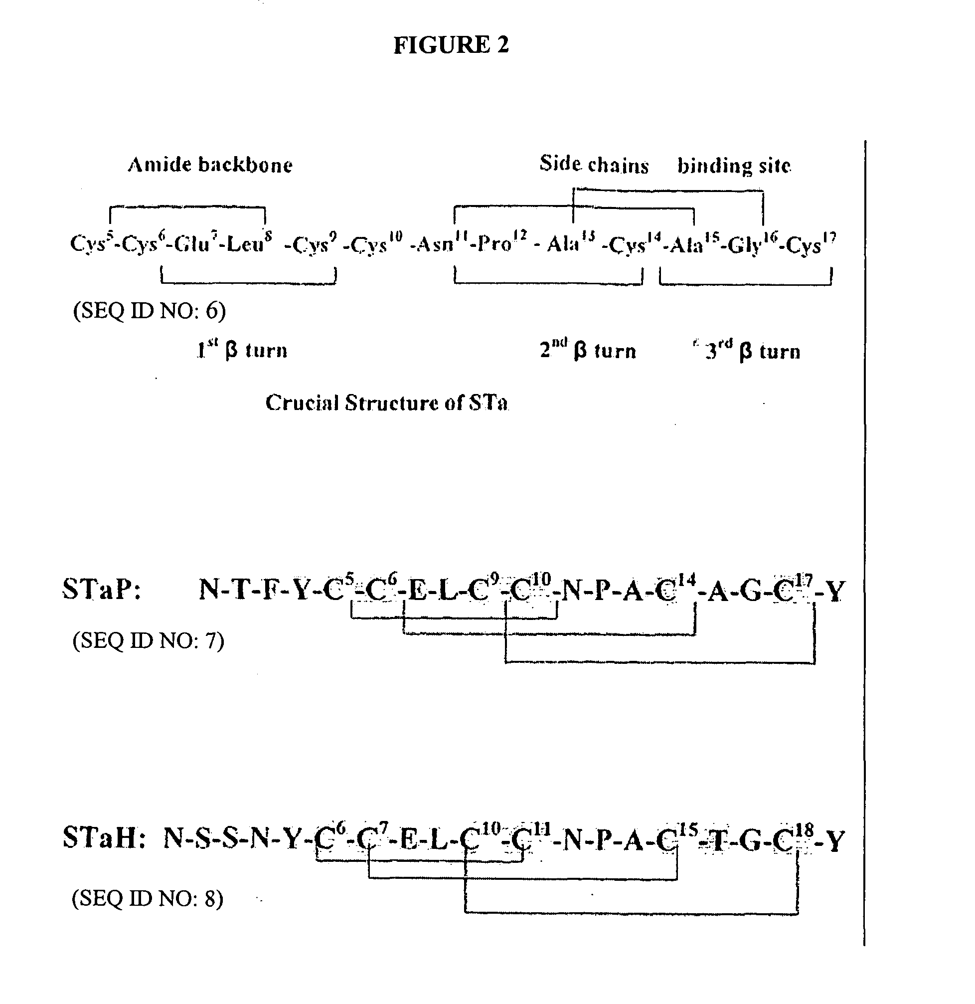
TABLE II No. Toxin and Amino host acids Sequence Reference STaH ETEC 19 N-S-S-N-Y-C-C-E-L-C-C-N-P-A-C-T-G-C-Y Aimoto et al (SEQ ID NO: 13) 1982 STaP ETEC 18 N-T-F-Y-C-C-E-L-C-C-N-P-A-C-A-G-C-Y Takao et al (SEQ ID NO: 12) 1983 STa ETEC 18 N-T-F-Y-C-C-E-L-C-C-N-P-A-C-A-G-C-Y Saeed et al (bovine) (SEQ ID NO: 11) 1984 Citrobacter 18 N-T-F-Y-C-C-E-L-C-C-N-P-A-C-A-G-C-Y Guarino et al Freundii (SEQ ID NO: 10) 1989 Yersinia 30 S-S-D-Y-D-C-C-D-Y-C-C-N-P-A-C-A-G-C Takao et al enterocolitica (SEQ ID NO: 9) 1985 V. cholera 17 I-D-C-C-E-I-C-C-N-P-A-C-F-G-C-L-N Yoshimura et al Non-O1 (SEQ ID NO: 19) 1986 V. cholera non- 18 L-I-D-C-C-E-I-C-C-N-P-A-C-F-G-C-L-N Arita et al O1 Hataka (SEQ ID NO: 18) 1991 V. mimicus 17 I-D-C-C-E-I-C-C-N-P-A-C-F-G-C-L-N Arita et al (SEQ ID NO: 17) 1991 E. coli EAST-1 38 ...A-S-S-Y-A-S-C-I-W-C-T---T-A-C-A-S-C-H-G Savarino et al (SEQ ID NO: 16) 1993 Conus 13 E-C-C-N-P-A-C-G-R-H-Y-S-C Gray et al geographus 13 (SEQ ID NO: 15) 1981 Guanylin 15 P---G-T-C-E-I-C-C-AY-A-A-C-T-G-C Greenberg et al (human) (SEQ ID NO: 14) 1997
TABLE III Base Primer Sequence (5-3) pair Reference (SEQ ID NO: 1) STa 5′-TCC GTG AAA CAA CAT GAC GG-3′ 224 Salvadori et al. (SEQ ID NO: 2) 5′-ATA ACA TCC AGC ACA GGC AG-3′ 2003 (SEQ ID NO: 3) STl 5′-TTA ATA GCA CCC GGT ACA AGC AGG-3′ 127 Olsvick and (SEQ ID NO: 4 5′-CTT GAC TCT TCA AAA GAG AAA ATT AC-3′ Strockbine 1993
TABLE IV Step Temperature (° C.) Time (min) Pre-denature 95 5 Denature 95 1 Annealing 60 1 Extension 72 1 Final extension 72 10 Storage 4 24 hours Number of PCR cycles 29 before storage
TABLE V Amount/ # of Amount/two rxns Component sample (μl) Samples Volume (μl) 10× buffer A 2 2.5 5 dNTP (10 mM) 0.4 2.5 1 Sta-F (20 μM) 0.4 2.5 1 Sta-R (20 μM) 0.4 2.5 1 25 mM MgCl2 0 0 0 Fisher Taq 0.1 2.5 1 polymerase Ultrapure water 14.7 2.5 36.75 Total volume 20 45 Template 2
TABLE VI NaCl 2.52 Na2SO4 0.14 Na acetate 10.00 MgSO4 0.05 K2HPO4•3H2O 8.12 MnCl2 1% 0.5 ml Asparagine 5.00 FeCl3 1% 0.5 ml
TABLE VII Protein Sp Ac Volume Total Conc/ MU × MED Purification Step ml Titer MU/107 mg 103/mg ng fold Cell Free 30 × 103 10−2 3 24,660 1.22 822 1 Filtrate Amberlite 120 10−4 1.2 1,378 8.70 114.9 7.13 XAD-2 BAC Acetone 60 10−5 6 676.8 88.7 11.28 72.67 Fractionation 60% MCl-gel F 30 10−5 3 267.6 112.10 8.92 91.88 RP-HPLC 80 10−6 80 90.4 8849.56 0.113 7253.28
TABLE VIII SuBSA/DMF HSBSA/PB SuBSA/Imidazole Conventional Method Method Method Method Carrier suBSA HS-BSA suBSA SuBSA Cross linker Organic soluble Water soluble carbodiimide Water soluble carbodiimide Water soluble carbodiimide (EDAC) (EDAC) carbodiimide (DCC) (EDAC) Medium N.N. DMF 5 mM PB Imidazole 0.1M MES (Na2HPO4/NaH2PO4) pH 7-9 7 7.2 5 Stating reactants 10 mg STa + 14 mg 5 mg STa + 12 mg HS-BSA 10 mg STa + 3.3 mg suBSA 2 mg STa + 2 mg suBSA + 6.5 mg p- suBSA NP Total STa MU 10 × 106 48 × 104 4 × 10⁶ 3 × 10³ Protein Assay 1.96 mg/ml 0.277 mg/ml 1.8 mg/ml 1.2 mg/ml Conjugation ratio 4-5:1 4:1 — — Conjugation ~52-64% ~26% ~40.6% ~30% efficiency (Lowery protein) Conjugation 100% ~20% 40% 30% efficiency (retained biological activity) Reference Atassi et al 1981 Fuentes et al 2005 Dean et al. 1990 Uptima, 2007
TABLE IX Amount Serial # Name RT pmole 1 Gln 1.78 5.983 2 Glu 20.63 549.781 3 Ser 25.61 147.557 4 Gly 27.42 329.482 5 His 29.29 174.214 6 Thr 30.47 311.339 7 Arg 31.57 219.833 8 Ala 31.886 507.974 9 Pro 33.18 377.778 10 Tyr 37.09 499.248 11 Cys 37.53 509.796 12 Val 37.85 297.137 13 Meth 39.214 38.214 14 Ile 43.65 118.619 15 Leu 44.80 576.721 16 Lys 46.87 333.965 17 Phe 38.22 386.2 18
TABLE X # of Total # of residue # of residue/ residue/ in STa-suBSA one BSA one STa Conjugation Residue conjugate sample molecule molecule Ratio Threonine 311.339/8.00 = 34 residues 1 38.9-34 = 38.9 4-5 Cysteine 509.796/8.00 = 35 residues 6 63.6-35 = 63.6 28.6/6 = 4-5 Leucine 576.721/8.00 = 72 65 residues 1 72-65 = 7 Alanine 507.974/8.00 = 48 residues 2 63.5-48 = 63.5 15.5/2 = 7-8
TABLE XI Gro- serum dilution up 10−3 10−4 10−5 10−6 G1 1.933 ± 0.013 1.923 ± 0.014 1.237 ± 0.568 0.379 ± 0.352 G2 1.964 ± 0.035 1.969 ± 0.031 1.550 ± 0.010 0.374 ± 0.173 G3 1.936 ± 0.006 1.656 ± 0.035 0.354 ± 0.015 0.040 ± 0.010 Base- 0.864 ± 0.318 0.232 ± 0.155 0.033 ± 0.048 0.039 ± 0.060 line
TABLE XII Anti-STa Response Neutralized Neutralization ELISA STa MU/ml Protein Assay Specific Bleeding Titer serum (Titer) mg/ml Activity Baseline 0 0 64.5 — Week 3 PI 0 0 66.8 — Week 6 PI 0 0 65.7 — Week 12 PI 10,000 2000 83.5 23.95 Week 15 PI 100,000 15,000 69.6 215.52 Week 20 PI 1,000,000 20,000 65.00 307.69 Week 24 PI 1,000,000 20,000 65.00 307.69 Week 28 PI 1,000,000 30,000 65.00 461.54
TABLE XIII Week Baseline Week 3 Week 6 12/17 Week 24 Avidity Index (A1) % after measurement OD at 405 nm Group 1 0.017 0.043 0.001 0.578 0.927 (4.70%) (8.91%) (0.03%) (25.94%) (48.21%) Group 2 0.043 0 0.022 0.027 0.762 (3%) (0%) (2.24%) (1.53%) (38.71%) Group 3 0.04 0 0.095 0.151 0.352 (0%) (0%) (11.36%) (12.05%) (21.30%)
TABLE XIV Neutralization Maximum Amount of capacity References neutralization titer SMU Total MU/ml serum Lowenadler et al. 1:55 30 1665 1991 Lockwood and 1:100 8 800 Robertson 1984 Frantz and 1:5000 to 1:10,000 1 5000 to 10,000 Robertson 1981 Alderete and 1:2,500 1 2,500 Robertson 1978 Pereira et al. 1:50 for 1 50 2001 immunogene using native STa fusion protein 1:4000 using 1 4,000 mutated STa fusion protein This study 1:30,000 1 30,000
TABLE XV STa neutralization Bird. G. weight/remaining Neutralization Avidity No. B. Weight Score Index (%) 1 0.073 ++ 76.44 2 0.078 + 66.70 3 0.056 ++++ 91.24 4 0.065 +++ 85.58 5 0.066 +++ 79.59 6 0.064 +++ 52.79 7 0.061 +++ 96.46 8 0.068 +++ 93.60 9 0.055 +++ 59.60 10 0.073 ++ 82.28 11 0.073 ++ 54.72 12 0.072 ++ 86.16 13 0.066 +++ 106.17 14 0.066 +++ 48.74 15 0.076 ++ 87.35 16 0.063 +++ 76.83 17 0.070 +++ 69.35 18 0.067 +++ 88.23 19 0.077 ++ 71.59 20 0.082 + 80.42 21 0.073 ++ 74.89 22 0.083 −/+ 70.72 23 0.085 −/+ 85.34 24 0.081 + 69.84
Login to view more PUM
Property | Measurement | Unit |
Temperature | 95.0 | °C |
Volume | 1.981E-15 | L |
Volume | 2.005E-15 | L |
tensile | MPa | |
Particle size | Pa | |
strength | 10 |
Description & Claims & Application Information
We can also present the details of the Description, Claims and Application information to help users get a comprehensive understanding of the technical details of the patent, such as background art, summary of invention, brief description of drawings, description of embodiments, and other original content. On the other hand, users can also determine the specific scope of protection of the technology through the list of claims; as well as understand the changes in the life cycle of the technology with the presentation of the patent timeline. Login to view more.
Login to view more Similar technology patents
Aliphatic polycarbonate macropolyol and aliphatic polycarbonate-co-aromatic polyester macropolyol
InactiveUS20160177027A1Low molecular weightIncrease possibilityPolyurea/polyurethane coatingsPolycarbonatePolyester
Owner:LOTTE CHEM CORP
Failure time estimation device, machine learning device, and failure time estimation method
ActiveUS20200282719A1High accuracyIncrease possibilityMachine learningOther printing apparatusImage representationMachine learning
Owner:SEIKO EPSON CORP
Delivery support system and delivery support program
PendingUS20210073726A1Increase possibilityCharacter and pattern recognitionPackagingEmbedded systemComputer hardware
Owner:AISIN CORP
Vehicle control device
PendingUS20220306111A1Increase possibilityScene recognitionExternal condition input parametersDriver/operatorVehicle control
Owner:TOYOTA JIDOSHA KK +1
Substrate processing apparatus and method of fabricating same
PendingUS20220139680A1Improve durabilityIncrease possibilityElectric discharge tubesSemiconductor/solid-state device manufacturingStructural engineeringElectrical and Electronics engineering
Owner:SEMES CO LTD
Classification and recommendation of technical efficacy words
- Increase possibility
Method, apparatus and system for directing access to content on a computer network
InactiveUS20010001863A1Increase possibilityCathode-ray tube indicatorsMultiple digital computer combinationsTraffic volumeTraffic capacity
Owner:INTELLECTUAL VENTURES I LLC +1
Resource allocation
ActiveUS20100069081A1Increase possibility of correct demodulationIncrease possibilityAssess restrictionOrthogonal multiplexData channelControl channel
Owner:NEC CORP
Image processing apparatus, image processing method, program, and recording medium
InactiveUS20070127785A1Increase possibilityAcquiring/recognising facial featuresImaging processingPrincipal component analysis
Owner:SONY CORP
Braking/driving control apparatus for vehicle
ActiveUS20110307154A1Reduce possibilityIncrease possibilityAnalogue computers for trafficAutomatic initiationsControl theoryAcceleration Unit
Owner:ADVICS CO LTD +1
Strength and power training system
ActiveUS7104936B2Increase possibilityReliable arrangementJumping apparatusWeightsStrength trainingGravitational force
Owner:KARLSTROM DAVID D