[0015]The present invention generally relates to a high-
throughput, high content screening (HCS)
cytometry device based on image-analysis and sorting of particles (i.e. cells) based on particle (i.e.
cell)
phenotype. Thus, the present invention relates to a high-
throughput, high content
cytometry screening for phenotypic sorting of particles (i.e. cells) in a sample. The methods and compositions described herein combine desirable features of
flow cytometry (FACS) and
microscope-based high-content screening (HCS) to permit sorting of particles based on phenotypic characteristics such as
asymmetry, shape,
fluorescence or
dye uptake / exclusion, and
intracellular information (e.g.,
organelle shape and area). The present invention further permits
intracellular localization assays to be performed, is
highly sensitive to detection of rare cells, and can be used for time-synchronized sampling, which is not possible using FACS or HCS alone. The present invention is advantageous in that it allows identification and distinguishing of particles, e.g., cells based on gross morphological characteristics using
line scan image analysis with a resolution which is high enough for distinguishing characteristics, but is of a resolution which does not significantly slow down the analysis, allowing for high throughput, high-content screening. One of the problems with 96- and 384-well plate-reader and plate-based
image analysis systems is that they are static systems, requiring time for the focusing of beam for accurate and clear image capturing. In contrast, the present
system, methods and apparatus performs image analysis on particles, e.g., cells, as they move or flow through a microchannel, therefore all particles, e.g., cells, are in focus allowing for rapid image capturing and accurate reading. Additionally, the present system, method and apparatus allows the particle, e.g., cell to be in the correct morphology, and reduces issues with
cell aggregation which can be an issue with static 96- and 384-well plate-reader and plate-based image analysis systems.
[0025]Unlike FACS sorting, which can only sort cells from a
single sample at one time and which requires cells from different samples to be sorted in sequential or subsequent FACS
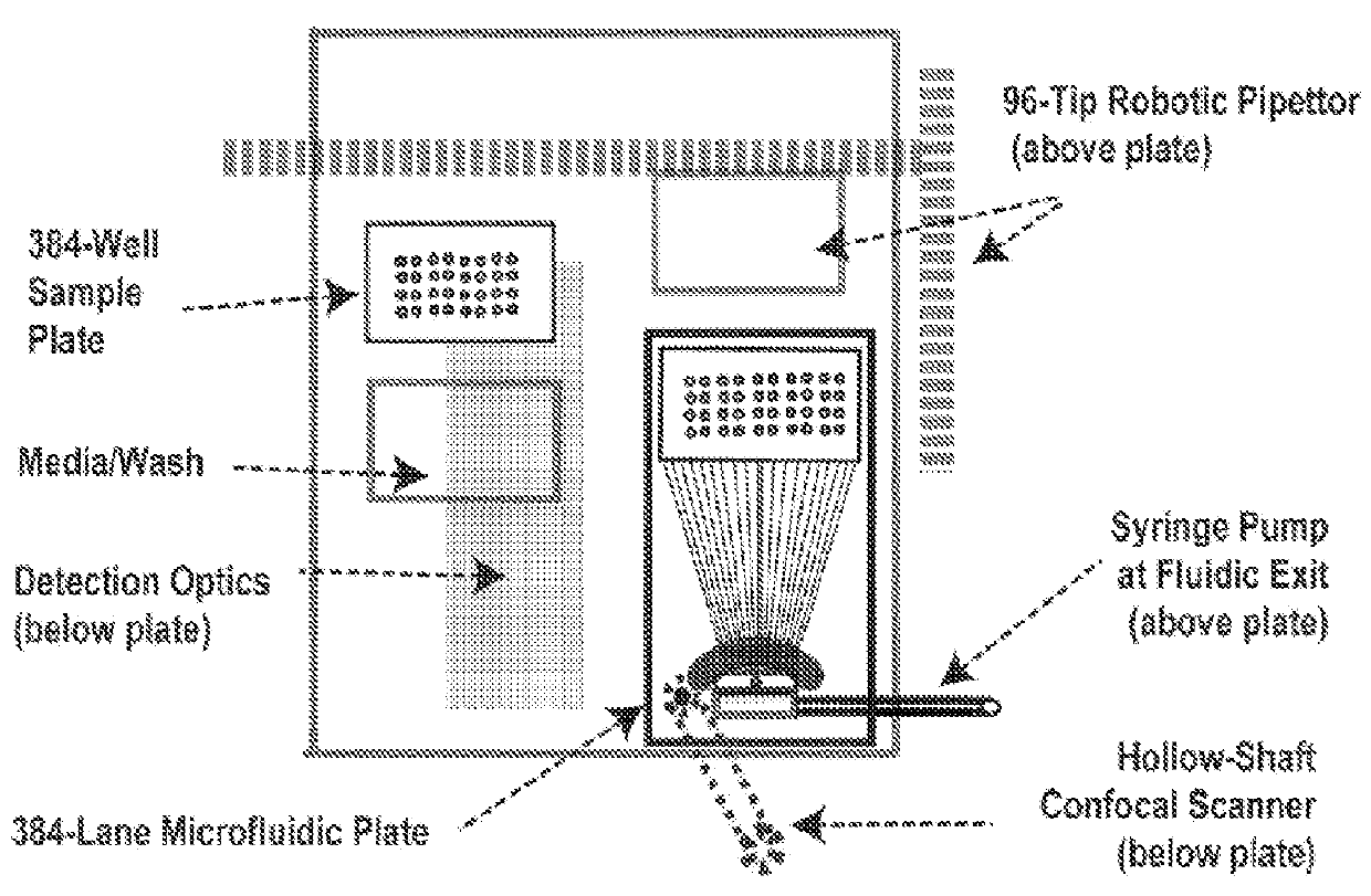
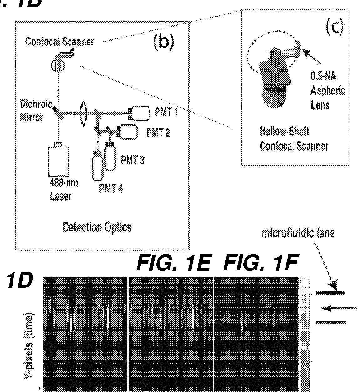
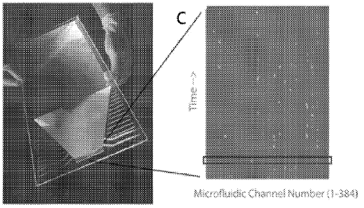
processing step, the present methods, apparatus, devices and composition allows for sorting of multiple different samples simultaneously, in real-time. As shown in FIG. 1, FIG. 2, and FIG. 18, in some embodiments, the present methods, apparatus, devices and composition allows for image analysis and subsequent sorting of 384 different samples simultaneously. In some embodiments, the multichannel architecture of the microfluidic device of the line-scan device as disclosed herein can be configured for line-image scanning of any number of different or unique samples. In some embodiments, the number of samples which can be line-scan imaged simultaneously can be about at least 16, or at least about at least 32, or at least about 96, or at least about 384, or at least about 500, or between about 500-1000, or multiples of 384, e.g., 768, or about 1152, or about 1536, or about 1536, or about 1920, or about 2304 etc. In some embodiments, the number of samples which are processed simultaneously is determined by one of ordinary skill in the art and can be determine in part, by the sample capacity of commercially available and conventional microwell plates (e.g., 16-well, 32-well, 96-well, 384-well plates etc). In another embodiment, one can scan a “sheet” of samples, and automate the process to prepare a new sheet of samples which can be simultaneously scanned or processed.
[0035]In some embodiments of the line-scan imager device, a large
laser spot size is used (e.g., 30×100 microns, or 30×30 microns). Such a large laser spot size can be used for high throughput sorting of larger cells, for example, based on
color ratio measurements without the need to resolve intracellular and
internal cell structure, and can be used, for example, for a high sensitivity to identify rare cells. In some embodiments of
line scan imaging device, a larger spot size will decrease the focus and
clarity of the image, but can be useful to identify larger cells and / or rare cells, and / or to distinguish cells on the basis of
light scattering, or the ratio of two or more different fluorescent markers. In some embodiments, the spatial resolution spot size can be determined by the user to identify a
cell type of a specific size. For example, in some embodiments, the spatial resolution spot size can identify cells within the range of 1-50 μm, for example a spatial resolution spot size of 3.5 μm can be used to identify cells of about 10 μm, or a spatial resolution spot size of about 4-5 μm in can be used to identify a
yeast cell of about 6 μm in
diameter, or a spatial resolution spot size of about 1-3.5 μm can be used to identify cells about 1 μm, such as bacterial cells, or a spatial resolution spot size of about 3.5-5 μm can be used to identify cells about 50 μm such as
heart cells and neurons. In some embodiments, the spatial resolution spot size can be adjusted accordingly to identify cells of about 3-15 μm in
diameter, or between about 3-20 μm in
diameter, or between about 1-10 μm in diameter, or cells about 20-50 μm in diameter, or cells greater than about 50 μm in diameter. In some embodiments, the spatial resolution spot size is adjusted so it allows enough resolution of the cell to see markers and / or internal structures of the cell and resolve details inside the cells yet being quick enough for quick
image capture for
high throughput analysis.
[0036]In some embodiments, the
detector for capturing the line-scan images is configured to capture images of the particle, e.g., a cell as it flows through a microchannel. Surprisingly, as the particles are in the microchannel, each particle, e.g., cell is always in focus. Accordingly, no time is needed for focusing the
image detector in the present system, which is a significant
advantage over plate-reader image analysis systems, where cells in a plate microwell are not in focus and time is required for focusing the
image detector for clear
image capture. In some embodiments, the line-scan imaging device comprises a microfluidic device comprising a plurality of microchannels, wherein each microchannel of the plurality of microchannels is at least 50 μm wide in diameter, or at least 100 μm wide in diameter. In some embodiments, the diameter of each microchannel (e.g., depth) is between 30 and 300 μm (e.g., between 30-175, 30-150, 30-125, 30-100, 30-75, 30-50, 30-40, 50-75, 50-100, 50-125, 50-150, 50-175, 50-200, 75-200, 100-200, 125-200, 150-200 or 175-200 μm, or about 200-300 μm). In some embodiments, the diameter of the width of a microchannel is not greater than 300 μm wide in diameter. In some embodiments, the microchannel diameter is configured so that the
detector does not need to focus to accurately and clearly capture the particles, e.g., cells as they flow the microchannel, or so that the
detector need only a narrowly-adjusted focus for focusing on particles as they flow through the microchannel.
[0040]In some embodiments, each microchannel of the plurality of microchannels on the microfluidic device is configured for focusing, such as vertical
hydrodynamic focusing. In some embodiments, vertical
hydrodynamic focusing is achieved by using multiple crossing junctions of each microchannel to flow into a single analytical microchannel, where a single analytical microchannel is a scanning window or analytical portion of the microchannel. In some embodiments, the vertical
hydrodynamic focusing locates cells to a zone of 20 μm or smaller (e.g., less than 18, less than 16, less than 14, less than 12, less than 10, less than 8, less than 6, less than 4, or less than 2 μm) in the direction of the optical focus. In some embodiments, vertical hydrodynamic focusing allows 4 μm to be detected with a 40×
microscope objective.
[0045]In some embodiments, the comparison module is configured to analyze at least one line-scan image or a plurality of line scan images from each cell by comparison with a
reference line scan image, and wherein if the line-scan image of the cell has substantially similar cell features, such as those in Table 1, as a
reference line scan, the comparison module sends instructions to the microfluidic device to trigger or de-select switching of the piezoelectric microswitch in the microchannel in which the cell was scanned, enabling selection of the cell.

