
Signal amplified biological detection with conjugated polymers

a polymer and conjugated technology, applied in the field of ultra sensitive detection methods for biomolecules, can solve the problems of limiting the conformation and adaptation ability of polymers to the shape of biologically-derived molecules, deleterious fluorescence self-quenching, and rigid rod structure of polymers, so as to reduce non-specific assay interactions and improve water solubility

Active Publication Date: 2012-10-04
NAT UNIV OF SINGAPORE +1
View PDF1 Cites 73 Cited by
  • Summary
  • Abstract
  • Description
  • Claims
  • Application Information

AI Technical Summary

Benefits of technology

[0008]The present invention relates to the design of novel CP materials adapted for biological detection in a variety of formats including immunoassays (flow cytometry, immunohistochemisry, ELISA, lateral flow, protein arrays, etc.) and nucleic acid assays (arrays, PCR, FISH, etc.). These include the incorporation of covalent conjugation sites and revised modifications to improve water solubility and reduce non-specific assay interactions.
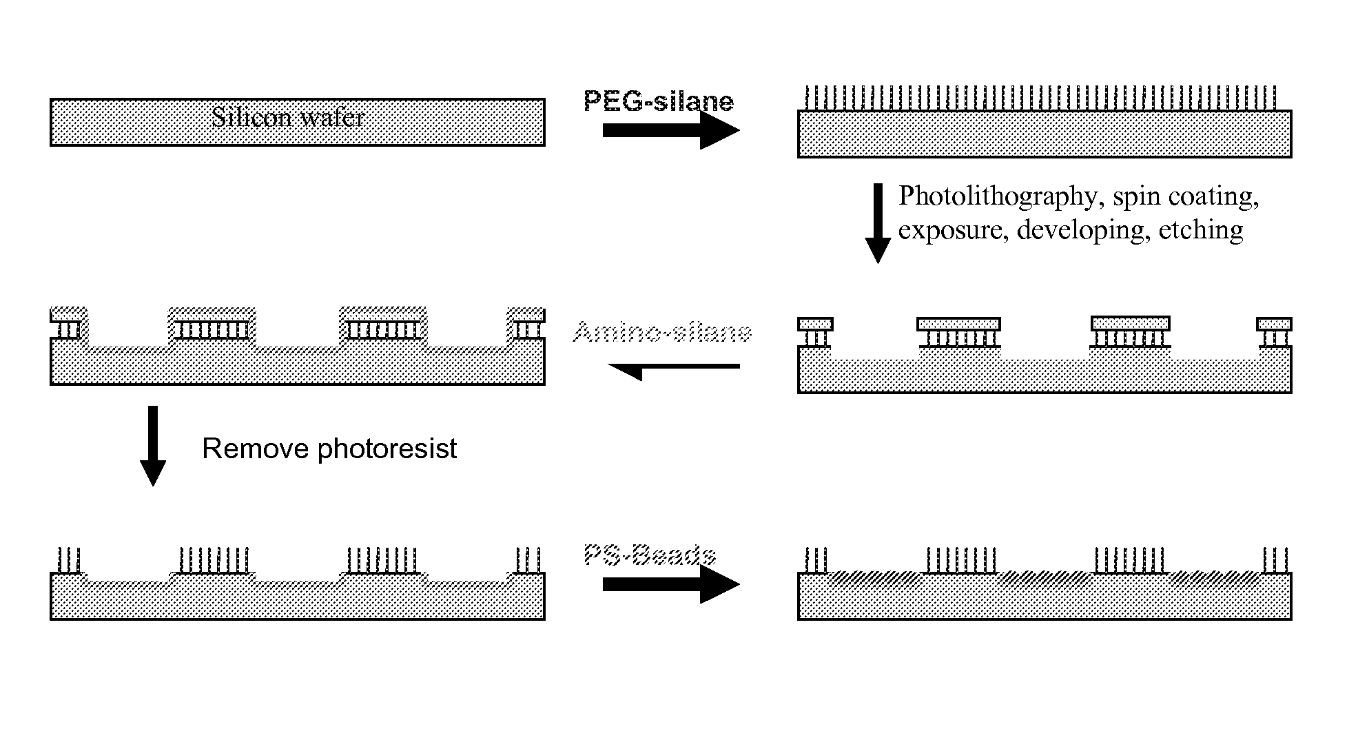
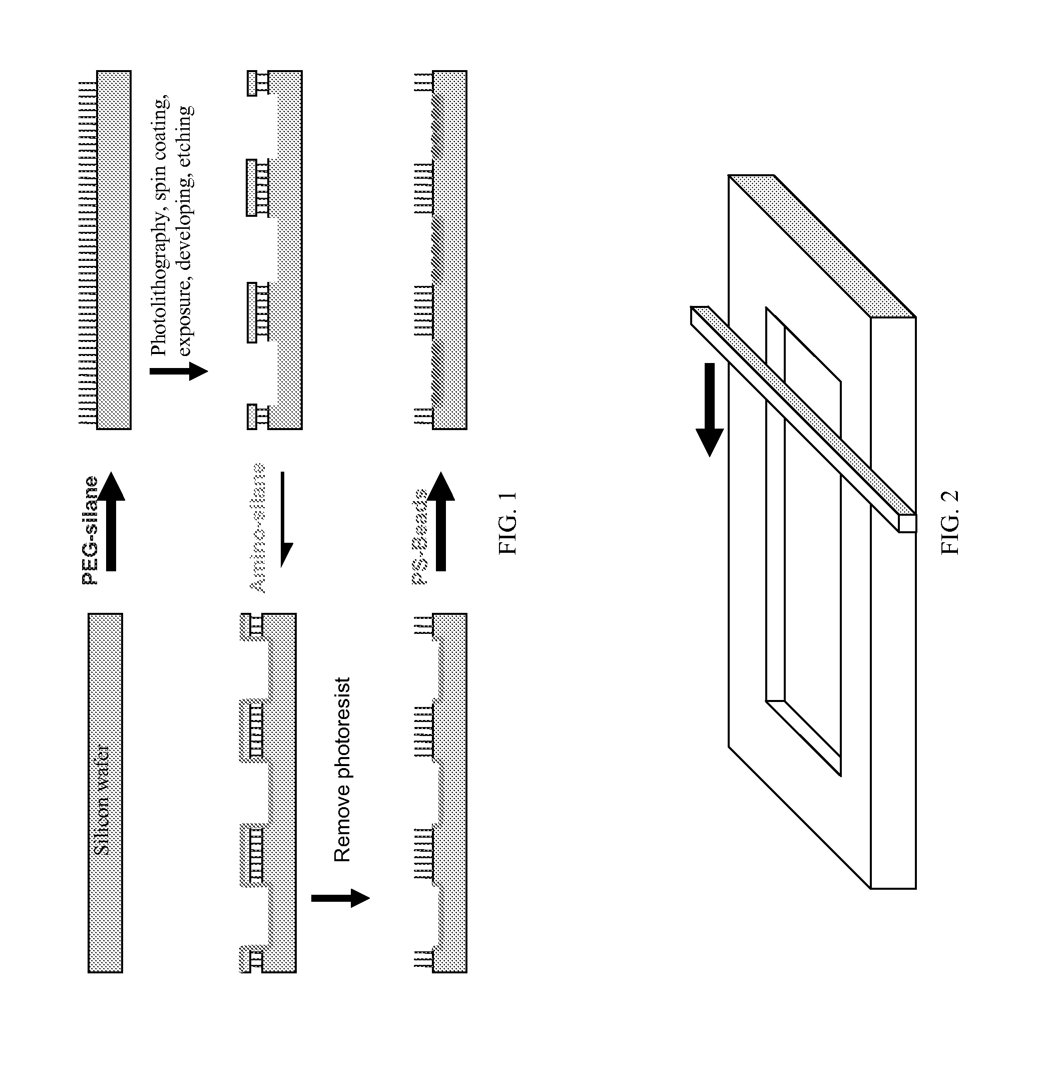
[0009]In some embodiments, a method for detecting nucleic acids with oligonucleotides attached to polystyrene (PS) beads and the application of conjugated polymers (CPs) as the signal reporter or signal amplifier is presented. The method provided by the present invention combines the micro-patterning of polystyrene beads in pre-fabricated microwells with conjugated polymers for label-free DNA detection. In addition, the developed method could also be used for labeled targets where the polymers are used to amplify the fluorescence signal from the label through fluorescence resonance energy transfer (FRET). The method of the present invention and products produced via this method will allow real-time detection of trace amount of nucleic acid, protein and other biomolecules with high sensitivity.
[0010]An objective of this invention is to develop an ultra sensitive detection method for DNA and other biomolecules. One of the key factors is to improve the specific detection sensitivity from the recognition event. In an array format this means increased signal output from the captured target (DNA, protein, analyte, etc.) and the reduced background signal. One embodiment of the present invention focuses on solving both problems simultaneously. First, patterned polystyrene beads were used instead of conventional planar microarrays. The patterned polystyrene beads provide high surface areas for immobilization of the probe DNA and therefore enable increased number of target binding to the capture DNA, which results in high signal output for both label-free and dye-labeled assays, upon addition of polymers. To reduce the background signal, new polymers have been designed that are compatible with the microarrays and have low cross talking between the excitation and emission wavelengths. In addition, these polymers are designed to match the excitation wavelength of the existing instruments.
[0013]Numerous examples exist which utilize non-specific interactions of CPs and biomolecules for detecting a biological recognition event (i.e. electrostatic interactions). Such methods are often indirect and control of unwanted interactions can be difficult. By conjugating directly to the biological probe the specificity of the assay is retained in the actual biological recognition event of interest, resulting in a more direct reporting event with the benefit of the collective optical properties of the CP. This further requires adaptation of the CP materials to ensure the polymers do not interact non-specifically with other components in the assay, allowing the specificity of the biomolecule to drive the interaction. New polymers were thus designed to improve solubility and reduce non-specific electrostatic interactions.

Problems solved by technology

Conjugated polymers frequently take the form of rigid-rod structures which have limited flexibility and consequently have a limited ability to adapt to particular three dimensional shapes, thus limiting their ability to conform to the shape of biologically-derived molecules.
A consequence of this torsional restriction is that the polymer has a “rigid rod” structure with limited conformations and ability to adapt to the secondary structures of bio-molecules.
Additionally, when cationic conjugated polymers are used as light-harvesting molecules, they can deleteriously exhibit fluorescence self-quenching when they cluster near negatively charged biomolecules.

Method used


Image

  • Signal amplified biological detection with conjugated polymers
  • Signal amplified biological detection with conjugated polymers
  • Signal amplified biological detection with conjugated polymers

Examples


example 1

Synthesis of monomer, 2,7-dibromo-9,9-di(2′,5′,8′,11′,14′,17′,23′,26′,29′,32′,35′-dodecaoxaoctatriacontan-38′-yl)fluorene (A) for subsequent polymerization

[0201]

Step 1: 2,7-dibromo-9,9-di(3′-hydroxypropanyl)fluorene

[0202]2,7-dibromofluorene (9.72 g, 30 mmol), tetrabutylammonium bromide (300 mg, 0.93 mmol), and DMSO (100 mL) were added to a 3-neck flask under nitrogen (g), followed by the addition of 50% NaOH (15 mL, 188 mmol) via syringe. The mixture was heated to 80° C., and 3-bromopropanol (6.70 mL, 77 mmol) was added dropwise via addition funnel, and the reaction mixture was stirred at 80° C. for another 2 hours. Upon completion, the mixture was cooled to room temperature and quenched with water (250 mL). The aqueous layer was extracted with ethyl acetate (3 150 mL portions). The organic layers were combined, washed with water, then dried over MgSO4, and filtered. The solvent was removed and the residual was recrystallized in chloroform to yield pale yellow needle crystals (9.20 ...

example 2

Synthesis of a branched monomer, 2,7-dibromo-9,9-bis(3,5-(2,5,8,11,14,17,20,23,26,29,32-undecaoxatetratriacontan-34-yl)benzyl)-9H-fluorene, for subsequent polymerization

[0205]

Step 1: 2,7-dibromo-9,9-bis(3,5-dimethoxybenzyl)-9H-fluorene

[0206]2,7-dibromofluorene (4.16 g, 12.8 mmol) and tetrabutylammonium bromide (362 mg, 1.12 mmol) were added to a round bottom flask charged with a Teflon stirbar. Next, 60 mL of dimethylsulfoxide was added to the flask and the mixture was stirred for 5 minutes. A portion of 50% NaOH aqueous solution (5.2 mL) was added followed immediately by 3,5-dimethoxybenzyl bromide (7.14 g, 31 mmol). Over the course of 2 hours the solution changes color from orange to blue. The reaction is stirred overnight. The resulting mixture is slowly poured into 200 mL of water and then extracted with three 100 mL portions of dichloromethane. The organic layers are combined and dried over magnesium sulfate and then filtered. The crude product is purified by column chromatogra...

example 3

Synthesis of a Neutral Base Polymer

[0209]

[0210]Polymerization

[0211]2,7-dibromo-9,9-di(2′,5′,8′,11′,14′,17′,23′,26′,29′,32′,35′-dodecaoxaoctatriacontan-38′-yl)fluorene (0.25 mmol), 1,4-divinylbenzene (32.3 mg, 0.25 mmol), palladium acetate (3 mg, 0.013 mmol), tri-ortho-tolylphosphine, (10 mg, 0.033 mmol), and potassium carbonate (162 mg, 1.2 mmol) are combined with 5 mL of DMF in a small round bottom flask charged with a Teflon coated stirbar. The flask is fitted with a needle valve and put in a Schlenk line. The solution is degassed by three cycles of freezing, pumping, and thawing. The mixture is then heated to 100° C. overnight. The polymer can be subsequently reacted with terminal linkers or capping units using similar (in situ) protocols to those provided in Example 5 or by modifying them post polymerization work up as a separate set of reactions.


PUM

PropertyMeasurementUnit
length×width×depthaaaaaaaaaa
pHaaaaaaaaaa
pHaaaaaaaaaa

Abstract

The devices and method are provided for detecting labeled and label-free analytes, such as nucleic acids and proteins, employing conjugated cationic, anionic and neutral polymers. The analytes can be immobilized on a solid support material. The solid support material can be disposed in the pre-fabricated patterns on a substrate. The chemical structures of the polymers employed by the various embodiments of the present invention are described herein.

Description

RELATED APPLICATION[0001]This application claims priority to U.S. Provisional Application No. 61 / 269,685 filed Jun. 26, 2009, which is incorporated herein by reference in its entirety.BACKGROUND OF THE INVENTION[0002]The present invention relates to ultra sensitive detection methods for biomolecules. Methods for the detection of biomolecules such as nucleic acids, proteins and cellular markers are highly significant not only in identifying specific targets, but also in understanding their basic function. Hybridization probe technologies in particular continue to be one of the most essential elements in the study of gene-related biomolecules. They are useful for a variety of both commercial and scientific applications, including the identification of genetic mutations or single nucleotide polymorphisms (SNP's), medical diagnostics, gene delivery, assessment of gene expression, and drug discovery. Heterogeneous formats for performing such hybridization probe assays have become increas...

Claims


Application Information

Patent Timeline
Patent Type & Authority Applications(United States)
IPC IPC(8): C08G65/00
CPCC08G61/02C08G2261/5222C08G2261/3422C08G2261/3142
Inventor LIU, BINWANG, CHUNZHAN, RUOYUBARTHOLOMEW, GLENN P.HONG, JANICE W.WHEELER, JEAN M.GAYLORD, BRENT S.
Owner NAT UNIV OF SINGAPORE
Learn More