Microgels for Encapsulation of Cells and Other Biologic Agents

a bioactive agent and microgel technology, applied in the field of microgels for encapsulation of cells and other biologic agents, can solve the problems of limited control of local cellular environment via incorporation of bioactive molecules (e.g., adhesive peptides), significant complexity of microfluidic devices, and translation of this work into covalent crosslinking of microgels within microfluidics, etc., to achieve the effect of enhancing cell adhesion and survival

Active Publication Date: 2015-03-12
GEORGIA TECH RES CORP
View PDF0 Cites 31 Cited by
  • Summary
  • Abstract
  • Description
  • Claims
  • Application Information

AI Technical Summary

Benefits of technology

[0017]In some embodiments, the macromer is functionalized prior to crosslinking In a particularly preferred embodiment, this is accomplished by reacting a bioactive active agent with a thiol group (e.g., a protein or peptide with cysteine) with a maleimide on the macromere prior to crosslinking In some examples, the functionalize moiety is a peptide including an RGD ligand for integrins to enhance cell adhesion and survival. In some embodiments, the bioactive agent is a therapeutic drug which can be released from the microgel by a protease or other enzymes in vivo.

Problems solved by technology

Control, 170:1 (2013)), even though the ability to control local cellular environment via incorporation of bioactive molecules (e.g., adhesive peptides) is limited.
However, translating this work into covalently crosslinking of microgels within microfluidic devices adds significant complexity because polymer precursors must be liquid while flowing through the focusing nozzle, but droplets must crosslink rapidly after being generated to prevent them from merging.
However, even for synthetic polymer encapsulation, control of cellular microenvironment by functionalization of polymers with bioactive molecules remains a significant challenge.
Most of these schemes require crosslinking using UV-based free radical polymerization, resulting in potentially cytotoxic effects on encapsulated cells.
Although cell encapsulation in synthetic microgels crosslinked without free radicals has been reported, the polymer cannot easily be functionalized with bioactive molecules (Rossow, et al., Journal of the American Chemical Society, 134(10):4983-9 (2012), Kesselman, et al., Small, 8(7):1092-8 (2012)).
This major limitation makes the maintenance of cells requiring adhesive ligands for viability and function difficult.
Microfluidic encapsulation of large clusters of cells, such as human islets, is more challenging than single cell encapsulation, because the larger particles tend to clog microfluidic channels.

Method used


Image

  • Microgels for Encapsulation of Cells and Other Biologic Agents
  • Microgels for Encapsulation of Cells and Other Biologic Agents
  • Microgels for Encapsulation of Cells and Other Biologic Agents

Examples


example 1

Design and Testing of a Microfluidic Strategy for Encapsulating Cells and other Biological Agents

Materials and Methods

[0121]Microfluidic Device Preparation

[0122]PDMS microfluidic flow focusing devices were cast using soft lithography from silicon and SU8 masters that were fabricated by the Stanford Microfluidics Foundry. Devices with 300 μm nozzles were bonded directly to glass slides after treatment with air plasma. 600 μm nozzle devices were manufactured by first bonding mirror-image PDMS channels, each with 300 μm depth, together to create a channel with 600 μm depth.

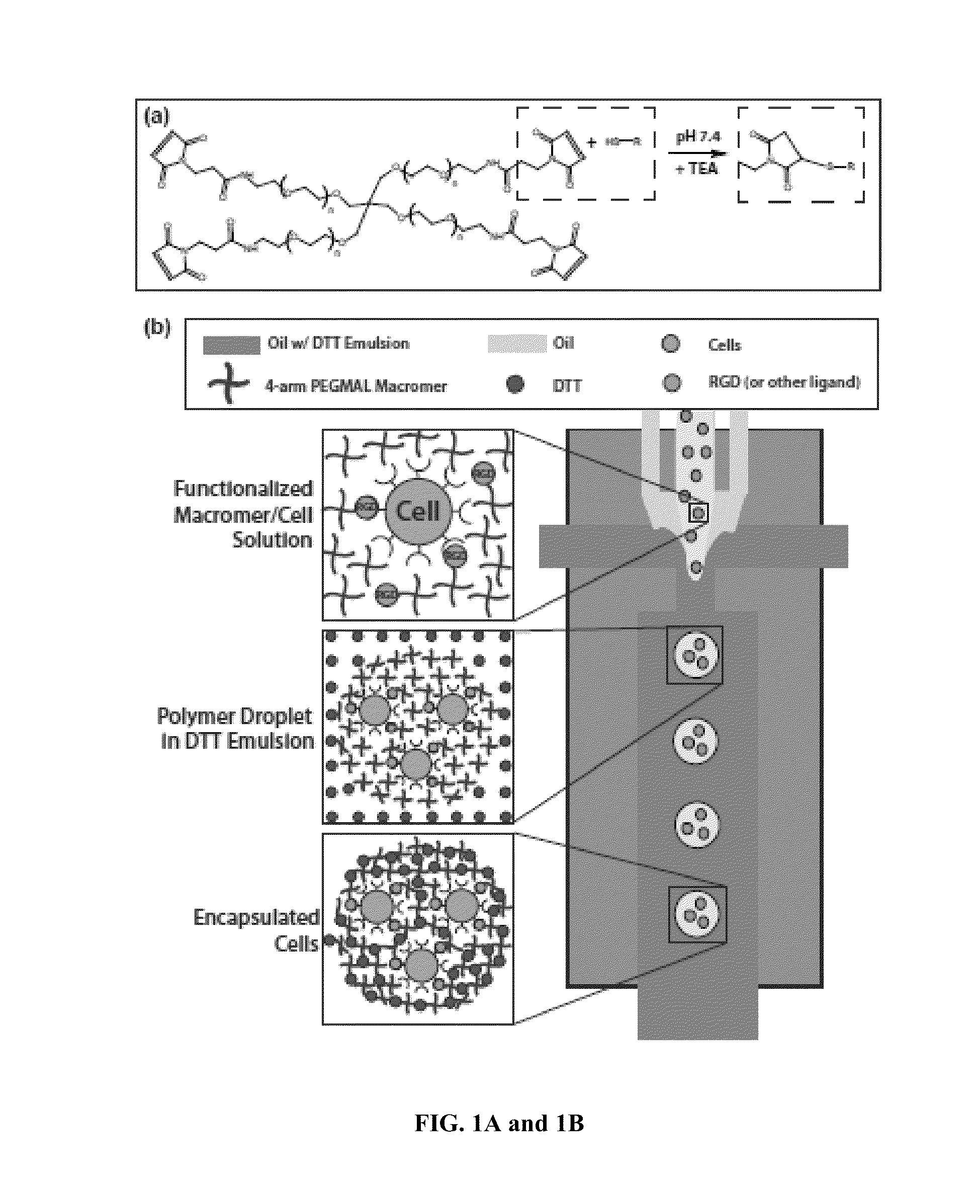
[0123]PEG-4MAL Microgel Formation and Particle Encapsulation

[0124]Flow-focusing microfluidic geometry was utilized to form polymer droplets. Both shielding and crosslinker phases consisted of light mineral oil (Sigma) with 2% SPAN80 (Sigma). The crosslinker phase also contained an emulsion, at a ratio of 1:15, of 20 mg / mL dithiothreitol (DTT) (Sigma) in PBS. A co-flowing shielding phase protected the macromer solutio...

example 2

Size and Dispersity of the Microgels are Tunable Materials and Methods

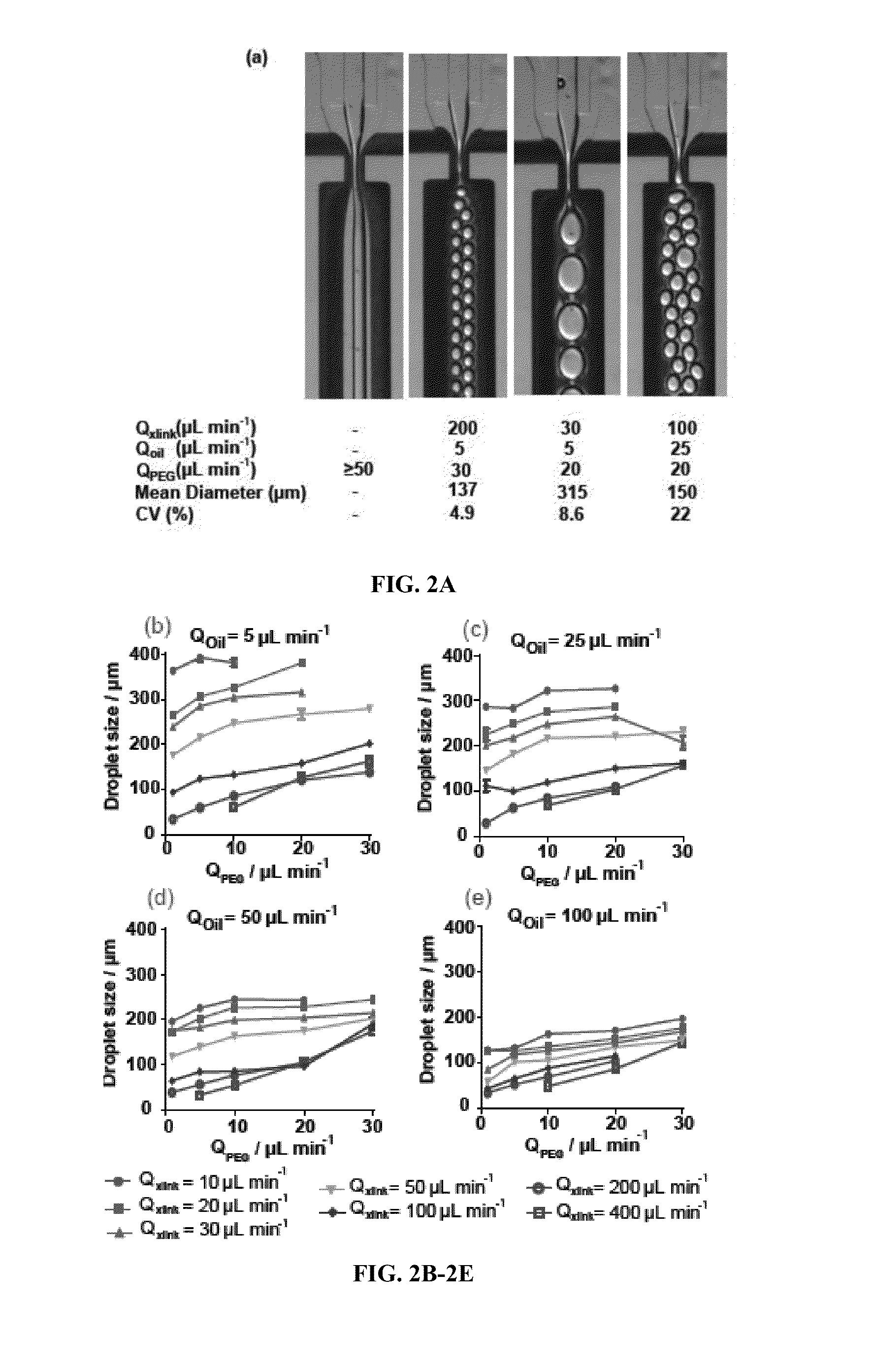
[0127]Microgel Size Control

[0128]To characterize the relationship between microgel size and the various macromer solution and continuous phase flow rates, hydrogel droplets were generated using computer-controlled syringe pumps, and were measured while still in the microfluidic chip. Harvard Apparatus Elite syringe pumps were computer controlled using FlowControl software to pump inlet solutions at various flow rates. Video was recorded during droplet generation using a Hammamatsu ORCA-ERA 1394 camera connected to a Nikon TE300 microscope. Droplet diameter was measured using ImageJ analysis software. The coefficient of variation (CV) was calculated for each flow rate combination by dividing the standard deviation of the sample by its mean. At least 30 microgels were measured for each flow rate combination.

Results

[0129]Precise control of particle size and monodispersity are critical for many applications of microge...

example 3

Permeability of the Microgels is Tunable

Materials and Methods

[0131]Protein Encapsulation and Release

[0132]AlexaFluor488-labeled IgG (goat anti-rabbit IgG, Life Technologies), bovine serum albumin-AlexaFluor488 conjugate (Life Technologies), 2-NBDglucose (Life Technologies) or insulin (Sigma) tagged with AlexaFluor488 was dissolved in a 5% PEG-4MAL (10 kDa or 20 kDa) solution before being microencapsulated by macromere droplet gelation. To prevent proteins from being crosslinked by the macromer, thiols were capped using aminoethylate reagent (Thermo Scientific) according to product instructions. Particles were washed and resuspended in PBS and divided into 5 replicates containing 2 mL total volume. 50 μL samples were taken of supernatant alone, as well as of supernatant containing well-mixed, protein-laden microgels. These samples were placed in a 96 well plate, and their fluorescent intensity was measured using a Perkin Elmer HTS 7000 plate reader. To generate release curves, supern...


PUM

PropertyMeasurementUnit
sizeaaaaaaaaaa
sizeaaaaaaaaaa
sizeaaaaaaaaaa

Abstract

Methods of encapsulating cargo in a microgel droplet, microgel droplets prepared according the provided methods, and methods of use thereof are disclosed. The methods of preparing cargo-encapsulated microgels generally include flowing through a flow-focusing nozzle of a microfluidic device a macromer phase, an oil phase, and a crosslinker phase to form microgel droplets by oil-water emulsion. The phases are pumped, injected, or flowed through the microfluidic device such that as the macromer phase approaches the flow focusing nozzle, the co-flowing oil phase shields the macromer from contact with the crosslinker phase until flow instability occurs and macromer phase droplets form. After flow instability occurs, the crosslinker diffuses from the crosslinker phase into the droplets in an effective amount to covalently crosslink the macromer into a microgel network encapsulating the cargo in the crosslinked macromer. Microgels prepared according to the disclosed methods and methods of use thereof are also provided.

Description

CROSS-REFERENCE TO RELATED APPLICATIONS[0001]This application claims priority to and benefit of U.S. Provisional Patent Application No. 61 / 875,287 filed on Sep. 9, 2013, and where permissible is incorporated by reference in its entirety.REFERENCE TO SEQUENCE LISTING[0002]The Sequence Listing submitted Sep. 9, 2014 as a text file named “GTRC—6467_ST25.txt,” created on Sep. 9, 2014, and having a size of 1,723 bytes is hereby incorporated by reference pursuant to 37 C.F.R. §1.52(e)(5).FIELD OF THE INVENTION[0003]The invention is generally directed to methods of encapsulating cells and other bioactive agents in microgel droplets, microgel droplet therefrom, and methods of use thereof.BACKGROUND OF THE INVENTION[0004]Hydrogel microencapsulation of cells is a promising strategy for immunoprotection after transplantation. Since the development of alginate-poly-L-lysine encapsulation by Lim and Sun in 1980 (Lim, et al., Science 210(4472):908-10 (1980)), their approach has remained the stand...

Claims


Application Information

Patent Timeline
Patent Type & Authority Applications(United States)
IPC IPC(8): A61K9/50A61K38/38A61K35/28A61K38/28A61K35/39A61K39/44A61K31/7004
CPCA61K9/5089A61K39/44A61K38/385A61K35/28A61K38/28A61K9/5026A61K35/39A61K31/7004A61K9/0024A61K9/5031A61K38/00
Inventor GARCIA, ANDRESHEADEN, DEVONPHELPS, EDWARD A.AUBRY, GUILLAUME A.LU, HANGGARCIA, CRISTINA GONZALEZ
Owner GEORGIA TECH RES CORP
Why Eureka
Try Eureka