High resolution imaging using near-infrared-ii fluorescence

a fluorescence and high-resolution technology, applied in the field of in vivo fluorescence-based optical imaging, can solve the problems of limited choice of nir-ii fluorophores, low fluorescence quantum yield, and no single modality provides adequate spatial and temporal resolution

Active Publication Date: 2015-10-22
THE BOARD OF TRUSTEES OF THE LELAND STANFORD JUNIOR UNIV
View PDF12 Cites 22 Cited by
  • Summary
  • Abstract
  • Description
  • Claims
  • Application Information

AI Technical Summary

Benefits of technology

[0025]In certain aspects, the solubilized nanostructures further comprise fluorophores that are organic small molecules fluorescing in the NIR-II region. The organic small molecules may be IR-1051(6-Chlor-2-[2-[3-[(6-chlor-l-ethyl-2H-benzo[cd]indol-2-yliden)-ethyliden]-2-phenyl-1-cyclopenten-l-yl]-ethenyl]-1-ethyl-benzo[cd]indolium tetrafluoroborate), IR-26 (4-(7-(2-phenyl-4H-1-

Problems solved by technology

However, current methods for assessing vasculature and hemodynamics in small vessels in vivo are suboptimal.
For imaging vascular structures, microscopic computed tomography (micro-CT) and magnetic resonance imaging (MRI) can resolve features down to ˜100 μm with deep penetration, but are limited by long scanning and post-processing time and difficulties in assessing vascular hemodynamics.
Current methodologies for physiological imaging of PADs are suboptimal in that no single modality provides adequate spatial and temporal resolutio

Method used


Image

  • High resolution imaging using near-infrared-ii fluorescence
  • High resolution imaging using near-infrared-ii fluorescence
  • High resolution imaging using near-infrared-ii fluorescence

Examples


examples 1-4

[0061]Described in these examples is the use of biocompatible, brightly fluorescent SWNTs as NIR-II imaging agents. They are shown to be suitable for imaging vascular structures down to ˜30 μm in mouse hindlimb using an epifluorescence imaging method with an indium-gallium-arsenide (InGaAs) imaging system. Compared to micro-CT, NIR-II fluorescence imaging attains a ˜3-fold improvement in spatial resolution. Further, NIR-II imaging allows for differentiation of arteries from veins through principal component analysis (PCA) to obtain dynamic contrast-enhanced imaging. This method can also quantify femoral artery blood flows in both normal and ischemic hindlimbs, and reveal the degree of occlusion due to ischemia. Thus, a single NIR-II imaging modality enables multi-functional imaging capable of accomplishing what is typically done by several traditional techniques including micro-CT, ultrasonography and MRI, making it a unique method that incorporates many desirable features such as h...

examples 5-9

[0066]These examples describe the use of a new NIR-II fluorophore, silver sulfide (Ag2S) QDs for imaging xenograft tumors and vascular structures with high fluorescence quantum yield (15.5%) of NIR-II emission at ˜1200 nm. The present QDs are free of heavy metals of Cd, Hg or Pb, and can be used to detect small tumors down to ·0.1 mm3 and distinguish arterial and venous hindlimb vessels with ˜2 mm depth of penetration

Definitions

[0067]Unless defined otherwise, all technical and scientific terms used herein have the same meaning as commonly understood by those of ordinary skill in the art to which this invention belongs. Although methods and materials similar or equivalent to those described herein can be used in the practice or testing of the present invention, the preferred methods and materials are described. Generally, nomenclatures utilized in connection with, and techniques of, cell and molecular biology and chemistry are those well known and commonly used in the art. Certain ex...

example 1

NIR-I and NIR-II Fluorescence Imaging of Vasculatures

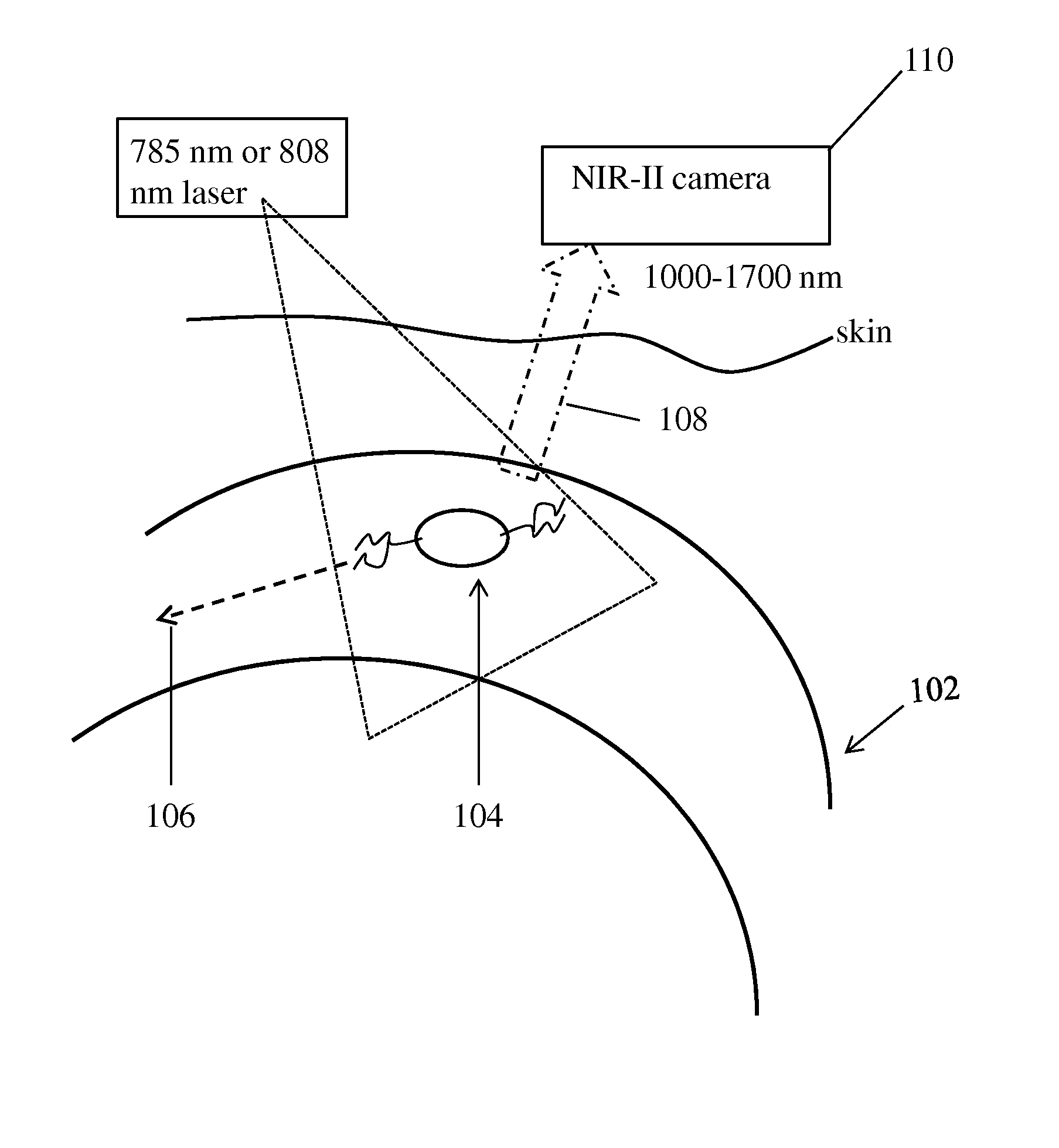
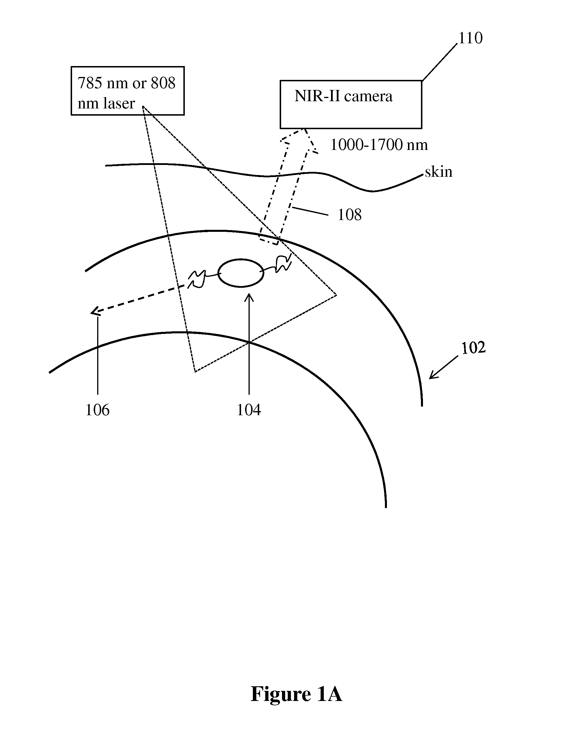
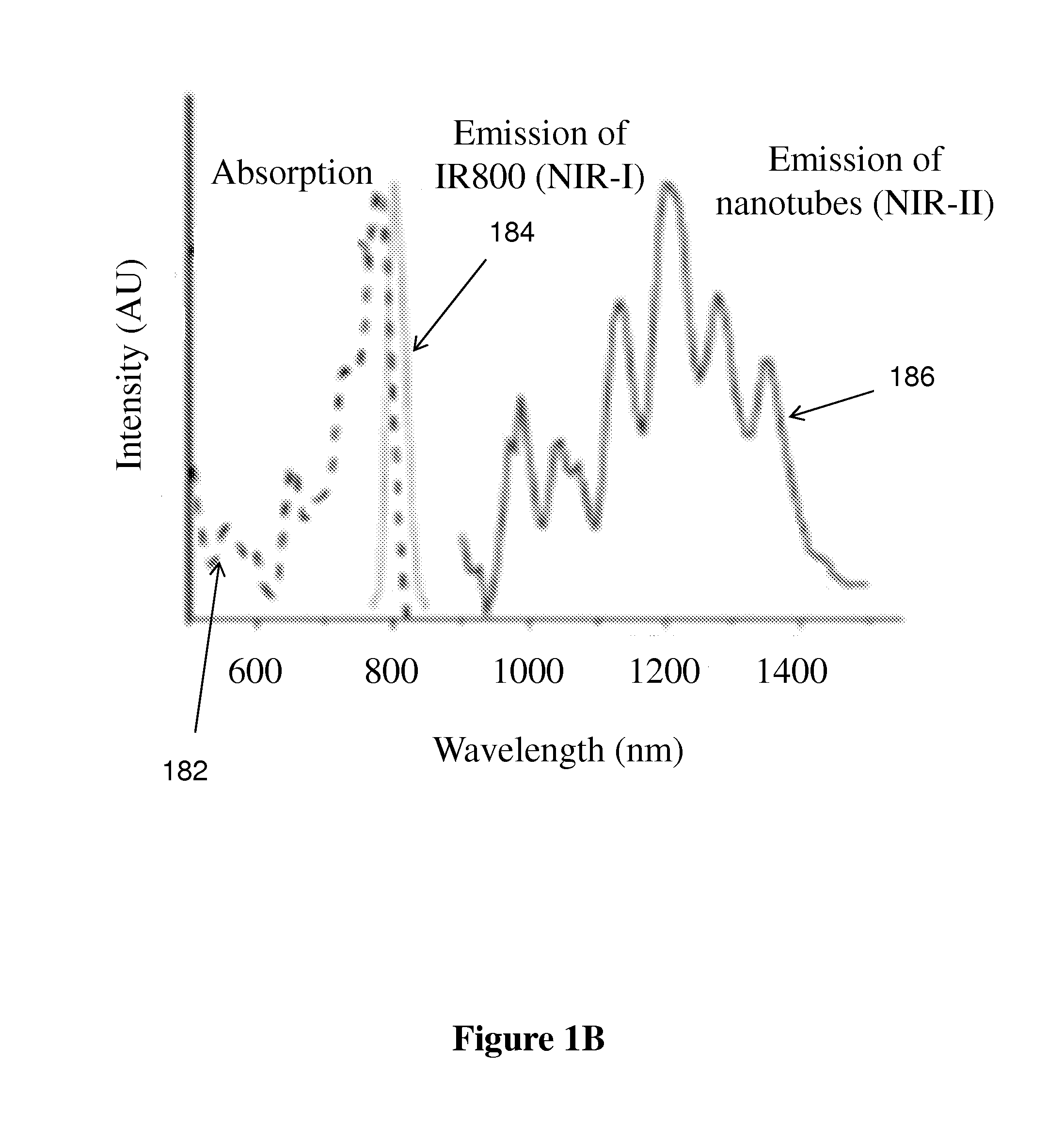
[0107]First, to glean the differences of in vivo NIR-I and NIR-II fluorescence imaging, we made biocompatible SWNT-IRDye-800 conjugates as dual-color imaging agents, where IRDye-800 (IRDye 800CW, LI-COR) was a commercial NIR-I fluorophore. High-pressure carbon monoxide conversion (HiPCO) SWNTs were stably suspended by biocompatible surfactants of 75% DSPE-mPEG (5kDa) and 25% DSPE-PEG(5kDa)-NH2 with amine groups covalently functionalized with IRDye-800 (FIGS. 1A and 5A-C). Both the SWNT and IRDye-800 label could be excited by a 785 nm laser, but exhibited different emissions. The IRDye-800 dye emitted at ˜800 nm in the NIR-I, while the SWNT emitted in the 1.1-1.4 m NIR-II region (FIG. 1B). The dual-color emission of SWNT-IRDye-800 conjugate ensured co-localization of SWNTs and IRDye-800 dyes, and enabled us to image the same tissue in two distinct spectral windows so as to evaluate the performance of photons in different wavelength...


PUM


Abstract

Disclosed are methods for imaging lumen-forming structures such as blood vessels using near-infrared fluorescence in the NIR-II region of 1000-1700 nm. The fluorescence is created by excitation of solubilized nano-structures that are delivered to the structures, such as carbon nanotubes, quantum dots or organic molecular fluorophores attached to hydrophilic polymers. These nanostructures fluoresce in the NIR-II region when illuminated through the skin and tissues. Fine anatomical vessel resolution down to −30 μm and high temporal resolution up to 5-10 frames per second is obtained for small-vessel imaging with up to 1 cm penetration depth in mouse hind limb, which compares favorably to tomographic imaging modalities such as CT and MRI with much higher spatial and temporal resolution, and compares favorably to scanning microscopic imaging techniques with much deeper penetration.

Description

STATEMENT OF GOVERNMENTAL SUPPORT[0001]This invention was made with Government support under contracts HL100397 and HL098688 awarded by the National Institutes of Health. The Government has certain rights in this invention.REFERENCE TO SEQUENCE LISTING, COMPUTER PROGRAM, OR COMPACT DISK[0002]None.BACKGROUND OF THE INVENTION[0003]1. Field of the Invention[0004]The present invention relates to the field of in vivo fluorescence-based optical imaging of lumen-forming structures such as blood vessels, bile ducts, etc. using near infrared emission in the NIR-II range and further to the use of nanostructures such as carbon nanotubes, quantum dots, etc. solubilized for in vivo use.[0005]2. Related Art[0006]Presented below is background information on certain aspects of the present invention as they may relate to technical features referred to in the detailed description, but not necessarily described in detail. The discussion below should not be construed as an admission as to the relevance...

Claims


Application Information

Patent Timeline
IPC IPC(8): A61B5/00A61B5/026A61K49/00A61B5/0275
CPCA61B5/0071A61B5/0275A61B5/0261A61B5/7278G01N21/6456A61K49/0019A61K49/0054A61K49/0093A61B5/4887A61B5/00A61B5/0075A61B5/0086A61K49/0041A61K49/0095G01N21/6428
Inventor HONG, GUOSONGLEE, JERRY CHUNG-YUHUANG, NGAN FONGCOOKE, JOHN P.DAI, HONGJIE
Owner THE BOARD OF TRUSTEES OF THE LELAND STANFORD JUNIOR UNIV
Why Eureka
Try Eureka