Hydrophilic electrospinning biological composite stent material used for tissue regeneration and preparation method and application thereof

Active Publication Date: 2016-09-29
SHANGHAI P & P BIOTECH
View PDF3 Cites 17 Cited by
  • Summary
  • Abstract
  • Description
  • Claims
  • Application Information

AI Technical Summary

Benefits of technology

[0040]According to specific embodiments of the present invention, soft tissue patches (such as pelvic floor patches, hernia patches, meninges patches, pleura patches, wound surface dressings, abdomen patches, small-caliber arteries and achilles tendons), tubular structure stents (such as arterial stents, external venous support, tracheal stents, esophageal stents, bladder, ureteral and urethral stents, and the like), ligaments, tendons and the like have been successfully prepared from the (super-) hydrophilic electrospinning filaments as mentioned above. Inducing-stent materials with respect to bones, cartilages and heart valves are being developed, and corresponding animal or human trials have been conducted. Related experiments indicate that the biological composite stent material of the present invention, after implantation in the body, may achieve the following technical effects: (1) within 1-2 weeks after implantation, the infiltration of neutrophils, monocytes and lymphocytes is

Problems solved by technology

It is difficult to obtain large quantities of products through the chemical method, and the physical methods are only limited to the preparation of L-polyactic acid nanofibers.
Therefore, the understanding of design and preparation of the electrospinning filaments and their in vivo or in vitro interaction with cells at molecular and cellular level is rather superficial.
Related studies have found that electrospinning of pure synthetic polymer has the following defects: (1) the filaments, compared with those prepared from natural polymers, lack cell recognition sites, and thus it is difficult for cells to adhere thereto; (2) the filaments inherently possess hydrophobicity and thus have poor hydrophilicity (for example, polycaprolactone (PCL) and polylactic acid-polycaprolactone (PLLA-CL) have a water contact angle of 109-120° and 109-133°, respectively), which seriously affects the adhesion of cells and subsequent cell activities; and (3) the degradation products of PLA and PGA are relatively strong acids (lactic acid or glycolic acid), and once these degradation products accumulates around a implant, delayed inflammatory reaction will appear in a few months or years.
Thus, although electrospinning of synthetic polymer has a porosity which is 1-2 orders of magnitudes higher than conventional methods (such as air bubbling method and freeze drying method) and have a very high specific surface area, the low hydrophilicity renders most pores empty, and thereby the three dimensional structure cannot be u

Method used


Image

  • Hydrophilic electrospinning biological composite stent material used for tissue regeneration and preparation method and application thereof
  • Hydrophilic electrospinning biological composite stent material used for tissue regeneration and preparation method and application thereof
  • Hydrophilic electrospinning biological composite stent material used for tissue regeneration and preparation method and application thereof

Examples


example 1

[0050]The samples in this example were divided into 3 groups:

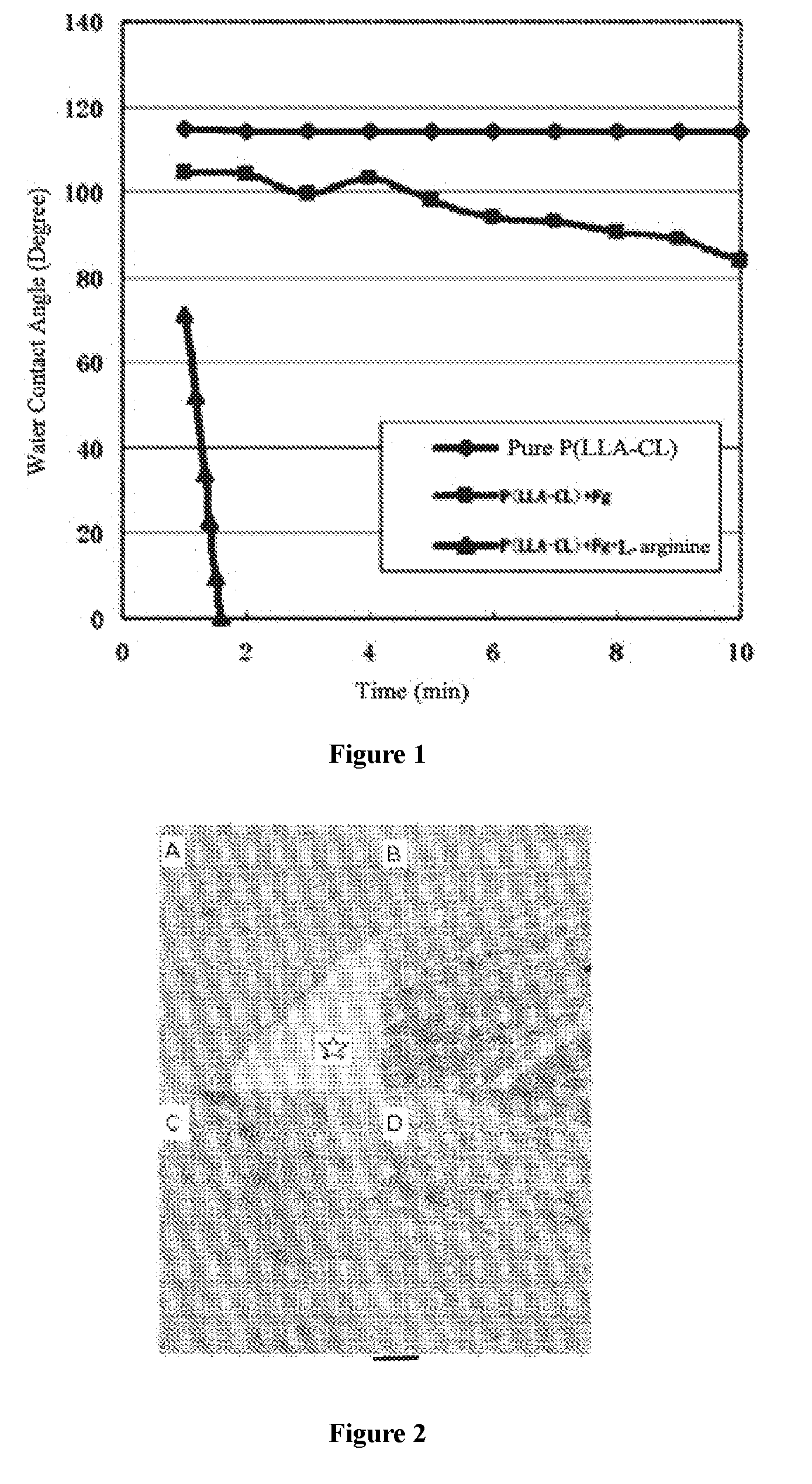
[0051](1) pure P(LLA-CL) group: 6 g P(LLA-CL) was dissolved in 100 ml hexafluoroisopropanol;

[0052](2) P(LLA-CL)+Fg group: Fg was dissolved in 20 ml saline to obtain solution 1; 6 g P(LLA-CL) was dissolved in 80 ml hexafluoroisopropanol to obtain solution 2; the solutions 1 and 2 were blended, such that there were 4g Fg and 6g P(LLA-CL) contained in 100 ml blend solution; and

[0053](3) P(LLA-CL)+Fg+protective agent group: Fg and L-arginine hydrochloride were dissolved in 20 ml saline to obtain solution 1; P(LLA-CL) was dissolved in 80 ml hexafluoroisopropanol to obtain solution 2; the solutions 1 and 2 were blended; such that there were 4 g Fg, 1 g L-arginine hydrochloride and 6 g P(LLA-CL) contained in 100 ml blend solution. The biological composite stent material was preapared by electrospinning. The parameters for an electrospinning machine were set as follows: electrospinning distance of 15 cm; electrospinning voltage of...

example 2

[0056]Bovine blood-derived Fg with different weights and 1 g L-arginine hydrochloride were dissolved in 20 ml saline to obtain solution 1, and the ratio of Fg / L-arginine hydrochloride of the solution is shown in Table 1; 6 g P(LLA-CL) was dissolved in 80 ml hexafluoroisopropanol to obtain solution 2; the solutions 1 and 2 were blended, and were divided into a total of 13 groups. An electrospinning machine from KATO TECH company (Japan) was used to prepare electrospinning membranes respectively. The parameters for the electrospinning machine were set as follows: electrospinning distance of 15 cm; electrospinning voltage of 15 KV; solution flow rate of 2 ml / h; and transverse speed of injection syringe of 10 cm / min. The thickness of the electrospinning membrane was about 200 μm.

[0057]The equilibrium contact angle of the electrospinning membranes of each group was measured by using an OCA20 optical contact angle measuring device (Germany). Results were shown in Table 1.

TABLE 1Dose-effec...

example 3

[0059]4 g Fg and 1 g L-arginine hydrochloride were dissolved in 20 ml saline to obtain solution 1; 6 g P(LLA-CL) was dissolved in 80 ml hexafluoroisopropanol to obtain solution 2; the solutions 1 and 2 were blended such that there were 4 g Fg, 1 g L-arginine hydrochloride and 6 g P(LLA-CL) contained in 100 ml blend solution; and the biological composite stent material was prepared by electrospinning. The parameters for an electrospinning machine were set as follows: electrospinning distance of 15 cm; electrospinning voltage of 15 KV; solution flow rate of 2 ml / h; and transverse speed of injection syringe of 10 cm / min. The thickness of the electrospinning membrane was 200-400 μm.

[0060]As a biological composite stent material, the prepared electrospinning membranes had a thickness of 200-400 μm, a water contact angle of less than 5° and a mechanical strength of 15-20 MPa. After sterilization using 25 KGy electron beam, the obtained material was used as swine abdominal defect patches.

[...


PUM

PropertyMeasurementUnit
Lengthaaaaaaaaaa
Lengthaaaaaaaaaa
Lengthaaaaaaaaaa

Abstract

A hydrophilic electrospinning biological composite stent material used for tissue regeneration and a preparation method and an application thereof are provided. An aqueous solution of Fibrinogen, L-arginine or hydrochloride thereof and a P(LLA-CL) solution are blended and an electrospinning technology is used to prepare the biological composite stent material. The biological composite stent material has an equilibrium contact angle that is less than 55°, is hydrophilic and has a good application prospect in repairing body tissue defects.

Description

TECHNICAL FIELD[0001]The present invention relates to a hydrophilic electrospinning biological composite stent material used for tissue regeneration and preparation method and application thereof, and specifically relates to a hydrophilic biological composite stent material prepared from fibrinogen, L-arginine (hydrochloride) and polylactic acid-polycaprolactone (P (LLA-CL)) as raw materials by using an electrospinning technique, and preparation method and application thereof in repairing body tissue defects. The present invention belongs to a field of medical health.BACKGROUND[0002]Tissue engineering is an emerging interdisciplinary which studies the tissues and organs of a biological organism or functional substitutes thereof using the principles and approaches of engineering science and bioscience. The basic principles and approaches of tissue engineering are that cells are absorbed in vivo or in vitro on a stent with good biocompatibility, which is prepared from synthetic polyme...

Claims


Application Information

Patent Timeline
IPC IPC(8): A61L31/04D01F1/10D01F6/62A61L31/16D01F6/84
CPCA61L31/046A61L31/041A61L31/16D01F6/84D01F6/625D01F1/10D10B2401/022A61L2430/06A61L2430/10A61L2430/22D01D5/0007D10B2509/06A61L2300/404C08L89/00C08L67/04
Inventor HE, HONGBINGSU, LINGLIU, ZHANGYANG, LI
Owner SHANGHAI P & P BIOTECH
Why Eureka
Try Eureka