
Biomimetic support for three-dimensional cell culturing, method for manufacturing same, and use thereof

a three-dimensional cell and biomimetic technology, applied in the field of biomimetic support for three-dimensional cell culturing, can solve the problems of difficult observation of interactions between different cell types, inability to fully mirror the cellular environment of living tissues in 2d co-culture, and many differences between two-dimensional and body tissue environment of cells, etc., to achieve excellent biomimetic function, improve 3d cell culture efficiency, and improve cell adhesion ability. , the effect of cell viability

Active Publication Date: 2017-07-20
PUSAN NAT UNIV IND UNIV COOPERATION FOUND
View PDF1 Cites 0 Cited by
  • Summary
  • Abstract
  • Description
  • Claims
  • Application Information

AI Technical Summary

Benefits of technology

The present invention relates to a fish collagen / synthetic polymer nanofiber scaffold and an agarose-collagen-alginate composite hydrogel support that can provide a better mechanical stability and a cell culture environment more similar to a living body which can improve 3D cell culture efficiency and biomimetic function. These supports can be used as a 3D cell culture model for research in drug sensitivity test, cancer cell formation, growth, metastasis and death, tissue engineering, and regenerative medicine. The composite hydrogel support is high transparency, easy observation of cells without cell staining during culture, and uses marine-derived collagen which has high cell affinity, biocompatibility, and low cytotoxicity. The biomimetic cell culture method using these supports can be used for biological activity analysis and cancer research. The patent aims to provide safe and efficient cell culture methods and evaluate the susceptibility of cells to anticancer drugs or develop therapeutic agents with excellent bioavailability.

Problems solved by technology

However, since a monolayer cell culture method of culturing on the 2D surface cannot accurately reflect the biotissue environment of the three-dimensionally grown cells attached to the extracellular matrix, there are many differences between two-dimensional and the body tissue environment of the cell.
In addition, because the same cell type is generally cultured in the conventional 2D cell culture, it is difficult to observe interactions between different cell types.
Although some attempts have been made to solve these problems partially through 2D co-culture, this 2D co-culture cannot fully mirror the cellular environment in living tissues.
However, to date, methods for the production of scaffolds most suitable for regenerating living tissues are not yet known.

Method used


Image

  • Biomimetic support for three-dimensional cell culturing, method for manufacturing same, and use thereof
  • Biomimetic support for three-dimensional cell culturing, method for manufacturing same, and use thereof
  • Biomimetic support for three-dimensional cell culturing, method for manufacturing same, and use thereof

Examples


example 2

Analysis of Cell Adhesion of PCL and Fish Collagen / PCL Nanofiber Support

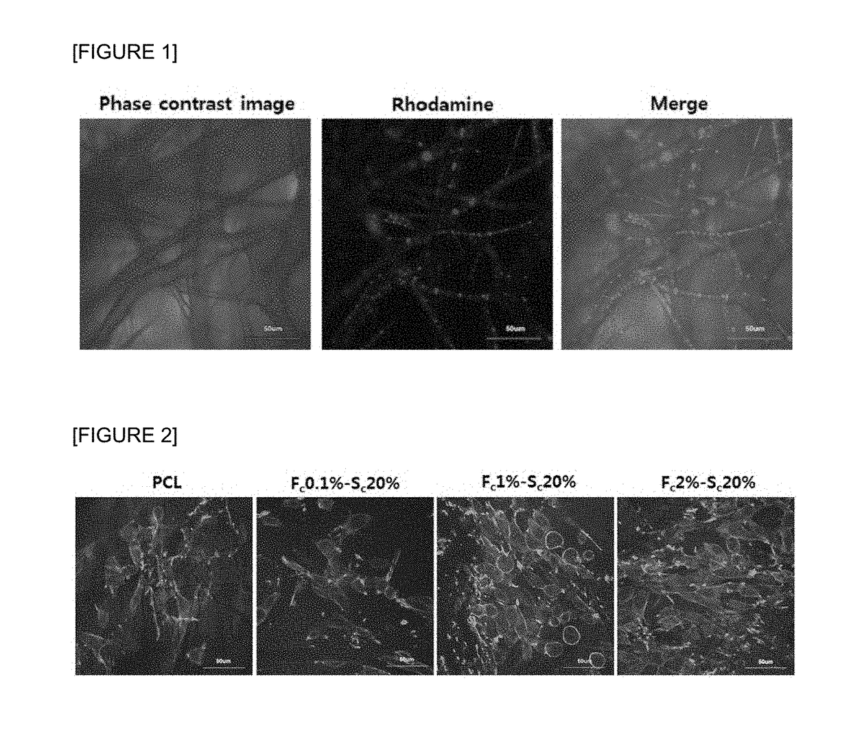
[0135]2-1. Cell Culture Using PCL and Fish Collagen / PCL Nanofiber Support

[0136]Thymic deep cortex or cortical reticular epithelial cell (hereafter, referred to as CREC), a type of normal mouse thymic epithelial cell (TEC) provided from Dr. Barbara B. Knowles (The Jackson Laboratory, Bar Harbor, Me., USA) was cultured and incubated in Dulbecco's Modified Eagle Medium (DMEM, HyClone, Logan, Utah, USA) containing 10% (v / v) fetal bovine serum (FBS; Gibco, Invitrogen), 100 IU / mL penicillin (Gibco, USA) and 100 μg / mL streptomycin (Gibco, Invitrogen) at 37° C. in a 5% CO2 atmosphere. The culture medium was replaced with new one every 2-3 days.

[0137]The PCL nanofiber support prepared in Example 1 and the fish collagen / PCL nanofiber support having final concentrations of 0.1, 1 and 2 wt % were placed in 96-well plates respectively and CREC cells were inoculated at 5×10⁴ cells per a well and cultured for 6 hours.

[0138]2-2...

example 3

Analysis of Optimized Concentration of Fish Collagen

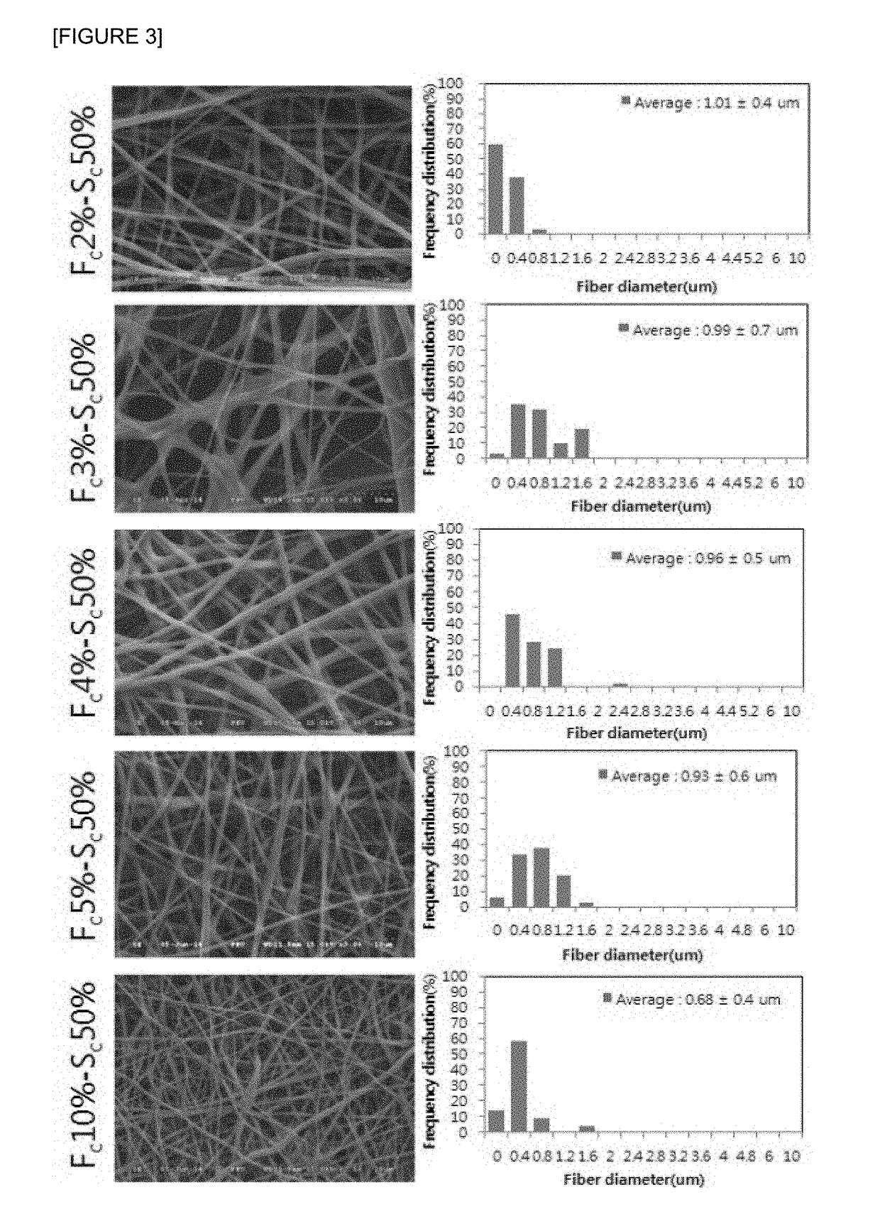
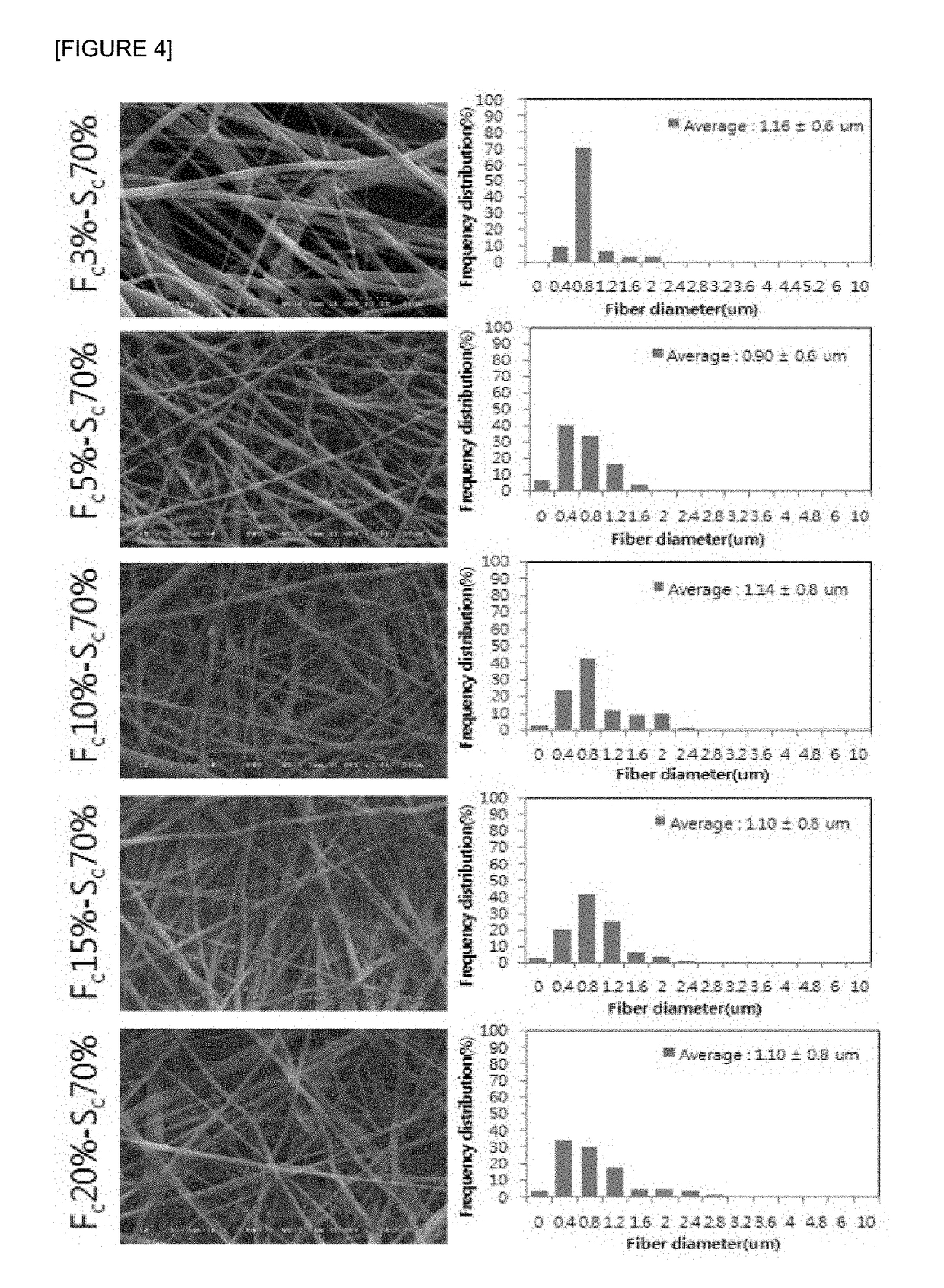
[0143]3-1. Production of Fish Collagen / PCL Nanofiber Support of a Concentration of 2 wt % or More

[0144]Fish collagen was added distilled water to prepare fish collagen stock solutions (Sc) having the concentrations of Sc 10 wt %, Sc 20 wt %, Sc 30 wt % Sc 40 wt % Sc 50 wt % Sc 60 wt % Sc 70 wt % Sc 80 wt % Sc 90 wt % and Sc 100 wt %, and were named as Sc 10%, Sc 20%, Sc 30% Sc 40% Sc 50% Sc 60% Sc 70% Sc 80% Sc 90% and Sc 100%. In the case of Sc 100%, the PCL cannot be included at all, so it is excluded from the selection condition (marked as X in Table 1), and the concentration of at least Sc 80% is excluded from the selection condition because the viscosity is too high to add the accurate content (marked as X in Table 1). In addition, in the case of high concentration of at least Sc 50%, Sc 60% was also excluded from the selection condition because there is no particular difference with Sc 50% or Sc 70%.

[0145]Accordingly, the fis...

example 4

Three-Dimensional Culture Characterization of Solid Cancer Cells According to Fish Collagen Concentration in Fish Collagen / PCL Nano / Micro Hybrid Fiber Support

[0156]4-1. Cell and Cell Culture

[0157]A human non-small cell lung cancer cells (NCI-H1703) were purchased from the American Type Culture Collection (ATCC, Manassas, Va., USA) and a human prostate cancer cell -145 were purchased from Korean Cell Line Bank. NCI-H1703 and DU-145 cell lines were cultured and maintained in RPMI-1640 medium (HyClone, Logan, Utah, USA) containing 10% (v / v) fetal bovine serum (FBS; Gibco, Invitrogen), 100 IU / mL penicillin (Gibco, Invitrogen) and 100 ug / mL streptomycin (Gibco, Invitrogen) at 37° C. under atmospheric conditions containing 5% CO2. The culture medium was changed with new one every 2-3 days.

[0158]4-2. Three-Dimensional Cell Culture Using Fish Collagen / PCL Nanofiber Support

[0159]In order to compare three-dimensional culture characteristics of fish collagen / PCL nanofiber support prepared acco...


PUM

PropertyMeasurementUnit
temperatureaaaaaaaaaa
thicknessaaaaaaaaaa

Abstract

Disclosed is a method for manufacturing a composite nanofiber support and a fish collagen / synthetic polymer nanofiber support manufactured by the method, wherein the method comprises: a first step for dissolving a synthetic polymer in an organic solvent; a second step for dissolving a fish collagen in water to prepare an aqueous fish collagen solution; a third step for adding the aqueous fish collagen solution prepared in the second step to the synthetic polymer solution prepared in the first step, followed by mixing; and a fourth step for electrospinning the mixture solution prepared in the third step to manufacture a nanofiber support.

Description

TECHNICAL FIELD[0001]The present invention relates to a method of preparing nanofiber scaffolds prepared by electrospinning in a mixture solution comprising hydrophilic natural polymers of fish collagen and hydrophobic synthetic polymers, as active ingredients and a use thereof, and a method of biomimetic three-dimensional cell culture using a hydrogel support comprising agarose, collagen and alginate, as active ingredients and a use thereof.BACKGROUND ART[0002]A tissue engineering has recently attracted an attention as a convergence technology which is intended to be used for clinical treatment by maintaining, enhancing, restoring or replacing vital functions through a combinatory use of cells, engineering materials and appropriate physiological / biochemical factors etc.[0003]The most fundamental technique of this tissue engineering is to attach cells separated from a biological tissue to porous scaffolds made of a biodegradable polymer and to transplant them in vivo or to culture t...

Claims


Application Information

Patent Timeline
Patent Type & Authority Applications(United States)
IPC IPC(8): C12N5/00G01N33/50
CPCC12N5/0062C12N2501/90C12N2501/998G01N33/5011C12M25/02C12M25/14C12N2533/54C12N2533/74C12N2533/76G01N33/54366
Inventor YOON, SIKKWAK, JONG-YOUNGJIN, SONG-WANJEONG, YOUNG HUNKIM, CHANG GUNCHOI, DA JEONGKANG, HAE YEONGFAZLI, SUBHANIKRAM, MUHAMAD
Owner PUSAN NAT UNIV IND UNIV COOPERATION FOUND
Features
  • Generate Ideas
  • Intellectual Property
  • Life Sciences
  • Materials
  • Tech Scout
  • Unparalleled Data Quality
  • Higher Quality Content
  • 60% Fewer Hallucinations
Learn More